J Korean Soc Radiol 2016;74(5):339-343 http://dx.doi.org/10.3348/jksr.2016.74.5.339
INTRODUCTION
Uterine artery embolization (UAE) as a method for control- ling postpartum hemorrhage was first introduced by Brown in 1979. Since then, the technique has become more widespread.
Several literatures have shown a high clinical success rate of UAE with a low complication rate, making it a valuable treatment op- tion for control of severe postpartum hemorrhage (1, 2). How- ever, failure of UAE may occur due to various causes. One of the important causes is the additional blood supply to the uterus via collaterals such as ovarian arteries (3). Thus, for performing suc- cessful UAE, it is essential that the operator should be aware of the anatomy and the variations of the ovarian artery.
Ovarian arteries usually originate directly from the anterior aspect of the aorta at the level of renal arteries or just below the
level of renal arteries (3). Anatomic variations of ovarian arter- ies are not rare. The most common anatomic variation is an ab- errant ovarian artery arising from the renal artery (4). In this report, we present a very rare case of an aberrant ovarian artery originating from the iliolumbar artery.
CASE REPORT
A 30-year-old multipara gave birth to a girl child weighing 3160 g at 38 weeks of gestation in a local hospital. After one hour, she was admitted to our emergency department with intracta- ble primary postpartum hemorrhage. Her vital signs were sta- ble, but her hematocrit value and hemoglobin level were below the normal range; they were 31% and 10.7 g/dL, respectively. She had no past medical history. Clinically, uterine atony was sus-
Aberrant Ovarian Artery Originating from the Iliolumbar Artery:
A Case Report
난소동맥의 엉덩허리동맥으로부터의 이상 기시: 증례 보고
Ji Eun Lee, MD, Jae Myeong Lee, MD*
Department of Radiology, Soonchunhyang University College of Medicine, Bucheon Hospital, Bucheon, Korea
Here, we report a case of a 30-year-old woman who presented with primary post- partum hemorrhage due to uterine atony. She received uterine artery embolization (UAE). During left internal iliac arteriography, an aberrant left ovarian artery origi- nating from the left iliolumbar artery was visualized. The aberrant left ovarian artery was connected to the left uterine artery via prominent collateral vessels. It supplied a significant amount of blood to the fundus of the uterus. Bilateral hypertrophied uterine arteries were embolized very carefully so that the embolic material did not reflux into the aberrant left ovarian artery. After the procedure, her vaginal bleeding was successfully controlled. Accurate understanding of anatomical variations of the ovarian artery is essential to avoid failure in controlling postpartum hemorrhage with UAE.
Index terms Ovary Uterine Artery
Postpartum Hemorrhage Uterine Artery Embolization
Received June 11, 2015 Revised October 6, 2015 Accepted January 28, 2016
*Corresponding author: Jae Myeong Lee, MD Department of Radiology, Bucheon Hospital, Soonchunhyang University College of Medicine, 170 Jomaru-ro, Wonmi-gu, Bucheon 14584, Korea.
Tel. 82-32-621-5851 Fax. 82-32-621-5874 E-mail: [email protected]
This is an Open Access article distributed under the terms of the Creative Commons Attribution Non-Commercial License (http://creativecommons.org/licenses/by-nc/3.0) which permits unrestricted non-commercial use, distri- bution, and reproduction in any medium, provided the original work is properly cited.
pected. After consultation with the emergency department and the gynecology department, UAE was considered. Embolization was performed via a single right femoral artery approach with a 5-Fr sheath (Radifocus; Terumo, Tokyo, Japan). First, selective angiography of the left internal iliac artery was performed with 5-Fr Cobra catheter (Cook, Bloomington, IN, USA). An hyper- trophied left uterine artery providing profound blood supply to the uterus body was visualized. From the left iliolumbar artery (the first branch originating from the posterior trunk of the in- ternal iliac artery), a small tortuous vessel with sinus course was opacified (Fig. 1A). Based on the typical tortuous appearance and characteristic ovarian blush, this vessel was identified as the aberrant left ovarian artery originating from the left iliolumbar artery. This aberrant ovarian artery showed communication with the left uterine artery. It provided rich perfusion to the en- larged uterine fundus (Fig. 1B). Selective angiography and care-
ful embolization of the left uterine artery was performed with PVA (355–500 µm, Contour; Boston-Scientific, Natick, MA, USA). We were cautious so that the embolic material did not re- flux into the aberrant left ovarian artery. After successful embo- lization of the left uterine artery, selective angiography of the aberrant left ovarian artery was performed using a 2.2-Fr mi- crocatheter (Progreat, Terumo, Tokyo, Japan). Persistent ovarian blush was observed without visualization of any uterine artery branch (Fig. 2). Selective angiography of the right internal iliac artery revealed a hypertrophied right uterine artery. However, no aberrant ovarian artery was seen. The right uterine artery was also embolized with PVA (355–500 µm, Contour; Boston- Scientific, Natick, MA, USA). After successful embolization of both uterine arteries, flush aortography was performed with a 5-Fr pigtail catheter (Cook) positioned just above the level of both renal arteries. There was no evidence of other aberrant
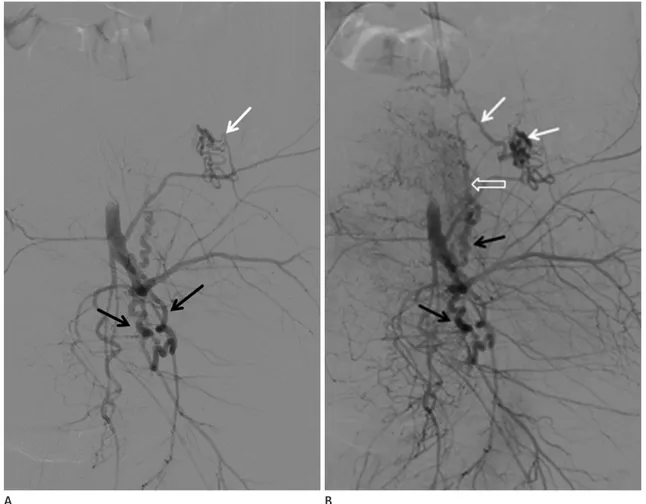
Fig. 1. Left internal iliac arteriogram shows hypertrophied left uterine arteries (black arrows) and an aberrant left ovarian artery originating from the left iliolumbar artery (white arrow) (A). This aberrant ovarian artery (white arrows) shows communication (open arrow) with hypertrophied left uterine arteries (black arrows) and provides prominent blood supply to the uterine fundus (B). There is no definite extravasation of contrast material into the uterine cavity in this arteriogram.
A B
ovarian arteries. The right ovarian artery was visualized and it originated from the typical position on the ventral aspect of the abdominal aorta close to the origin of the inferior mesenteric artery. It showed the characteristic tortuous course with the typi- cal ovarian blush. There was no evidence of blood flow from the right ovarian artery to the uterus. After the procedure, her vagi- nal bleeding was successfully controlled. The patient was dis- charged from our institution a few days later after full recovery.
DISCUSSION
Postpartum hemorrhage is one of the most important causes of maternal mortality, despite the progress in medical and surgi- cal treatment (5). Occasionally, life-threatening obstetric com- plication can occur shortly (several hours to days) after the de- livery. Primary postpartum hemorrhage is generally defined as more than 500 mL of vaginal bleeding during the first 24 hours after delivery. The most common cause is uterine atony as ob- served in our case, followed by lower genital tract injury, placen- tal retention, abnormal placentation, or coagulopathy (5). Treat- ment options for controlling postpartum bleeding include con-
servative treatment such as vaginal packing and administration of uterotonic drugs. With respect to intractable postpartum hem- orrhage, more invasive procedures such as vascular ligation or hysterectomy can be performed (5). The advantages of UAE over surgical treatment include less invasiveness, potential to preserve the uterus and fertility, and an overall shorter hospitalization period. Thus, in recent years, UAE has become one of the major alternative treatments for controlling postpartum hemorrhage.
In spite of its many merits, clinical failure of UAE is an issue that must be confronted. One of the major causes is the presence of blood supply to the uterus by ovarian arteries (3). This em- phasizes the importance of being aware of the normal and ana- tomic variation in the origin of ovarian arteries. The ovarian ar- teries normally originate directly from the anterior circumfer- ence of the aorta at the level of the renal artery or just below the level of the renal artery (3). The ovarian artery descends into the pelvic cavity up to the level of the ovarian hilum, where it pro- vides blood supply to the ipsilateral ovary and fallopian tube. It forms an anastomosis with the corresponding distal branches of the uterine arteries (6). Angiographically, normal ovarian ar- teries appear as tortuous vessels (corkscrew appearance) with a small caliber and the typical ovarian blush (6).
Variations in the origin of ovarian arteries are common. In 6–12% of the cases, they arise from the renal artery, more com- monly from the right renal artery (3). When accessory renal ar- teries are present, the ovarian arteries tend to arise more com- monly from these arteries rather than from the aorta (7). Few case reports have demonstrated more rare locations of the aber- rant ovarian artery origin, including the proximal portion of the common iliac artery, external and internal iliac artery, and infe- rior mesenteric artery (4, 7-9). To the best of our knowledge, this is the first report of an aberrant ovarian artery originating from the iliolumbar artery, which is the first branch of the posterior trunk of the internal iliac artery.
In addition to gaining knowledge of the vascular anatomy of the ovarian artery, it is important to avoid unwanted emboliza- tion of the ovarian artery. Ovarian artery embolization is associ- ated with increased risk of ovarian failure with symptoms such as hot flashes and amenorrhea (10). However, postpartum hem- orrhage is one of the most dramatic and acute hemostatic dis- orders with high maternal mortality. Thus, if angiography shows a hypertrophied ovarian artery providing profound blood sup- Fig. 2. Selective angiography of the aberrant left ovarian artery (black
arrows). Left ovarian staining is demonstrated (white arrow) without visualization of the uterine artery branch.
ply to the enlarged uterus, the issue of fertility becomes second- ary. In previously published case reports, all aberrant ovarian arteries (4, 7-9) were embolized. They supplied a significant amount of blood to either uterine fibroids or the enlarged uter- us itself.
In our case, embolization of the left uterine artery was per- formed first, taking care careful that the PVA particles did not enter the ovarian circulation via anastomotic channels. Then, selective angiography of the aberrant left ovarian artery was per- formed, and it showed no evidence of persistent vascular flow from the aberrant ovarian artery to the left uterine artery branch- es. Thus, unlike the previously published case reports (4, 7-9) embolization of the ovarian artery was not necessary, and the left ovarian blood supply could be preserved.
In conclusion, here, we report a rare case of an aberrant left ovarian artery originating from the left iliolumbar artery. Knowl- edge of main anatomical variations in the origin of ovarian ar- teries is essential because ovarian arterial supply to the uterus could be a major cause of UAE failure.
REFERENCES
1. Ganguli S, Stecker MS, Pyne D, Baum RA, Fan CM. Uterine artery embolization in the treatment of postpartum uter- ine hemorrhage. J Vasc Interv Radiol 2011;22:169-176 2. Kirby JM, Kachura JR, Rajan DK, Sniderman KW, Simons ME,
Windrim RC, et al. Arterial embolization for primary post- partum hemorrhage. J Vasc Interv Radiol 2009;20:1036- 1045
3. Matson M, Nicholson A, Belli AM. Anastomoses of the ovar- ian and uterine arteries: a potential pitfall and cause of
failure of uterine embolization. Cardiovasc Intervent Radiol 2000;23:393-396
4. Smoger DL, Kancherla V, Shlansky-Goldberg RD. Uterine fundal blood supply from an aberrant left ovarian artery originating from the inferior mesenteric artery: implications for uterine artery embolization. J Vasc Interv Radiol 2010;
21:941-944
5. Deux JF, Bazot M, Le Blanche AF, Tassart M, Khalil A, Ber- kane N, et al. Is selective embolization of uterine arteries a safe alternative to hysterectomy in patients with postpar- tum hemorrhage? AJR Am J Roentgenol 2001;177:145-149 6. Pelage JP, Walker WJ, Le Dref O, Rymer R. Ovarian artery: an- giographic appearance, embolization and relevance to uter- ine fibroid embolization. Cardiovasc Intervent Radiol 2003;
26:227-233
7. Kwon JH, Kim MD, Lee KH, Lee M, Lee MS, Won JY, et al.
Aberrant ovarian collateral originating from external iliac artery during uterine artery embolization. Cardiovasc Inter- vent Radiol 2013;36:269-271
8. Kim WK, Yang SB, Goo DE, Kim YJ, Chang YW, Lee JM. Ab- errant ovarian artery arising from the common iliac artery:
case report. Korean J Radiol 2013;14:91-93
9. Reed RA, McLucas B. Aberrant right ovarian artery from proximal internal iliac artery in uterine artery embolization patient. Open J Radiology 2012;2:117-119
10. Wang MQ, Liu FY, Duan F, Wang ZJ, Song P, Song L. Ovarian artery embolization supplementing hypogastric-uterine ar- tery embolization for control of severe postpartum hemor- rhage: report of eight cases. J Vasc Interv Radiol 2009;20:
971-976
난소동맥의 엉덩허리동맥으로부터의 이상 기시: 증례 보고
이지은 · 이재명*
분만 후 자궁이완증으로 발생한 일차성 산후출혈로 30세 여자 환자가 내원하였다. 자궁동맥색전술을 위해 시행한 좌속엉 덩동맥조영술에서 좌엉덩허리동맥으로부터 이상 기시하는 좌난소동맥의 증례를 경험하였다. 이 좌난소동맥은 측부혈관을 통해 좌자궁동맥과 연결되어 있었고 자궁저로 많은 양의 혈류를 공급하고 있었다. 저자들은 색전물질이 난소동맥으로 역 류되지 않도록 주의하며 양측 자궁동맥에 대한 혈관색전술을 시행하였고 환자는 보존적 치료 후 합병증 없이 회복되었다.
자궁동맥색전술은 산후출혈의 치료로서 보편적으로 이용되고 있으며, 성공적인 치료를 위해 측부혈관 중 하나인 난소동맥 으로부터의 곁순환 유무을 확인하고 해부학적 변이를 이해하는 것이 중요하다.
순천향대학교 의과대학 부천병원 영상의학과