91
Korean J Radiol 14(1), Jan/Feb 2013
kjronline.org
INTRODUCTION
Uterine artery embolization (UAE) is a safe and effective procedure for the control of postpartum hemorrhage (1).
Detailed knowledge of uterine vascularization and ovarian artery supply to the uterus is necessary for the management of postpartum hemorrhage. The ovarian arteries usually arise from the anterior circumference of the abdominal aorta at the level of the renal arteries (2). Anatomic variations of the ovarian arteries are relatively common (2), and awareness regarding these variants is essential for
Aberrant Ovarian Artery Arising from the Common Iliac Artery: Case Report
Won Kyung Kim, MD
1, Seung Boo Yang, MD
2, Dong Erk Goo, MD
3, Yong Jae Kim, MD
3, Yun-Woo Chang, MD
3, Jae Myeong Lee, MD
21Department of Radiology, Soonchunhyang University Bucheon Hospital, Soonchunhyang University College of Medicine, Bucheon 420-767, Korea;
2Department of Radiology, Soonchunhyang University Gumi Hospital, Soonchunhyang University College of Medicine, Gumi 730-904, Korea;
3Department of Radiology, Soonchunhyang University Seoul Hospital, Soonchunhyang University College of Medicine, Seoul 140-743, Korea
A 46-year-old Vietnamese woman received embolization therapy in order to control postpartum hemorrhage. Angiography revealed an aberrant ovarian artery arising from the right common iliac artery. Superselective catheterization and subsequent embolization of the aberrant ovarian artery and bilateral uterine arteries were performed. Precise knowledge of the anatomic variations of the ovarian artery is important for successful embolization.
Index terms: Aberrant ovarian artery; Angiography; Uterine artery angiography; Interventional procedure; Postpartum hemorrhage
Received October 30, 2011; accepted after revision February 1, 2012.
Corresponding author: Seung Boo Yang, MD, Department of Radiology, Soonchunhyang University Gumi Hospital, Soonchunhyang University College of Medicine, 250 Gongdan- dong, Gumi 730-904, Korea.
• Tel: (8254) 468-9391 • Fax: (8254) 464-9300
• E-mail: [email protected]
This is an Open Access article distributed under the terms of the Creative Commons Attribution Non-Commercial License (http://creativecommons.org/licenses/by-nc/3.0) which permits unrestricted non-commercial use, distribution, and reproduction in any medium, provided the original work is properly cited.
Case Report
Korean J Radiol 2013;14(1):91-93
successful UAE. Few studies have reported the abnormal origin or course of ovarian arteries (2, 3). Here, we present a very rare case of an aberrant ovarian artery arising from the common iliac artery.
CASE REPORT
A 46-year-old pregnant woman at 31 weeks gestation was referred to the gynecology department of our hospital for premature membrane rupture with uterine atony and placenta accreta. She had no significant past medical history, except a previous caesarean section. After an emergency caesarean section, the patient presented severe postpartum hemorrhage secondary to uterine atony and was referred to our department (interventional radiology).
Embolization was performed using the single femoral artery approach with insertion of a 5-Fr introducer sheath (Terumo, Tokyo, Japan). At the beginning of the procedure, flush aortography (Fig. 1A) with a 5-Fr pigtail catheter (Cook, Bloomington, IN) positioned at the level of the celiac trunk showed a single aberrant artery arising from the right common iliac artery. Selective angiography of
http://dx.doi.org/10.3348/kjr.2013.14.1.91pISSN 1229-6929 · eISSN 2005-8330
Korean J Radiol 14(1), Jan/Feb 2013
kjronline.org 92
Kim et al.
the aberrant artery using a 5-Fr Yashiro catheter (Terumo, Tokyo, Japan) revealed that it had a sinuous course and was tortuous (Fig. 1B). This artery showed the typical ovarian blush and abundant collateral perfusion to the markedly enlarged uterine fundus (Fig. 1C).
Both ovarian arteries could not be observed on pelvic aortography, and anastomosis with the uterine artery was not clearly defined. The diameter of the aberrant artery was larger than the diameters of both uterine arteries.
Selective angiography of both the internal iliac arteries and both uterine arteries showed markedly increased uterus vascularity. The aberrant ovarian artery (Fig. 1D) and both uterine arteries (Fig. 1E, F) were embolized with 1 x 1 mm gelfoam pledgets (Johnson & Johnson, Skipton, UK) by using a 2.7-Fr microcatheter (Progreat®, Terumo,
Tokyo, Japan). Vaginal bleeding ceased after this successful embolization procedure.
Three days after embolization, the patient underwent a contrast-enhanced abdominal-pelvic computed tomography (CT) scan, and no associated unusual findings such as anomalies of the kidney or renal arteries were noted.
The patient’s condition rapidly stabilized after the embolization procedure, and she was discharged in a good condition.
DISCUSSION
Anatomic variations in the origin of ovarian arteries are relatively common (2), and awareness regarding these variants is essential to ensure successful UAE. Few cases
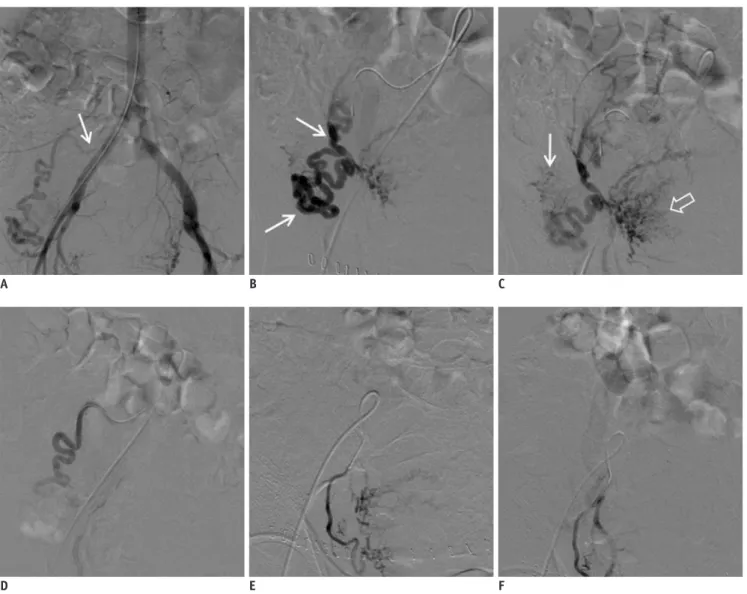
A B C
Fig. 1. Images of 46-year-old female patient with postpartum hemorrhage.
Flush aortography (A) showing aberrant ovarian artery arising from right common iliac artery (arrow). Selective angiography (B) showing that artery has sinuous course and is tortuous (arrows). This artery shows typical ovarian blush (arrow) and abundant collateral perfusion to markedly enlarged uterine fundus (open arrow) (C). Embolization of aberrant ovarian artery demonstrating occlusion of uterine branches of artery (D).
Selective angiography of right uterine artery (E) and post-embolization state of this artery (F).
D E F
Korean J Radiol 14(1), Jan/Feb 2013
kjronline.org 93
Aberrant Ovarian Artery Arising from Common Iliac Artery
REFERENCES
1. Gonsalves M, Belli A. The role of interventional radiology in obstetric hemorrhage. Cardiovasc Intervent Radiol 2010;33:887-895
2. Smoger DL, Kancherla V, Shlansky-Goldberg RD. Uterine fundal blood supply from an aberrant left ovarian artery originating from the inferior mesenteric artery: implications for uterine artery embolization. J Vasc Interv Radiol 2010;21:941-944 3. Bensalah J, Dumousset E, Niro J, Cassagnes L, Ravel A, Gallot
D, et al. Aberrant ovarian and uterine feeding from the renal artery at the end of gestation: two cases. J Vasc Interv Radiol 2010;21:1911-1912
4. Ambekar A, Vogelzang RL. Aberrant uterine artery as a cause of uterine artery embolization treatment failure. Int J Gynaecol Obstet 2001;74:59-60
5. Horton AW, Patel U, Belli AM. An unusual arterial supply to the uterus. A case report and review of anatomy-implications for uterine artery embolization. Clin Radiol 2010;65:1038- 1042
6. Wi JY, Kim HC, Chung JW, Jun JK, Jae HJ, Park JH.
Importance of angiographic visualization of round ligament arteries in women evaluated for intractable vaginal bleeding after uterine artery embolization. J Vasc Interv Radiol 2009;20:1031-1035