J Korean Soc Radiol 2017;76(6):420-424 https://doi.org/10.3348/jksr.2017.76.6.420
INTRODUCTION
Developmental venous anomaly (DVA) is a common congen- ital malformation characterized by dilated medullary veins (1, 2) configured in the caput medusa and a draining vein. The in- cidence of DVAs is bigh, but they are benign anatomic variations and rarely display symptoms. Here, we report findings by com- puted tomography (CT) and magnetic resonance imaging (MRI) with perfusion images of acute infarction from underlying DVA in a 63-year-old female patient who presented with acute onset of neurologic symptoms and recovered without any deficit re- maining.
DVAs are quite common with a reported incidence between 0.7 and 2.56% in the general population (3) but unlike other con- genital malformations, they are rarely symptomatic and in most cases diagnosed by chance. The reasons why they rarely cause
symptoms can be found ubeither mechanical compression to ad- jacent intracranial structures or misbalance of the blood flow (4).
Venous infarction resulting from DVA is infrequent and all of the reported cases were associated with thrombosis in the draining vein (5, 6). Here, we present CT and MR findings with perfusion images of a venous infarction case with DVA.
CASE REPORT
A 63-year-old woman showing up in an emergency room com- plained about an abrupt onset of right side motor weakness and sensory changes. These symptoms were followed by dysarthria the day before her visit. However, all the symptoms disappeared spontaneously by the time she arrived at the emergency room and at the time of the initial physical examination, which included motor and sensory grades andwere within normal limits.
Venous Infarction of Developmental Venous Anomaly:
A Case Report with Perfusion Imaging
대뇌정맥발달기형의 정맥경색: 관류영상소견을 포함한 증례 보고
Jung Youn Kim, MD
1, Hye Jeong Kim, MD
1*, Eun Soo Kim, MD
2, Su-Jeong Hyun, MD
1, Hee Yeong Kim, MD
1, Han Myun Kim, MD
1, Ji-Young Hwang, MD
1, Hye Suk Hong, MD
1, Ji Young Woo, MD
1, Ik Yang, MD
11Department of Radiology, Kangnam Sacred Heart Hospital, Hallym University College of Medicine, Seoul, Korea
2Department of Radiology, Hallym University Sacred Heart Hospital, Hallym University College of Medicine, Anyang, Korea
Developmental venous anomaly (DVA) is a common congenital venous malformation characterized by dilated medullary veins in caput medusa configuration and a drain- ing vein. Despite the high incidence of DVAs, they are benign anatomic variations and rarely cause symptoms. Here, we report computed tomography and magnetic reso- nance imaging findings with perfusion images of acute infarction from underlying DVA in a 63-year-old female patient who presented with acute onset of neurologic symptoms and recovered without any neurologic deficit.
Index terms
Central Nervous System Venous Angioma Infarction
Magnetic Resonance Imaging Perfusion Imaging
Received June 16, 2016 Revised August 26, 2016 Accepted September 24, 2016
*Corresponding author: Hye Jeong Kim, MD Department of Radiology, Kangnam Sacred Heart Hospital, Hallym University College of Medicine, 1 Singil-ro, Yeongdeungpo-gu, Seoul 07441, Korea.
Tel. 82-2-829-5241 Fax. 82-2-832-1845 E-mail: [email protected]
This is an Open Access article distributed under the terms of the Creative Commons Attribution Non-Commercial License (http://creativecommons.org/licenses/by-nc/4.0) which permits unrestricted non-commercial use, distri- bution, and reproduction in any medium, provided the original work is properly cited.
She was in good health prior to the event and had no past medical history. Neither was there anything noteworthy about her family history. Further evaluation included a conventional echocardiogram. The following additional laboratory studies were either negative or within normal range: antinuclear and anticardiolin antibodies, antithrombin III, factor V Leiden mu- tation, protein C, protein S, activated protein C resistance, and homocysteine.
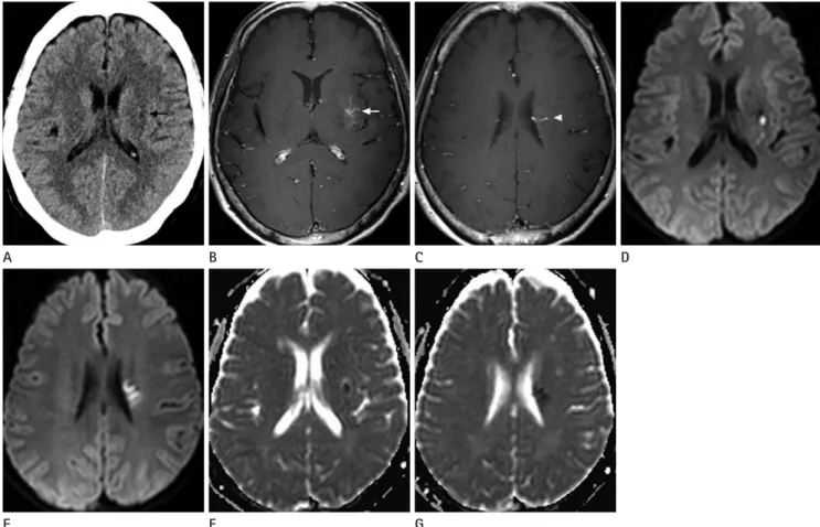
Initial ordinary CT images of the brain displayed a subtle hy- podense area in the left basal ganglia, but no mass effect or acute hemorrhagic focus was identified (Fig. 1A).
On the following day, MRI was conducted and gadolinium- enhanced T1-weighted images showed growing medullary veins at the left putamen in a characteristic ‘caput medusa’ configura- tion with a collector vein draining to the surface of the left lateral
ventricle, which confirmed the DVA diagnosis (Fig. 1B, C). This location corresponded to the hypodense area noted on the CT image. Diffusion weighted images (DWI) revealed the presence of an area of bright signal intensity (SI) in the left basal ganglia and periventricular white matter nearby the draining vein, which demonstrated decreased SI on an diffusion coefficient (ADC) map (Fig. 1D-G). There was no discernible hemorrhagic trans- formation on susceptibility weighted images. Magnetic reso- nance angiography demonstrated no significant stenosis or in- tracranial artery occlusion. There was no increased focal mean flow velocity as shown by a transcranial doppler.
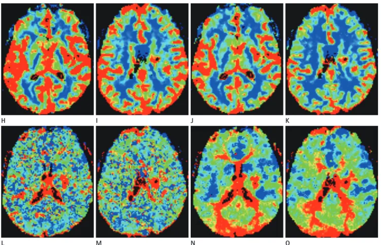
The perfusion study showed relatively increased cerebral blood volumes (rCBV) and cerebral blood flows (rCBF) with an asso- ciated prolonged mean transit time (MTT) and time to peak (TTP), as compared to contralateral normal parenchyma, repre-
Fig. 1. Venous infarction of developmental venous anomaly in a 63-year-old woman who presented with an acute onset of right side motor weakness and sensory changes.
Axial, non-enhanced (conventional) computed tomography (A) revealed a subtle hypodense area (black arrow) in the left basal ganglia compared to the contralateral side. Gadolinium-enhanced T1-weighted axial images displayed the enhancement of medullary veins (white arrow, B) at the left putamen in the ‘caput medusa’ configuration with a collector vein (white arrowhead, C) draining to the eft lateral ventricle’s surface. Diffu- sion weighted images (D, E) showed hyperintense lesions in left basal ganglia and periventricular white matter nearby the draining vein. A corre- sponding diffusion coefficient map (F, G) demonstrated diffusion restrictions, indicating acute infarction.
A B C D
E F G
senting venous congestion (Fig. 1H-O).
The patient’s symptoms resolved spontaneously within 24 hours after onset without neurologic deficits. The patient contin- ued to be symptom-free for at least 11 months following initial presentation.
DISCUSSION
Currently, DVAs are regarded as a primary dysplasia of capil- laries and small transcerebral veins or a compensatory mecha- nism caused by an arrest of normal venous development during- embryogenesis (7). They are thought to represent variations of parenchymatas’ venous drainage and surgical resection should be avoided, because when they are drained no other normal ve- nous discharging system exists (1, 2).
Despite the DVAs’ high incidence, they are benign anatomic variations and rarely cause symptoms due to either mechanical
compression to adjacent intracranial structures, which may re- sult in hydrocephalus, tinnitus, hemifacial spasms, and trigemi- nal neuralgia or a misbalance of the blood flow (4). Flow related complications can be characterized as a misbalance of the blood’s in- and outflow in the DVA system, which raises the pressure in the DVAs, either due to an increase of the former or a restriction of the latter (4). They may also give rise to acute symptoms which can mimic arterial stroke due to the draining vein’s restricted out- flow and venous congestion, which might cause an increase of complications such as hemorrhages or infarctions. Ruíz et al. (8) reported complications in 19 cases with symptomatic DVAs (ve- nous ischemic infarction. 53%, parenchymal hemorrhage, 37%, and subarachnoid and intraventricular hemorrhages, 5%).
DVAs also frequently coexist with other types of vascular mal- formations, such as cavernous ones and they may be associated with a higher hemorrhagic risk. There are several reported cas- es of DVAs with thrombotic obstruction of draining veins, re-
H I J K
L M N O
Fig. 1. Venous infarction of developmental venous anomaly in a 63-year-old woman who presented with an acute onset of right side motor weakness and sensory changes.
Perfusion images revealed a regional increase of relative cerebral blood flow (H, I) and of cerebral blood volume (J, K), surrounding the develop- mental venous anomaly, with a prolongation of mean transit time (L, M) and time to peak (N, O).
sulting in venous infarction with or without parenchymal hem- orrhage (5, 6, 8). Thrombosis of an DVA’s draining vein leading to venous brain infarction is a rare complication and an infarc- tion remained nonhemorrhagic, if early recanalization is ach- ieved (9).
According to Pereira et al. (4), mechanical and increased in- flow types of flow-related symptomatic DVA can be excluded in our case, since there was no intracranial structure causing me- chanical compression or cerebral arteriovenous malformation which may also result in an increased inflow of DVA. In our case, there was no discernible venous thrombosis as found in the im- aging studies and we assumed there a possibility of venous in- farction with early recanalization of thrombosed vein might exist;
this implies a decreased outflow type of flow-related symptomat- ic DVA, considering that there is no hemorrhage associated with infarction and that the acute onset of the patient’s symptoms hap- pened without a significant past medical history.
DVAs are thought to lack smooth muscle cells and elastic con- nective tissue resulting in less flexibility to changes of hemody- namic disturbances (5, 6). Truwit (1) reported that a focal steno- sis of the draining vein is possible at the point where it penetrates the dura which can cause lessened compliance, increased resis- tance to flow, and reduced capacity of the vessel for pressure change adjustments. In addition, a relative volume overload, previous hemorrhages, and chronic cerebral ischemia or ve- nous hypertension can result when a large parenchymal territo- ry is drained by a DVA. As a result, it has been suggested that imaging abnormalities of high SI on T2 FLAIR images, higher ADC values, and perfusion alterations in the DVAs’ vicinity can be explained by chronic cerebral ischemia, resulting from ve- nous congestion (10). The pattern of perfusion abnormalities in our case was similar to previously reported results, e.g., an in- creased rCBV and rCBF with prolonged MTT and TTP, impli- cating venous congestion with delayed perfusion (10).
Regarding DWI and ADC maps, focal SI change of diffusion restriction may be indistinguishable from arterial infarction, but underlying DVA can be identified by enhanced T1-weighted im- ages. In addition, DVA-associated perfusion abnormalities may represent its role regarding venous congestion.
Treatment principles of acute infarctions related to DVA have not been established, but a number of case series has reported fa- vorable outcomes after anticoagulation therapy, particularly in
instances of the draing vein associated with thrombus (5-7). Due to differences in the pathophysiology of venous and arterial stroke, venous infarction may not have the same prognostic value as the arterial infarct and it may be completely reversible (6).
In conclusion, even though DVAs commonly encounter insig- nificant anatomic variants, they rarely cause venous infarction with acute neurologic symptoms; characteristics of imaging fea- tures by perfusion studies may provide a clue to diagnosis. The understanding of and familiarity with imaging findings of com- plicated DVAs is essential for accurate diagnoses with different kinds of management of and prognoses about arterial infarction.
REFERENCES
1. Truwit CL. Venous angioma of the brain: history, signifi- cance, and imaging findings. AJR Am J Roentgenol 1992;
159:1299-1307
2. Lasjaunias P, Burrows P, Planet C. Developmental venous anomalies (DVA): the so-called venous angioma. Neurosurg Rev 1986;9:233-242
3. Töpper R, Jürgens E, Reul J, Thron A. Clinical significance of intracranial developmental venous anomalies. J Neurol Neurosurg Psychiatry 1999;67:234-238
4. Pereira VM, Geibprasert S, Krings T, Aurboonyawat T, Ozanne A, Toulgoat F, et al. Pathomechanisms of symptomatic de- velopmental venous anomalies. Stroke 2008;39:3201-3215 5. Griffiths D, Newey A, Faulder K, Steinfort B, Krause M.
Thrombosis of a developmental venous anomaly causing venous infarction and pontine hemorrhage. J Stroke Cere- brovasc Dis 2013;22:e653-e655
6. Masson C, Godefroy O, Leclerc X, Colombani JM, Leys D. Ce- rebral venous infarction following thrombosis of the drain- ing vein of a venous angioma (developmental abnormality).
Cerebrovasc Dis 2000;10:235-238
7. Field LR, Russell EJ. Spontaneous hemorrhage from a cere- bral venous malformation related to thrombosis of the cen- tral draining vein: demonstration with angiography and se- rial MR. AJNR Am J Neuroradiol 1995;16:1885-1888 8. Ruíz DS, Yilmaz H, Gailloud P. Cerebral developmental ve-
nous anomalies: current concepts. Ann Neurol 2009;66:
271-283
9. Konan AV, Raymond J, Bourgouin P, Lesage J, Milot G, Roy
D. Cerebellar infarct caused by spontaneous thrombosis of a developmental venous anomaly of the posterior fossa.
AJNR Am J Neuroradiol 1999;20:256-258
10. Jung HN, Kim ST, Cha J, Kim HJ, Byun HS, Jeon P, et al. Dif-
fusion and perfusion MRI findings of the signal-intensity abnormalities of brain associated with developmental ve- nous anomaly. AJNR Am J Neuroradiol 2014;35:1539-1542
대뇌정맥발달기형의 정맥경색: 관류영상소견을 포함한 증례 보고
김정윤
1· 김혜정
1* · 김은수
2· 현수정
1· 김희영
1· 김한면
1· 황지영
1· 홍혜숙
1· 우지영
1· 양 익
1대뇌정맥발달기형은 흔한 선천성 정맥기형으로 메두사머리 형태의 확장된 속질정맥과 배액정맥으로 특징지어진다. 대뇌 정맥발달기형은 높은 발생 비율에도 불구하고 양성 해부학적 변이로 드물게 증상을 초래한다. 저자들은 갑자기 발생한 신경학적 증상으로 내원하여 신경학적 후유증 없이 회복한 63세 여자에서 급성 경색을 일으킨 기저 대뇌정맥발달기형의 컴퓨터단층촬영과 자기공명영상 소견을 뇌관류영상소견과 함께 보고하고자 한다.
1한림대학교 의과대학 강남성심병원 영상의학과, 2한림대학교 의과대학 한림대학교성심병원 영상의학과