Ann Clin Microbiol Vol. 21, No. 3, September, 2018 https://doi.org/10.5145/ACM.2018.21.3.64
pISSN 2288-0585⋅eISSN 2288-6850
Nocardia abscessus Cutaneous Abscess:
A Case Report and Review of the Literature
Hee Sue Park1, Bo Ra Son2, Min Suk Song3, Kyeong Seob Shin2
1Department of Laboratory Medicine, Chungbuk National University Hospital, Departments of 2Laboratory Medicine and 3Microbiology, Chungbuk National University College of Medicine, Cheongju, Korea
We describe a cutaneous abscess caused by Nocardia abscessus in a previously healthy woman. A 74- year-old woman presented with recurrent bullae on her left forearm that developed 1 week prior and was initially suspected to be a cutaneous infection with Mycobacteria or Tinea corporis. Histopathologically, the skin lesion formed an abscess. A smear revealed a few branched Gram-positive filamentous micro- organisms that formed a creamy white colony on a blood agar plate after incubation for 3 days. The col- ony tested negative on acid-fast bacilli (AFB) stain- ing, but was positive on modified AFB staining. The
isolate was confirmed to be N. abscessus by 16S rRNA sequencing analysis. The isolate was suscep- tible to trimethoprim-sulfamethoxazole, amikacin, ce- fotaxime and erythromycin but resistant to penicillin.
The patient was treated with clarithromycin but sub- sequently lost to follow-up. To the best of our knowl- edge, this is the first report of a human cutaneous infection with N. abscessus in Korea. (Ann Clin Microbiol 2018;21:64-67)
Key Words: Cutaneous abscess, Nocardia abscessus, 16S rRNA sequence
64
Received 12 March, 2018, Revised 8 June, 2018, Accepted 9 June, 2018
Correspondence: Kyeong Seob Shin, Department of Laboratory Medicine, Chungbuk National University College of Medicine, 1 Chungdaero, Seowon-gu, Cheongju 28644, Korea. (Tel) 82-43-269-6240, (Fax) 82-43-271-5243, (E-mail) [email protected]
ⓒ The Korean Society of Clinical Microbiology.
This is an Open Access article distributed under the terms of the Creative Commons Attribution Non-Commercial License (http://creativecommons.org/licenses/by-nc/4.0) which permits unrestricted non-commercial use, distribution, and reproduction in any medium, provided the original work is properly cited.
INTRODUCTION
Nocardia are aerobic, branched, Gram-positive bacteria that are ubiquitous in the soil, and that cause various forms of dis- ease in humans, including pulmonary, systemic, extra-pulmonary, cutaneous, and central nervous system nocardiosis [1]. Infection with Nocardia species usually occurs through inhalation or di- rect cutaneous inoculation of the organism. Cutaneous infection acquired by direct inoculation typically presents as a localized nodular process in immunocompetent hosts [2]. However, sys- temic infection is frequently observed in immunocompromised hosts and is associated with a high mortality rate. Nocardia ab- scessus, previously known as Nocardia asteroides type 1, re- portedly causes human pulmonary infection, brain abscess [3], pericarditis and soft tissue infection [4]. To the best of our knowledge, only one case of human infection in a 29-year-old female having infected with N. asteroids type I isolated from her lung abscess has been reported in Korea [5]. However, no cutaneous infection by N. abscessus has been reported in Korea.
We describe a forearm abscess due to N. abscessus in im- munocompetent patient, which was initially suspected to be cu- taneous infection with Mycobacterium or Tinea corporis.
CASE REPORT
A previously healthy 74-year-old woman presented with re- current forearm bullae that had developed 1 week prior. Her vi- tal signs were stable, with the exception of her body temper- ature (37.2°C). Laboratory data tests indicated a white blood cell count of 7,910/μL with 82.3% neutrophil, a hemoglobin level of 8.4 g/dL, and a platelet count of 223,000/μL. The level of C-reactive protein (CRP) was 0.28 mg/dL. Renal and liver blood chemistry tests were within reference ranges. Her chest radiography revealed no active lesion.
The culture for bacteria and fungi and skin biopsy at the le- sion site in left forearm ware carried out. Histopathologically, an abscess had formed, but staining results (Gram, periodic acid-Schiff, and acid-fast-bacilli [AFB]) did not indicate the
Hee Sue Park, et al. : Cutaneous Abscess by Nocardia abscessus
65
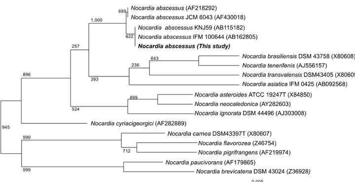
Fig. 1. Phylogenetic tree of the current isolate (CBU 05/1969: 1,362 bp) and Nocardia species. The 16S rRNA gene sequences of Nocardia species available in GenBank were aligned using CLUSTAL V and the phylogenetic tree was generated by the neighbor-joining method. Bootstrap values (%) are shown near their corresponding branches; ‘0.1’ indicates 0.1 nucleotide substitutions per site.
presence of microorganisms. However, microscopic examination of the smear revealed filamentous to rod-shaped bacteria stained with gram-positive bacteria; these stained partially positive in modified Ziehl-Neelson acid-fast staining. Creamy white colo- nies with white aerial hyphae developed after incubation on Blood Agar Plate (BAP) agar for 3 days. This suggested the presence of Nocardia species. To identify the microorganism to species level, we carried out 16S rRNA sequencing (1,362 bp), which indicated 100% similarity with N. abscessus strains (GenBank accession number AB115182, AB162805). The phy- logenetic relationships of isolate CBU 05/1969 with other re- lated Nocardia strains based on 16S rRNA sequence (27F:
AGA GTT TGA TCM TGG CTC AG, 1492R: TAC GGY TAC CTT GTT ACG ACT T) are shown in Fig. 1. The minimal in- hibitory concentration was 1.5 μg/mL for trimethoprim/sulfa- methoxazole (SXT), 0.125 μg/mL for amikacin, 1.5 μg/mL for cefotaxime, 0.25 μg/mL for erythromycin, 0.032 μg/mL for imipenem [6], 1 μg/mL for penicillin, 24 μg/mL for vancomy- cin as determined in E-test (bioMérieux Inc., Durham, NC, USA). The patient was initially treated with clarithromycin and subsequently lost to follow-up.
DISCUSSION
Nocardia species are aerosolized in dust; consequently, the respiratory tract is the main portal of entry [7]. However, direct inoculation of the skin and subcutaneous tissues can cause pri- mary cutaneous infection, which typically presents as a lo- calized nodular process with abscess formation. The course of infection is closely related to the immune competence of the host; infections in immunocompetent hosts are mostly chronic and localized to a single organ or region. Primary cutaneous no- cardiosis usually occurs following traumatic introduction into the skin by a thorn, puncture wound, or animal scratch [1]. The cause of the infection was not clear in this case, but direct in- oculation by traumatic introduction such as garden or farm work likely caused the infection. Although a few cutaneous infections by N. asteroides, Nocardia otitidiscaviarum, and N. abscessus have been reported, most cases are caused by Nocardia brasiliensis. In Korea, a few cases of cutaneous infections by N.
astheroides or N. brasiliensis, but none by N. abscessus, have been reported (Table 1) [8-14]. Cutaneous norcardiosis is under- diagnosed because of the relatively slow growth of the organ- ism, leading to failure of isolation [1]. Moreover, N. abscessus was only recently classified in the year 2000 [15], and cuta- neous infection by this organism may be rare. Indeed, primary
66
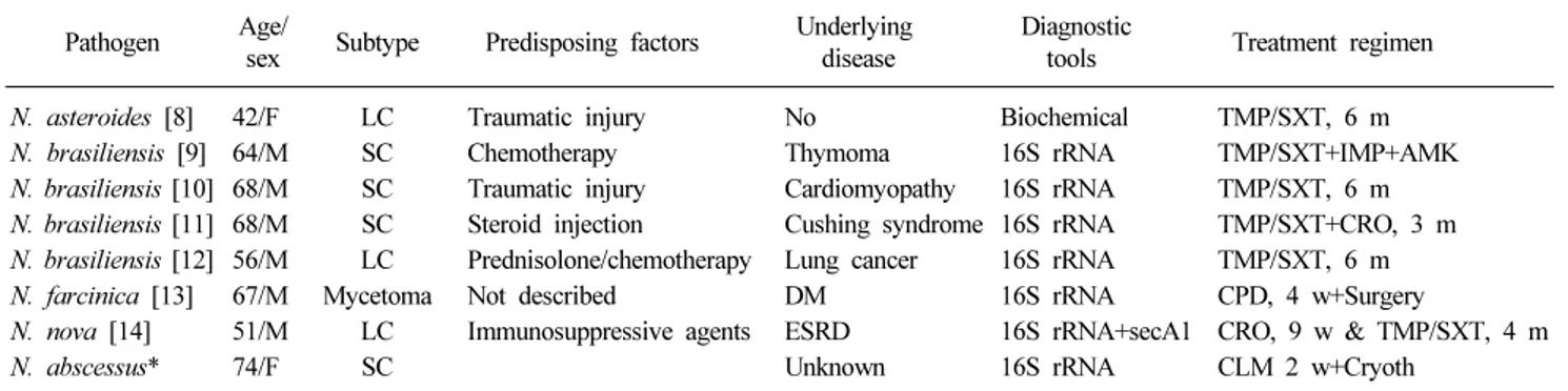
Ann Clin Microbiol 2018;21(3):64-67Table 1. Clinical characteristics and diagnostic tools for the patients with primary cutaneous Nocardiosis in Korea
Pathogen Age/
sex Subtype Predisposing factors Underlying disease
Diagnostic
tools Treatment regimen
N. asteroides [8] 42/F LC Traumatic injury No Biochemical TMP/SXT, 6 m
N. brasiliensis [9] 64/M SC Chemotherapy Thymoma 16S rRNA TMP/SXT+IMP+AMK
N. brasiliensis [10] 68/M SC Traumatic injury Cardiomyopathy 16S rRNA TMP/SXT, 6 m N. brasiliensis [11] 68/M SC Steroid injection Cushing syndrome 16S rRNA TMP/SXT+CRO, 3 m N. brasiliensis [12] 56/M LC Prednisolone/chemotherapy Lung cancer 16S rRNA TMP/SXT, 6 m
N. farcinica [13] 67/M Mycetoma Not described DM 16S rRNA CPD, 4 w+Surgery
N. nova [14] 51/M LC Immunosuppressive agents ESRD 16S rRNA+secA1 CRO, 9 w & TMP/SXT, 4 m
N. abscessus* 74/F SC Unknown 16S rRNA CLM 2 w+Cryoth
Abbreviations: M, male; F, female; LC, lymphocutaneous; SC, superficial cutaneous; DM, diabetes mellitus; ESRD, end stage of renal disease;
16S rRNA, 16S ribosomal RNA sequencing analysis; secA1, secA1 sequence; TXP/SXT, trimethoprim/sulfamethoxazole; IMP, imipenem; AMK, amikacin; CRO, ceftriaxone; CPD, cefpodoxime; CLM, clarithromycin; Cryoth, Cryotherapy; m, month; w, week.
*Present study.
cutaneous norcardiosis is likely considerably more common than is generally appreciated [1].
The clinical findings of nocardiosis, including cutaneous cas- es, are nonspecific, and cases may be mistaken for other bacte- rial infections including actinomycosis and tuberculosis as well as fungal infections and malignancies that affect multiple systems. Awareness of the possibility of nocardiosis can ex- pedite the diagnostic work-up, particularly in patients with pre- disposing factors or who are immunocompromised. Modified acid-fast and Gram staining are particularly important for a rap- id presumptive diagnosis [16]. Most Nocardia species are Gram-positive branched rods that stain positive in acid-fast tests if a weak acid is used. Mycobacteria do not stain well with gram stain and modified acid-fast stain. Similarly, Actinomyces are not stained by modified acid-fast stains. Typical colonies are usually seen after 3 to 5 days and have a chalky white or cotton ball appearance because of the abundant aerial filaments [17].
Initial species identification can be performed based on bio- chemical reactions but this is not useful for differentiating Nocardia species. In some cases, species must be confirmed us- ing a molecular technique such as 16S rRNA sequencing or PCR, which may change the initial biochemical identification.
Identification of Nocardia to the species level is important for adjusting the antibiotic therapy, as resistance profiles differ among species [18].
As nocardiosis is rare, the most appropriate therapeutic agent, administration route, and treatment duration are unclear, but sul- fonamide has been the agent of choice for more than 60 years [2]. For patients with disseminated or severe nocardiosis, combi- nation therapy with two or more active agents (e.g., ceftriaxone, imipenem, amikacin) is usually used [2]. In primary cutaneous
nocardiosis, SXT monotherapy may be adequate or used in combination with a fluoroquinolone for deep infection or myce- toma [19]. The duration of therapy depends on the site of the lesion and the patient’s immune status. Primary cutaneous no- cardiosis should be treated for 1-3 months. However, cases with pulmonary and CNS involvement should be treated for ≥6 months due to the risk of recurrence [17].
In conclusion, we described a cutaneous abscess due to N.
abscessus infection in an immunocompetent patient, which was initially suspected to be due to cutaneous Mycobacterium or T.
corporis infection. To the best of our knowledge, this is first re- port of a human infection by N. abscessus in Korea. 16S rRNA sequencing is essential for identification of Nocardia to the spe- cies level. Accurate diagnosis may facilitate development of an effective treatment for infections by Nocardia species.
REFERENCES
1. Beaman BL and Beaman L. Nocardia species: host-parasite relationships. Clin Microbiol Rev 1994;7:213-64.
2. Ambrosioni J, Lew D, Garbino J. Nocardiosis: updated clinical review and experience at a tertiary center. Infection 2010;38:
89-97.
3. Al Tawfiq JA, Mayman T, Memish ZA. Nocardia abscessus brain abscess in an immunocompetent host. J Infect Public Health 2013;6:158-61.
4. Horre R, Schumacher G, Marklein G, Stratmann H, Wardelmann E, Gilges S, et al. Mycetoma due to Pseudallescheria boydii and co-isolation of Nocardia abscessus in a patient injured in road accident. Med Mycobiol 2002;40:525-7.
5. Choe WH, Kang JO, Pai HJ, Choi TY. A case of Norcardia asteroids type I induced pneumonia. Ann Lab Med 2005;25:324-8.
6. CLSI. Susceptibility testing of mycobacteria, nocardia, and other aerobic actinomycetes; approved standard. CLSI document M24-A.
Wayne, PA: Clinical and Laboratory Standards Institute; 2003.
Hee Sue Park, et al. : Cutaneous Abscess by Nocardia abscessus
67
7. Ferrer A, Llorenc V, Codina G, de Gracia-Roldan J. Nocardiosis and bronchiectasis. An uncommon association? Enferm Infect Microbiol Clin 2005;23:62-6.
8. Lee SH, Suh CW, Choi JH, Sung KJ, Moon KC, Koh JK. A case of primary cutaneous sporotrichoid nocaardiosis caused by Nocardia asteroides. Ann Dermatol 1999;11:90-3.
9. Shin JU, Kwon YS, Kim HJ, Park Y, Lee K, Lee KH. A case of disseminated cutaneous nocardiosis due to Nocardia brasiliensis diagnosed by fine needle aspiration biopsy and 16S ribosomal RNA sequencing. Korean J Dermatol 2009;47:1024-8.
10. Kang GS, Kim DM, Lee MH, Suh MK, Ha GY, Jang TJ, et al.
Primary cutaneous nocardiosis caused by Nocardia brasiliensis.
Korean J Dermatol 2011;49:730-4.
11. Yun NR, Lee HJ, Hong SJ, Lee J, Kim DM, Jang SJ, et al. A case of disseminated nocardiosis by Nocardia brasiliensis after steroid injection. Infect Chemother 2011;43:367-71.
12. Ryu HW, Lee KS, Rhyoo NH, Cho JW. Primary cutaneous nocardiosis with sporotrichoid pattern by N. brasiliensis in lung cancer patient. Korean J Dermatol 2012;50:468-71.
13. Park SD, Kim HJ, Jang IH, Uh Y, Kim J, Yoon KJ, et al. First
report of Nocardia farcinica bursitis in a patient with diabetes mellitus. Ann Lab Med 2014;34:252-5.
14. Kim YK, Oh JR, Choi HK, Kim HY, Park SD, Uh, Y. Primary cutaneous nocardiosis caused by Nocardia nova in a kidney transplant recipient. J Med Microbiol 2014;63:140-3.
15. Yassin AF, Rainey FA, Mendrock U, Brzezinka H, Schaal KP.
Nocardia abscessus sp. nov. Int J Syst Evol Microbiol 2000;50:
1487-93.
16. Martínez R, Reyes S, Menéndez R. Pulmonary nocardiosis: risk factors, clinical features, diagnosis and prognosis. Curr Opin Pulm Med 2008;14:219-27.
17. Corti ME and Villafane-Fioti MF. Nocardiosis: a review. Int J Infect Dis 2003;7:243-50.
18. McNeil MM and Brown JM. The medically important aerobic actinomycetes: epidemiology and microbiology. Clin Microbiol Rev 1994;7:357-417.
19. Negroni R, López Daneri G, Arechavala A, Bianchi MH, Robles AM. Clinical and microbiological study of mycetomas at the Muñiz hospital of Buenos Aires between 1989 and 2004. Rev Argent Microbiol 2006;38:13-8.
=국문초록=
Nocardia abscessus 에 의해 발생한 피부 궤양: 증례보고 및 문헌고찰
1충북대학교병원 진단검사의학과, 충북대학교 의과대학 2진단검사의학교실, 3미생물학교실
박희수1, 손보라2, 송민석3, 신경섭2
저자들은 이전에 건강하였던 74세 여자환자에서 Nocardia abscessus에 의해 발생한 피부감염을 국내에서 최초로 보고하 고자 한다. 환자는 1주일 전부터 왼쪽 전완부에 반복적인 수포가 발생하여 마이코박테리아에 의한 감염 또는 체부백선 을 의심하였다. 피부 병변의 조직검사에서 궤양의 형태를 보였으며 도말검사에서 사상형 그람양성균이 관찰되었다. 3일 후 우유 빛의 백색 균집락이 관찰되었으며 AFB 염색에 음성이었으나 modified AFB에 양성 결과를 나타냈다. 16S rRNA 염기서열 검사에서 N. abscessus와 일치하였으며 trimethoprim-sulfamethoxazole, amikacin, cefotaxime, erythromycin에 감수 성이었다. Clarithromycin으로 치료를 시작하였으나 이후 본 병원을 방문하지 않았다. 저자의 확인에 의하면 이 보고는 N. abscessus에 의한 사람에서 피부감염의 국내 최초의 예이다. [Ann Clin Microbiol 2018;21:64-67]
교신저자 : 신경섭, 28644, 충북 청주시 서원구 충대로 1 충북대학교 의과대학 진단검사의학교실 Tel: 043-269-6240, Fax: 043-271-5243 E-mail: [email protected]