∙Address for correspondence Ho-Seung Lee, M.D.
388-1 Pungnap-2dong, Songpa-gu, Seoul 138-736, Korea, Department of Orthopaedic Surgery, Asan Medical Center, College of Medicine, Ulsan University,
Tel:+82-2-3010-3530 Fax:+82-2-488-7877 E-mail:hosng@amc.seoul.kr
*본 논문의 요지는 2003년 제 47차 대한정형외과학회 추계학술대회에 서 구연되었음.
족관절 원위 경비 인대 결합의 급성 파열에 대한 초음파 검사의 진단적 의의
울산대학교 의과대학 서울아산병원 정형외과학교실, 방사선과학교실*
이호승․박수성․김지완․신명진*․김성문*․이상훈*․이상민*
Diagnostic Value of Ultrasonography for Acute Tear of Tibiofibular Syndesmosis in Ankle
Ho-Seung Lee, M.D., Soo-Sung Park M.D., Ji-Wan Kim M.D., Myung-Jin Shin, M.D.*, Sung-Moon Kim, M.D.*, Sang-Hoon Lee, M.D.*, Sang-Min Lee, M.D.*
Departments of Orthopaedic Surgery and Radiology*, Asan Medical Center, College of Medicine, Ulsan University, Seoul, Korea
=Abstract=
Purpose: We studied the diagnostic value of ultrasonography to confirm the presence of the syndesmosis injuries in the ankle.
Materials and Methods: Pre-operative ultrasonography were taken in 42 cases of ankle fractures to determine injury of syndesmosis between February 2000 and August 2003. Tear of syndesmosis was confirmed when they met the following criteria; widening of distal tibiofibular clear space greater than 7 mm on pre-operative AP X-rays, leakage of contrast dye during intra-operative arthrography, and operative findings revealing syndesmosis rupture.
We compared these diagnoses with those made by pre-operative ultrasonography.
Results: Syndesmosis injuries were confirmed in 23 cases among the total 42 cases. Ultrasonography revealed syndesmosis tear in 25 cases, with 20 cases corresponding with confirmed tear and 5 cases without tear in confirm. Syndesmoses were found to be intact in 17 cases of ultrasonograph, but 3 cases were actually confirmed tear. In ankle ultrasonography, the sensitivity was 90.9%, the specificity was 75.0%.
Conclusion: Preoperative ultrasonography is a non-invasive and useful study to determine the syndesmosis injury in ankle fractures.
Key Words: Ankle, Syndesmosis, Acute tear, Diagnosis, Ultrasonography
서 론
족관절 원위 경비 인대 결합은 족관절의 기능과 안정성 에 중요한 역할을 하여 족관절 손상시 족관절 원위 경비 인 대의 손상 유무는 치료 및 예후에 중요한 영향을 미치지만 이를 정확히 진단하기에는 어려움이 있다.
원위 경비 인대 파열은 족관절 전후면 촬영 및 격자상 촬
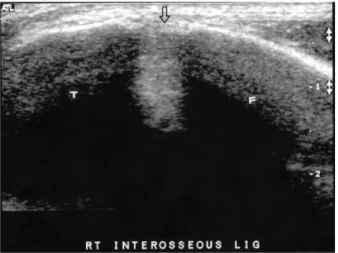
Figure 1. sonographic findings of syndesmosis injury.
*: discontinuity of the ligament, fluid collection. arrow: anteroinferior tibio-fibular ligament. T, tibia; F, fibula.
Figure 2. Ultrasonograph of ankle. Sonography can show interosseous ligament. T; tibia, F; fibula.
영상의 원위 경비골 간격의 이개 정도에 따라 진단하지만, 진단의 정확도는 높지 않다9,18,22). 최근 자기 공명 영상이나 관절경검사 방법을 통해 비교적 정확한 진단을 할 수 있다 고 소개되고 있으나8,11,24,27), 고가의 검사이거나 침습적인 검사라는 문제점을 안고 있다. 이에 본 연구에서는 비교적 경제적이면서도 비침습적인 초음파 검사를 시행하여 원위 경비 인대 결합 손상 여부에 대한 진단적 가치와 유용성을 알아보고자 하였다.
대상 및 방법
2000년 2월 16일부터 2003년 8월 29일까지 족관절 골절 로 내원하여 원위 경비 인대 결합 손상을 확인하기 위하여
수술 전 초음파 검사를 시행한 42예를 대상으로 하였다. 남 자 29명, 여자 13명이었고, 평균 연령은 40.4세(12-67세) 였다. Lauge-Hansen 분류상 회외-외회전형이 18예, 회외 -내전형이 1예, 회내-외회전형 6예, 회내-외전형 5예였 고, Lauge-Hansen 분류에 속하지 않는 경우가 12예였다.
초음파 검사는 수상 후 평균 2.1일(1-6일)에 시행하였 으며, 5-12 MHz의 초고해상도의 탐침(probe)을 사용하였 다. 환자를 앙와위에서 슬관절을 신전하고 족관절은 중립 위치에서 원위 경비 인대의 파열 여부에 대한 초음파 검사 를 시행하여 반대편 족관절의 검사 소견과 비교하였다. 족 관절 족배 굴곡과 외회전 상태에서 검사를 시행하면 파열된 인대를 관찰하기 쉬우나, 수상 직후의 환자가 심한 통증을 호소하여 중립위에서 시행하였다. 초음파 검사 상 파열의 진단은 반대편과 비교했을 때 인대의 연속성이 없을 때와 혈종이나 부종에 의한 저신호 영상이 보이면서 인대 구조물 이 관찰되지 않을 때로 하였다14)(Fig. 1). 초음파 검사 시 관 찰할 수 있는 구조물은 전하 경비 인대와 골간 인대이고, 후 면에 위치한 후하 경비 인대와 하횡 인대는 관찰할 수 없었 다(Fig. 2).
원위 경비 인대 파열의 확진은 수술 전 전후면 사진상 원 위부 경골-비골 간격이 7 mm 이상인 경우 및 수술 중 관절 조영술상 조영제의 누출이 있거나 또는 수술 소견상 외측과 골절의 정복을 위한 외측 절개부위에서 전방으로 박리를 조 금 더 하여 전하 경비 인대의 부분 파열 및 연속성이 없는 소견이 확인되는 경우에 인대 결합이 파열된 것으로 판정하 였다. 대부분의 환자에서 관절 조영술을 시행하였고, 관절 조영술을 시행하지 않은 경우에는 수술 소견으로 확인하였 다. 이와 같은 방법으로 확진된 원위 경비 인대 파열에 대하 여 술 전 초음파 검사상의 손상 유무에 대한 검사 결과와 비 교하여 민감도, 특이도, 정확도를 알아보았다.
결 과
전체 42예의 대상 중 원위 경비 인대가 파열된 것으로 확 진된 경우는 23예였다.
초음파 검사에서 원위 경비 인대가 파열된 것으로 진단 된 경우는 모두 25예로 그 중 20예는 확진 소견과 일치하였 고 나머지 5예는 초음파 검사에서 인대 손상이 의심되었지 만 파열을 확진할 수 없었던 위양성 경우였다. 위양성을 보 였던 5예 중 2예는 초음파 검사에서 원위 경비 인대 결합의 손상이 의심되는 경우였고, 1예는 전하 경비 인대의 부분적 인 인대 손상으로 진단되었던 경우이다.
그리고 초음파 검사에서 원위 경비 인대의 파열 소견을
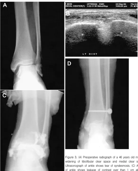
Figure 3. (A) Preoperative radiograph of a 46 years old man shows widening of tibiofibular clear space and medial clear space. (B) Ultrasonograph of ankle shows tear of syndesmosis. (C) Arthrogram of ankle shows leakage of contrast over than 1 cm above tibia plafond. (D) Postoperative radiograph shows internal fixation using transfixing screw.
관찰할 수 없었던 경우는 17예로 그 중 14예서는 원위 경비 인대가 파열되지 않았고, 나머지 3예서는 파열되었다 (Table 1). 위음성을 보였던 3예 중 1예는 적절하지 못한 시 야를 보여 부적절한 검사였다.
이상의 결과로 족관절 원위 경비 인대 파열에 대한 초음 파 검사의 민감도는 90.9%, 특이도는 75.0%, 정확도 (accuracy)는 83.3%였다.
Ultrasonographic diagnosis
+ -
Confirmative Diagnosis
+ -
20 5
3 14 +; syndesmosis tear, -; intact syndesmosis
Table 1. Result of Ultrasonographic Diagnosis for Syndesmosis Tear (Cases)
Lauge-Hansen 분류상 회외-외회전형 18예 중 6예에서 원위 경비 인대 결합의 손상이 있었고, 회내-외회전형 6예 모두와, 회내-외전형에서는 5예 모두 손상이 있었다. 회외 -내전형 1예도 원위 경비 인대 결합의 손상이 관찰되었다.
외측 과의 골절만 단독으로 있는 경우, 혹은 내측 과의 견열 골절이나 삼각 인대의 파열이 있고, 전하 경비 인대의 파열 이 있으나 비골 골절이 없는 경우와 같이 Lauge-Hansen 분류를 따를 수 없었던 12예 중 5예에서 원위 경비 인대 결 합의 손상이 있었다.
증례 보고
46세 남자 환자로 내원 당일 등산하다 발목을 접질려 발 생한 좌측 족관절 동통을 주소로 내원하였다. 족관절 전후 면 촬영 사진에서 내측 관절 간격이 4.8 mm로 삼각 인대의 파열이 의심되었고, 원위부 경골-비골 간격이 9.1 mm로 원위 경비 인대 결합의 파열이 의심되어서(Fig. 3A), 근위 부 비골 골절이 동반된 Maisonneuve 골절로 진단하였다.
수상 1일 후 시행한 족관절 초음파 검사에서 좌측 전하 경비 인대 및 골간 인대, 삼각 인대의 파열 소견을 보였다(Fig.
3B). 수술 직전 시행한 관절조영술상 조영제의 유출 소견을 보여(Fig. 3C) 1개의 횡나사를 삽입하여 원위 경비 간격 이 개를 교정하였다(Fig. 3D).
고 찰
원위 경비 인대 결합은 전하 경비 인대(anteroinferior tibiofibular ligament), 후하 경비 인대(posteroinferior tibiofibular ligament), 골간 인대(interosseous ligament), 하횡인대(transverse ligament)로 이루어진다6). Oglivie- Harris와 Reed15)는 전하 경비 인대가 35%의 안정성을 제공 하고, 골간 인대가 22%를, 후하 경비 인대가 33%를 하횡 인대가 9%의 안정성을 제공한다고 하였으며, Bonnin1)은 전하 경비 인대의 파열은 경비골을 4 mm까지 분리시킬 수 있고 골간 인대가 추가로 파열될 경우 1 cm까지 분리된다 고 하였다. Xenos 등29)은 사체 연구에서 전하 경비 인대를 절개하면 경비골이 2.3 mm 분리되고, 골간 인대의 원위부 8 cm을 잘랐을 경우 추가로 2.2 mm 더 분리되고 후하 경비 인대까지 절개하였을 경우 총 7.3 mm까지 분리가 일어난 다고 하였다. Close4)는 원위 경비 인대 결합에 파열이 있는 경우 경비 관절이 벌어지면서 거골의 외측 전이가 발생한다 고 하였는데, Ramsey와 Hamilton19)은 거골의 외측 전위가 1 mm일 경우 경거골 관절 접촉면의 42%가 감소한다고 하
였으며, Burns 등3)은 삼각인대의 파열과 원위 경비 인대 결 합의 완전 파열은 경거골 관절 접촉면을 40% 감소시키고 경거골 접촉 압력을 36% 증가시킨다고 하였다.
이와 같이 족관절 원위 경비 인대 결합은 족관절의 기능 과 안정성에 중요한 역할을 하여 족관절 손상시 족관절 원 위 경비 인대의 손상 유무는 치료 및 예후에 중요한 영향을 미친다. 원위 경비 인대 결합 손상의 발생 기전은 주로 외회 전 손상에 의해 야기되는데19,20), Lauge-Hansen의 족관절 분류에서는 원위 경비 인대 파열은 회외-외회전형, 회내- 외전형, 회내-외회전형의 골절에서 발생한다고 하였다12). 원위 경비 인대의 손상시 경비골을 관통하는 나사를 삽입 할 경우 손상된 원위 경비 인대 결합의 복구에는 도움이 되 나 족관절의 배굴 제한이 남기 쉬워 관통나사의 삽입 여부 를 결정하기 위하여는 인대의 손상 유무를 정확히 진단하여 야 한다.
원위 경비 인대 결합 손상의 진단 방법으로는 족관절 방 사선 사진, 관절 조영술, 자기 공명 영상, 관절경 검사 등이 있다. 족관절 방사선 사진은 경비 인대 결합 손상이 의심될 때 시행하는 기본적인 검사로서 족관절 전후면 사진, 격자 상 촬영을 하여 내측 관절 간격, 경골-비골 간격, 경골-비 골 중복 정도를 측정하여 인대 결합 손상 판정의 기준을 삼 는다. 이 중 경골-비골 간격이 가장 정확한 지표로 이용되 지만5), 방사선 촬영시 촬영 각도와 측정자에 따라 측정치의 차이가 커서 원위 경비 인대 결합 손상을 진단하기 어렵다.
저자에 따라 원위 경비골 간격의 정상 범위는 다양하게 제 시하였는데 Ramsey13), Sclafani23)는 3-6 mm로, Harper 와 Keller7)는 6 mm 이하라고 하였고, Ostrum17)은 남자에 서는 6.5 mm 이하, 여자에서는 5.2 mm 이하를 정상이라 하였다. 본 연구에서는 진단율을 높이기 위하여 경비골 간 격이 7 mm 이상인 경우 원위 경비 인대가 파열된 것으로 확진하였다.
족관절의 관절 조영술도 진단에 이용되는데2,10,28), 조영 제가 경골 천장부(tibia plafond) 상방 1 cm 이상 유출되면 원위 경비 인대 결합의 손상을 의심하지만21), 족관절을 외 회전하여야 조영제의 유출을 확인할 수 있다. 마취가 필요 하며, 수상 후 일주일 이상 경과된 환자에 있어서는 유용하 지 못하다는 단점이 있다2).
자기 공명 영상도 이용할 수 있는데8,11,24,27), 인대의 파열 이 있어도 영상의 절단면에 따라 나타나지 않을 수도 있고 고가의 검사라는 단점이 있다. 또한 손상 직후 골절로 인한 혈종과 주변 조직 손상으로 인한 신호 강도의 전반적인 변 화로 원위 경비 인대의 손상을 진단하는데 어려움이 있다.
관절경 검사도 인대 손상을 가시적으로 확인할 수 있다
는 장점이 있지만16,25,26), 침습적인 검사인데다 급성기에는 골절편 사이로 수액이 유출되어 부종을 유발할 수 있고 전 하 경비 인대와 후하 경비 인대를 관찰할 수 있는 반면 골간 인대의 손상은 확인하기가 어렵다는 단점이 있다.
초음파 검사는 검사자에 따라 진단의 차이가 날 수가 있 고, 후하 경비 인대를 관찰하기 어려운 단점이 있지만 비교 적 경제적이면서도 비침습적인 검사로 인대 손상을 진단하 는데 유용하게 이용될 수 있다. 그러나 원위 경비 인대 결합 근처의 인대 파열 없이 골절이 동반되어 혈종이 생긴 경우 오진의 가능성이 있어 주의를 요한다.
본 연구에서 족관절 원위 경비 인대 결합의 손상을 진단 하기 위한 초음파 검사는 민감도 90.9%, 특이도 75.0%, 정 확도 83.3%를 보여 원위 경비 인대 결합의 파열에 있어서 비교적 유용한 검사라 할 것이다. 향후 초음파 검사가 원위 경비 인대 손상 정도를 정량적으로 진단할 수 있다면 원위 경비 인대 손상시 치료 방법을 결정하는데 도움이 될 수 있 을 것이다.
결 론
초음파 검사는 원위 경비 인대 결합 손상을 알아보는 방 법으로 비침습적이면서 비교적 유용한 검사이다.
REFERENCES
1. Bonnin JG: Injuries to the ankle, Darien, Conn, Hafner:
147-185, 1970.
2. Brostroem L, Liljedahl SO and Lindvall N: Sprained ankles.Ⅱ. Arthrographic diagnosis of recent ligament ruptures, Acta Chir Scand, 129: 485-499, 1965.
3. Burns WC II, Prakash K, Adelaar R, Beaudoin A and Krause W: Tibiotalar joint dynamics: indications for syndesmotic screw: a cadaver study. Foot Ankle, 14:
153-158, 1993.
4. Close JR: Some applications of the functional anatomy of the ankle joint. J Bone Joint Surg, 69-A: 596-604, 1987.
5. Garbarino JL, Clancy M, Harcke HT, et al.: Congenital diastasis of the inferior tibiofibular joint: a review of the literature and report of two cases. J Pediatr Orthop, 5:
225-228, 1985.
6. Grath GB: Widening of the ankle mortise: A clinical and experimental study. Acta Chir Scand. suppl, 263: 1-88, 1960.
7. Harper MC and Keller TS: A radiographic evaluation of the tibiofibular syndesmosis. Foot Ankle, 10: 156-160, 1989.
8. Jaas MH: Clinical significance of magnetic resonance
imaging of the ankle and foot. Magn Reson Imaging Clin North Am, 2: 29-38, 1994.
9. Joy G, Patzakis MJ and Harvey JP: Precise evaluation of the reduction of severe ankle fractures: Technique and correlation with end results. J Bone Joint Surg, 56-A:
979-993, 1974.
10. Katznelson A, Lin E and Militiano J: Ruptures of the ligaments about the tibio-fibular syndesmosis. Injury, 15:
170-172, 1983.
11. Kerr R, Forrester DM and Kingston S: Magnetic resonance imaging of foot and ankle trauma. Orthop Clin North Am, 21: 591-601, 1990.
12. Lauge-Hansen N: Fractures of the ankle: II. Combined experimental-surgical and experimental-roentgenologic investi- gations. Arch Surg, 60: 957-985, 1950.
13. Leeds HC and Ehrlich MG: Instability of the distal tibiofibular syndesmosis after bimalleolar and trimalleolar ankle fractures. J Bone Joint Surg, 66-A: 490-503, 1984.
14. Milz P, Milz S, Steinborn M, Mittlmeier T, Putz R and Reiser M: Lateral ankle and ligaments and tibiofibular syndesmosis. 13-MHz high-frequency sonography and MRI compared in 20 patients. 69(1): 51-55, 1998.
15. Oglivie-Harris DJ, Reed SC and Hdemn TP: Disruption of the ankle syndesmosis: biomechanical study of the ligamentous restraints. Arthroscopy, 10: 558-560, 1994.
16. Oglivie-Harris DJ and Reed SC: Disruption of the ankle syndesmosis: Diagnosis and treatment by arthroscopic surgery. Arthroscopy, 10: 561-568, 1994.
17. Ostrum RF, De Meo P and Subramanian R: A critical analysis of the anterior-posterior radiographic anatomy of the ankle syndesmosis. Foot Ankle int, 16: 128-131, 1995.
18. Pettrone FA, Gail M, Pee D, Fitzpatrick T and Van Herpe LB: Quantitative criteria for prediction of the results after displaced fracture of the ankle. J Bone Joint Surg, 65-A: 667-677, 1983.
19. Ramsey PL and Hamilton W: Changes in tibiotalar area of contact caused by lateral talar shift. J Bone Joint Surg, 58-A: 356-357, 1976.
20. Rasmussen O: Stability of the ankle joint. Analysis of the function and traumatology of the ankle ligaments. Acta Orthop Scand, Suppl, 211, 1985.
21. Mann RA and Coughlim MJ: Surgery of the foot and ankle. 6th ed. St. Louis, Mosby-Year Book Inc: 1148, 1993.
22. Sarkisian JS and Cody GW: Closed treatment of ankle fractures; A new criteria for evaluation-A review of 250 cases. J Trauma, 16: 323-326, 1976.
23. Sclafani SJ: Ligamentous injury of the lower tibiofibular syndesmosis: radiopgraphic evidence. Radiology, 156:
21-27, 1985.
24. Steinbronn DJ, Bennett GL and Kay DB: The use of magnetic resonance imaging in the diagnosis of stress fractures of the foot and ankle:four case reports. Foot Ankle Int, 15: 80-83, 1994.
25. Takao M, Ochi M, Naito K, et al.: Arthroscopic diagnosis of tibiofibular syndesmosis disruption. Arthros- copy, 17: 836-843, 2001.
26. Takao M, Ochi M, Oae K, Naito K and Uchio Y:
Diagnosis of a tear of the tibiofibular syndesmosis. The role of arthroscopy of the ankle. J Bone Joint Surg, 85-B:
324-329, 2003.
27. Vogl TJ, Hochmuth K, Diebold T, et al.: Magnetic resonance imaging in the diagnosis of acute injured distal
tibiofibular syndesmosis. Invest Radio, 32: 401-409, 1997.
28. Woolson ST, Dev P, Fellingham LL, et al.: Three- dimensional imaging of the ankle joint from computerized tomography. Foot Ankle, 6: 2-6, 1985.
29. Xenos JS, Hopkinson WJ, Mulligan ME, et al.: The tibiofibular syndesmosis: Evaluation of the ligamentous structures, methods of fixation, and radiolographic assessment. J Bone Joint Surg, 77-A: 847-856, 1995.