Received April 19, 2010 Accepted May 13, 2010
∙Gab-Lae Kim, M.D.
Gangdong Sacred Heart Hospital, 445 Gil-dong, Gangdong-gu, Seoul 134-701, Korea
Tel: +82-2-2225-2706 Fax: +82-2-789-4391 E-mail: [email protected]
최소 침습적 정복술 및 금속강선 고정술을 이용한 전위된 관절내 종골 골절의 치료
한림대학교 강동성심병원 정형외과, 강릉동인병원 정형외과*
이진영⋅박인헌*⋅김갑래⋅김태화⋅오범석
Minimally Invasive Reduction and Pin Fixation Treatment for Displaced Intraarticular Calcaneal Fracture
Jin-Young Lee, M.D., In-Heon Park, M.D.*, Gab-Lae Kim, M.D., Tae-Hwa Kim, M.D., Bum-Suk Oh, M.D.
Department of Orthopedic Surgery, Kangdong Sacred Heart Hospital, Hallym University School of Medicine, Seoul, Korea Department of Orthopedic Surgery, Gang-neung Dong-in General Hospital, Gangneung, Korea*
=Abstract=
Purpose: To evaluate the clinical efficacy of the minimally invasive posterior approach for the surgical treatment of intraarticular fracture of calcaneus.
Materials and Methods: From March 2006 to October 2008, we studied retrospectively 45 patients, 56 cases who were treated with minimally invasive reduction and pin fixation treatment for displaced intraarticular calcaneal fracture and were followed up for more than 1 year. The clinical results were evaluated with Creighton-Nebraska score and AOFAS score, circle draw test after 1 year. We checked simple AP, lateral, axial and Broden’s view preoperatively and 1 year after surgery, and compared Böhler angle and Gissane angle.
Results: By Creighton-Nebraska score, Sanders type 1 was 81, type 2 was 75, type 3 was 69, type 4 was 61. By AOFAS score, Sanders type 1 was 88, type 2 was 82, type 3 was 78, type 4 was 63. And by circle draw test, type 1 was 8.8 cm, type 2 was 8.5 cm, type 3 was 8 cm, type 4 was 6.6 cm. Preoperative Böhler angle and Gissane angle were 7.2°, 98°, and it increased to 21.2°, 116°
after postoperative 1 year.
Conclusion: Minimally invasive reduction and pin fixation treatment for displaced intraarticular calcaneal fracture was considered to be an effective treatment modality.
Key Words: Calcaneal intraarticular fracture, Minimally invasive posterior approach
서 론
전위된 관절내 종골 골절의 치료는 최근까지도 그 치료 법에 논란이 많았다. 관절내 종골 골절의 치료는 얇은 골피 질과 해면골로 이루어진 복잡한 구조 및 얇은 연부 조직으 로 인해서 50년 전까지만 해도 대증적 치료가 가장 대중적 인 치료였으나1,2), 이후 관혈적 정복술 및 내고정술이 보다 나은 치료 성과로 인하여 표준 치료로 선택되었다3,4). 그러
A B
Figure 1. (A) Incisional site on bullae, but operation was submitted.
(B) The patient was placed at the operating table on the prone position with pneumatic tourniquet at 400 mmHg pressure on upper thigh and hanging the ankle out of the operating table.
A B
Figure 2. Straight skin incision was put on near the lateral border of the achilles tendon and carried down to subcutaneous tissue.
The red line is incisional line.
Table 1. Patient Demographics
Factor %
Age 40.8 years
(32~62) Sex
Male 37 82.2%
Female 8 17.8%
Sanders classification
Type 1 1 1.8%
Type 2 19 33.9%
Type 3 32 57.4%
Type 4 4 7.2%
Essex-Lopresti classification
Joint depression 32 57.1%
Tongue type 24 42.9%
나 많은 정형외과 의사들이 수술적 치료를 함에 있어서 개 개인의 치료 경험의 부족 및 종골의 골절의 3차원적 구조에 대한 이해의 부족으로 인해 수술하기 매우 힘든 골절의 하 나로 생각되고 있다5,6). 특히 전위된 관절내 종골 골절은 수 술적 치료 후 수술 창상 부위의 괴사 및 염증, 외상성 거골 하 골관절염과 비복 신경의 손상 등 합병증의 발생 빈도가
높다2,7,8). 저자들이 최소 침습적 골정복술을 이용하여 관혈
적 정복술 및 금속내 고정술을 시행한 임상적 및 방사선학 적 결과를 분석함으로써 이러한 치료법의 임상적 유용성을 확인하고자 한다.
대상 및 방법
1. 연구 대상
2006년 3월부터 2008년 10월까지 관절내 종골 골절에 대해 최소 침습적 정복술 및 금속강선 고정술을 시행한 후 1년 이상 추시가 가능하였던 총 45명, 56예를 대상으로 후 향적 분석을 시행하였다. 성별은 남자가 37명(82.2%)이었 고 여자가 8명(17.8%)이었으며, 평균 연령은 40.8세였다.
골절의 분류는 전산화단층촬영을 이용한 Sanders 분류9)를 사용하였으며 1형은 1예(1.8%), 2형은 19예(33.9%), 3형은 32 예(57.4%), 4형은 4예(7.2%)였다. Essex-Lopresti 분류10)에서 관절함몰형은 32예(57.1%), 설상형은 18예(42.9%)였다. 평균 추시 기간은 평균 13.5개월이었다(Table 1).
2. 수술 방법
수술적 방법은 모든 예에서 최소 침습적 정복술을 사용 하였다. 수술 부위의 부종과 관계없이 바로 수술을 시행하 였으며, 부종이 심하여 수포가 생긴 경우에도 수술을 시행 하였으나 단 수술 절개 부위에 감염 소견이 있는 경우에는 수술을 지연하였다. 수술은 수상 후 평균 2.4일 후에 시행 되었다.
경막외 마취를 시행한 후 환자를 엎드린 위치로 준비하 며 족부가 수술대 아래쪽으로 약 20~30 cm 나오도록 준비 하였으며, 영상 증폭 투시기를 수술대 아래쪽에 준비하여 수술 중에도 언제나 족관절의 전후면, 측면, 종골의 축상 및 Broden’s 촬영11)이 가능하게 하였다(Fig. 1).
피부 절개는 종골건의 외측 1 cm 부위에서 1~1.5 cm정 도 종절개를 시행하였고, 비복 신경이 손상되지 않도록 확 인하며 피하지방 조직을 절개하였다(Fig. 2).
절개 부위로 Crego 골막 감자를 골절 간격으로 삽입하여 종골 전방의 시상 골절편이나 재거 돌기 또는 조면 골절편 을 들어올리면서 정복하여 Gissane 각을 예각에서 둔각으 로 회복하였고, Bone hook을 이용하여 전족부를 굴곡하면 서 회전시켜 거골하 관절을 정복한 다음 영상 증폭 투시기 를 이용하여 Broden’s 촬영 및 측면 사진을 실시간으로 확 인하였다(Fig. 3, 4).
Figure 3. After inserting Crego periosteal elevator & bone hook retractor at intervals of fragment, thalamic fragment, anterior calcaneus, sustentaculum tali or fragment of calcaneal tuberosity was lifted up and reduced observing joints directly.
Figure 4. Compression was applied in bilateral dirction with large hand-maded compressor on calcaneus. Reduced angulation deformity of calcaneus with lateral compression. We can check reducibility on axial view instantly.
정복 후에는 금속 강선으로 고정하였다. 금속 강선의 방 향은 족관절 외과의 하방 1 cm, 후방 2 cm에서 전내측, 즉 종골 종축의 약 45도 방향으로 하여 재거 돌기 방향으로 향 하며 골절면에 거의 직각으로 통과하게 하였다(Fig. 5).
또한 Steinmann pin을 조면 골절편과 재거 돌기를 통과 시켜 종골의 높이를 회복시키면서 고정하면 1차 골절도 해 결될 수 있으나 경우에 따라 내고정이 충분하면 Steinmann pin은 필요 없었다. 그러나 종골 전방의 분쇄가 심하며 골 절이 종입방 관절을 포함하는 경우에는 Steinmann pin을 종골 후방에서 삽입하여 입방골까지 고정하였다. 그리고 외
측으로 돌출된 골절편은 조면 골절편과 시상 골절편이 정 복되면서 충분한 공간이 발생하여 도수 정복 및 집게 모양 의 기구로 압박하면 쉽게 정복이 가능하다. 외측으로 돌출 된 골절편은 집게 모양의 도구로 종골의 전방부에서부터 후방부로 차근차근 압박해주며 특히 재거 돌기 부위 및 중 간 관절면 부근을 잘 압박해 준다. 모아진 골절편은 종골의 후상부 직하방에서 steinmann pin을 이용하여 고정한다.
정복 및 내고정 시행 후 골이식은 시행하지 않았으며, 창 상 봉합 후 추가적인 석고 고정을 하지 않았다. 체중 부하 는 수술 후 2주까지 금하였지만 수술 직후부터 환 운동
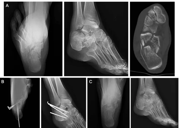
A
B C
Figure 6. Preoperative Broden’s 30° view and calcaneus axial radiograph, CT axial view (A) of a 40-year-old female patient fell from 2.5 m height show intraarticular calcaneal fracture (Sander’s classification IIIAC). Postoperative radiography. (B) shows after open reduction and internal fixation with K-wires and Steinmann pins. Thirteen months follow-up radiography. (C) shows good alignment of subtalar joint, subtalar joint motion and excellent fuctional outcome.
Figure 5. After opened reduction of displaced subtalar joint, K-wire
& steinmann pin fixation. The fluoroscope shows anatomical reduction and clearly demonstrated well restoration of the subtalar joint.
(circle draw excercise)12)을 시행하였다. 수술 후 2주에서 4 주 이후에 전족부 부분 체중 부하를 시행하였고, 수술 후 4 주에서 6주 이후에 약 20 kg의 부분 체중 부하를 시행하였 으며, 6주째 강선 제거 후 체중 부하를 50% 시행 후 8주 이후에 완전 체중 부하를 시작하였다(Fig. 6).
3. 연구 방법
치료에 대한 임상적 평가는 Creighton-Nebraska 평가13)와 미국정형외과 족부족관절학회 평가표(AOFAS score)14)에 따라 분류하였고, 환 운동 검사(Circle draw test)15)를 이용 하여 수술 후 1년 이상 경과하였을 때 평가하였다.
Creighton-Nebraska 평가는 종골 골절 후의 동통, 운동 범 위 이외에 직무 능력, 신발 교체 여부 등의 항목에 대해 분 석하는 방법이며13), 환 운동 검사는 환자가 편안한 자세로 앉아 있는 상태에서 반대편 무릎에 족부를 올린 후 발목을 고정하여 전족부가 그릴 수 있는 최대한의 원을 능동적으 로 그리게 한 후 지름을 측정하여 거골하 관절의 운동 범위 를 측정하는 방법으로 원의 크기가 10 cm 이상을 매우 우 수, 8에서 10 cm 정도를 우수, 5에서 8 cm을 양호, 5 cm 미만을 불량으로 분류하였다(Table 3). 또한 방사선학적 평 가는 족관절의 전후면, 측면, 종골의 축상 및 Broden’s 촬
Table 2. After 1 year later, Operation Result by Creighton-Nebraska scrore & AOFAS score & Circle Draw Test Creighton-Nebraska score (score) AOFAS score
(score) Circle draw test (cm)
Sanders classification Preoperation 1 year later Preoperation 1 year later 1 year later
Type 1 70 81 72 88 8.8
Type 2 69 75 67 82 8.5
Type 3 66 69 60 78 8.0
Type 4 53 61 51 63 6.6
Table 3. Subtalar Motion by Circle Draw Exercise
Result Diameter Number %
Excellent >10 cm 15 26.8
Good 8∼10 cm 28 50.0
Fair 5∼8 cm 11 19.6
Poor <5 cm 2 3.6
Table 4. Adequacy & Maintenance of Reduction of Minimally Invasive Posterior Approach
Preoperation Immediate post-operation After 1 year
Average of Böhler’s angle 7.20±3.42 21.89±7.82* 21.20±6.82†
Average of Gissane’s angle 98.00±10.02 117.96±11.16‡ 116.21±8.94§
Calcaneal width∥ 44.10±1.70 40.58±2.59¶ 39.52±2.86**
(*; p=0.028, †; p=0.091, ‡; p=0.053, §; p=0.071, ∥; Calcaneal width was defined as longest width below sustentaculum tali on plain calcaneal axial view (mm), ¶; p<0.05 **; p<0.05).
영을 수술 전 후 및 수술 후 1년 이상 경과하였을 때 시행 하여 Böhler 각 및 Gissane 각을 비교하였으며, 수술 전 종 골의 축상에서 재거 돌기 이하에서 가장 넓은 종골의 너비 를 측정하고, 같은 부위를 수술 직후 및 수술 후 1년 경과 시 측정하여 비교하였다. 통계 처리는 SPSS 12.0K for window를 사용하여 분석하였다.
결 과
수술 1년 이후 경과가 진행하여 시행한 Creighton- Nebraska 평가에 따른 임상적 평가에서는 Sanders 분류 제 1형은 평균 81점, 제 2형은 평균 75점, 제 3형은 평균 69점, 제 4형은 평균 61점이었다. 또한 AOFAS score는 Sanders 분류 제 1형은 평균 88점, 제 2형은 평균 82점, 제 3형은 평균 78점, 제 4형은 평균 63점이었다(Table. 2).
환 운동 검사의 측정치는 Sanders 분류 제 1형은 평균 8.8 cm, 제 2형은 평균 8.5 cm, 제 3형은 평균 8.0 cm, 제 4형은 평균 6.6 cm였다. 술 전후 평균 후족부 관절 운동은 15예(26.8%)에서 매우 우수, 28예(50.0%)에서 우수, 11예에 서 양호(19.6%), 2예(3.6%)에서 불량으로 나왔다(Table 2, 3). 54예에서 만족스러운 결과를 보였다. 불량으로 나온 2 예의 경우 한 예는 중간 추시가 불가능하였으나 최종 추시
에 합류하여 본 연구에 포함되었으며 거골하 관절염이 발 생하여 근막을 이용한 관절 성형술로 치료하였고, 한 예는 순응도가 떨어서 수술 후 재활에 적극적으로 동참하지 않 았으며, 불량한 결과를 보였다.
모든 예에서 수술 전 및 수술 후 1년 이상 경과한 후 방 사선 사진을 촬영하여 Böhler 각과 Gissane 각을 측정하였 고, Böhler 각은 수술 전 평균 7.2도였으나, 수술 후 1년 경 과 후에는 평균 21.2도 로 평균 14도 증가하였다. Gissane 각은 수술 전 평균 98도였으나, 수술 후 1년 경과 후에는 평균 116도로 평균 18도 증가하였다. 또한 종골의 너비는 수술 전 평균 44.1 cm에서 수술 직후 40.6 cm로 감소하였 고, 1년 후 추시에서 39.5 cm로 감소하였다(Table 4).
합병증으로 심부 감염이나 비복 신경의 손상은 발생하지 않으며, 족관절 및 거골하 관절의 강직을 나타내는 예는 없 었다. 3명에서 거골하 관절염이 발생하였으나, 지방 및 근 막 이식 관절 성형술을 이용한 수술로 치료하였다.
고 찰
종골은 족부에서 가장 큰 뼈로서 족부의 종 아치 및 외측 기둥(lateral column)의 후방을 구성하는 중요한 구조물이 며, 형태는 모두 4개의 관절면을 가진 불규칙한 장방형이
다15,16,17). 종골은 체중을 지지하고 보행 시 체중 이동을 원 활히 해주는 역할을 한다. 종골 골절은 전체 골절의 1~2%
를 차지하고 족근 골절 중에서는 약 60%, 그 중 관절내 골 절이 60~75%를 차지한다고 알려져 있다18,19).고에너지 손 상으로 인해 분쇄상 골절이 흔하고, 해부학적 구조와 골절 양상이 복잡하여 치료가 어렵고 수술적 치료 또한 얇은 골 피질과 해면골로 이루어진 복잡한 구조 및 얇은 연부조직 등의 원인으로 정복 및 내고정에 어려움이 있어 치료 방법 에 많은 논란이 있었다20,21). 과거 Pozo 등과 Miller의 연구 에 의하면 관절내 종골 골절의 치료로 도수 정복만으로도 좋은 결과를 얻었다고 하였으나, 이후 Sanders 등과 Palmer 등은 관혈적 정복술 및 금속 내고정술의 필요성을 주장하
였다22,23). 또한 Stromsoe 등과 Stephenson이 관혈적 정복술
및 금속 내고정술 후 만족할 만한 결과를 발표하면서24,25), 현재는 관절내 종골 골절의 치료로써 관혈적 정복술 및 금 속 내고정술이 널리 사용되고 있지만 수술 시의 도달법에 대해서는 논란의 여지가 있다3,5). Whittaker, McReynolds 는 내측 도달법을26,27), Palmer, Hazlett은 외측 도달법
을22,28), Stephenson은 양측 도달법을23), Letounel은 확장
외측 도달법을 주장하였다5,23,29).
내측 도달법은 종골 내측면을 정복하고 고정하기 전에 전위된 거골하 골절도 정복하는 방법으로 해부학적으로 정복해야 할 거골하 관절 및 골절을 거의 볼 수 없다는 점과 신경 혈관총이 절개 부위에 인접하여 있는 단점이 있
다26,27,29). 외측 도달법은 비복신경 및 비골근건의 감압술이
용이하며 거골하 관절을 직접 관찰하고 정복할 수 있어 좋 은 방법이지만 종골 외측 피질골이 얇으며 분쇄 골절이 흔 하여 내고정이 힘들고 견고하지 못하다는 단점이 있다22,28). 양측 도달법은 해부학적 정복을 위하여 사용되지만 광범위 한 연부조직 손상 등의 합병증이 발생할 수 있다는 단점이
있다23,29). 확장 외측 도달법은 비복 신경의 손상을 피할 수
있는 장점이 있으나 혈관 손상으로 인한 조직 괴사 및 창상 감염의 위험이 있는 단점이 있다30).
제한적 후방 도달법의 경우 중요 구조물의 손상 없이도 거골하 관절을 거의 전부 볼 수 있으며, 함몰된 골편을 직 접 관찰하면서 관혈적 정복이 가능하지만 거골하 관절의 전방 1/3과 종골의 전방 부위는 간접적 정복술을 이용할 시 방사선 노출량이 증가되는 단점이 있다. 또한 후족부의 부 종이 심하거나, 수포가 생길 경우 사용할 수 없거나, 창상 부위의 합병증이 발생할 확률이 높다. 본 연구에서 사용한 최소 침습적 정복술은 거골하 관절 유합술 시 사용되던 제 한적 후방 도달 방법을 단순화한 것으로 관혈적 정복에 필 요한 최소한의 절개를 가하여 종골 후방에 접근하여 거골
하 관절을 정복함으로써 심한 부종이나 수포가 있을 경우 에도 수술이 가능하며, 창상 부위의 합병증을 최소화할 수 있는 장점이 있는 반면에, 직접 거골하 관절면을 관찰할 수 가 없는 단점이 있으며, 숙련되지 않을 경우 정복에 어려움 을 겪을 수 있다.
임상적으로 수술 후 1년 이상 경과하여 시행한 미국정형 외과 족부족관절학회 평가표에 따른 임상적 평가에서는 Sanders 분류 제 1형은 88점, 제 2형은 평균 82점, 제 3형은 평균 78점, 제 4형은 평균 63점이다. 또한 활 운동 검사의 측 정치는 Sanders 분류 제 1형은 8.8 cm, 제 2형은 평균 8.5 cm, 제 3형은 평균 8 cm, 제 4형은 평균 6.6 cm로 수술 후 관절 기능이 상당히 유지되고 있는 것을 알 수 있다. 따라 서 Sanders 분류 제 4형을 제외한 종골 골절에서 최소 침습 적 정복술을 이용한 관절내 골절의 치료 결과가 만족할 만 하다 하겠다.
분쇄 정도가 심한 Sanders type 4의 관절내 골절의 경우 분 쇄가 심하지 않은 다른 군들에 비하여 Creighton- Nebraska 평가 및 미국정형외과 족부족관절학회 평가표에 따른 임상 적 평가, 환 운동 검사 치에서 낮은 결과를 보이고 있는데, 저자들도 분쇄의 정도가 심하나, 외부 부종이 심하지 않고, 외부 상처가 없는 경우에는 제한적 후방 도달법을 사용하 여 관절면을 노출시킨 상태에서 수술하는 것이 더 나은 방 법이라고 생각하고 있다. 다만 본 연구에서는 부위 골절과 동반하여 부종이나, 수포 등으로 후족부의 상처가 있음에도 응급 수술로 같이 시행하여야 할 경우에 최소 침습적 도달 법을 사용하여 수술하였으며, 결과적으로 관절내 분쇄가 심 하지 않은 군에 비하여 낮은 결과를 보였다.
치료의 합병증으로 최소 침습적 정복술을 사용한 경우 창상의 1 cm 정도의 종절개 이외에는 중요 혈관의 손상이 없기 때문에 창상의 괴사 및 심부 감염의 발생 예가 없었으 며 피판 이식이 필요한 경우는 없었다. 또한 종절개 부위가 비복 신경의 주행을 횡단하지 않기 때문에 비복 신경의 손 상 또한 발생하지 않았으며 합병증을 최소화할 수 있었다. 심부 감염의 발생 예가 없어 피판 이식이 필요한 경우는 없 었으며, 비복신경의 손상 또한 발생하지 않아서 다른 도달 법으로 관혈적 정복술을 시행할 수 없는 경우 시행할 수 있 는 치료법이라 할 수 있겠다.
결 론
관절내 분쇄 정도가 심하지 않은 관절내 골절에서 최소 침습적 정복술을 사용하여 관절내 종골 골절을 치료함으로 써 만족할 만한 임상적 결과를 얻었으며, 다른 도달법을 사
용했을 때 발생할 수 있는 수술 창상 부위 괴사 및 염증, 비 복신경의 손상 등의 합병증을 감소시킬 수 있다.
REFERENCES
1. Barnard L. Nonoperative treatment of fractures of the calcaneus. J Bone joint Surg. 1963;45:865-7.
2. Folk JW, Starr AJ, Early JS. Early wound complications of operative treatment of calcaneus fractures: analysis of 190 fractures. J Orthop Trauma. 1999;13:369-72.
3. Benirschke SK, Sangeorzan BJ. Extensive intraarticular fractures of the foot. Surgical management of calcaneal fractures. Clin Orthop. 1993;292:128-34.
4. Leung KS, Yuen KM, Chan WS. Operative treatment of displaced intra-articular fracture of the calcaneum. Medium- term results. J Bone Joint Surg. 1993;75:196-201.
5. Kim ES, Seo HM, Lee KM, et al. Result of surgical treatment of intra-articular fractures of the calcaneus. Based on CT classification and open reduction and internal fixation. J Korean Foot Ankle Soc. 2003;7:238-49.
6. Sung CH, Park BM, Song KS, Kim HG, Kim JM, Kim TE.
Operative treatment of intraarticular calcaneal fracture- Comparison of outcomes between open reduction and closed reduction. J Korean Fracture Soc. 2005;18:170-5.
7. Buckley R, Tough S, McCormack R, et al. Operative compared with nonoperative treatment of displaced intra- articular calcaneal fractures: a prospective, randomized, controlled multicenter trial. J Bone Joint Surg. 2002;84:
1733-44.
8. Sanders R, Fortin P, DiPasquale T, Walling A. Operative treatment in 120 displaced intraarticular calcaneal fractures.
Results using a prognostic computed tomography scan classification. Clin Orthop Relat Res. 1993;290:87-95.
9. Sanders R. Displaced intra-articular fractures of the calcaneus. J Bone Joint Surg. 2000;82-A:225-50.
10. Essex-Lopresti P. The mechanism, reduction technique and results in fractures of the os calcis. Br J Surg. 1952;39:
395-419.
11. Lowery RB, Calhoun JH. Fracture of calcaneus. Foot Ankle.
1996;17:230.
12. Song KW, Lee GL, Lee JY, Lee KN, Seo EH. Result of the early exercise and rehabilitation after limited posterior operative treatment of the calcaneal fractures. J Korean Foot Ankle Soc. 2008;12:93-9.
13. Kim MH, Jung HG, Seo JB, Kim YJ, Yu JW. Surgical treatment of displaced intra-articular calcaneal fractures:
Minimum of 2-year Follow-up. J Korean Fracture Soc.
2006;19:201-7.
14. Heckman JD. Fracture and dislocations of the foot. In fractures in adult. Rockwood and Green. 3rd ed. Philadelphia:
JB Lippincott Co; 1984. 2103-31.
15. Park IH, Lee KB, Song KW, Lee JY, Lee EJ, Park RS.
Correlationship between degree of displacement and range of motion of the subtalar joint after calcaneal fracture. J Korean Foot Ankle Soc. 1998;2:19-29.
16. Maskill JD, Bohay DR, Anderson JG. Calcaneus fractures: a review article. Foot Ankle Clin. 2005;10:463-89.
17. Myers DB, Marshall RN, Palmer DG. Morphological and biochemical comparison of convex and concave articular surfaces from adult subtalar and midtarsal joints. Scand J Rheumatol. 1983;12:119-23.
18. Cave EF. Fractures of the Os Calcis: The problem in general.
Clin Orthop. 1963;30:64-6.
19. Guyer BH, Levinson EM, Fredricksson BE, Bailey GL, Formikel lM. Computed-tomography of calcaneal fractures:
Anatomy, pathology dosimetry, and clinical relevance.
American Journal of Roentgenology. 1985;145:911-9.
20. Choi JC, Lee KS, Kim BS, Park BY, Cha JH. Open reduction and internal fixation of intraarticular calcaneal fractures by the extended lateral approach. J Korean Orthop Assoc.
1997;32:370-5.
21. Park BM, Kim NH, Han DY, Oh DS. A clinical study on the fractures of the calcaneus. J Korean Orthop Assoc. 1982;17:
697-703.
22. Palmer I. The mechanism and treatment for the fractures of calcaneus. Open reduction with use of cancellous grafts. J Bone Joint Surg. 1948;30:2-8.
23. Stephenson JR. Treatment of displaced intra-articular fractures of the calcaneus using medial and lateral approaches, internal fixation, and early motion. J Bone Joint Surg. 1987;69:115-30.
24. Stephenson JR. Surgical treatment of displaced intraarticular fractures of the calcaneus. A combined lateral and medial approach. Clin Orthop Relat Res. 1993;290:68-75.
25. Stromsoe K, Mork E, Hem ES. Open reduction and internal fixation in 46 displaced intraarticular calcaneal fractures.
Injury. 1998;29:313-6.
26. McReynolds IS. The case for operative treatment of fractures of the Os Calcis. In: Controversies in Orthopedic Surgery.
Philadelphia: Saunders; 1982. 232-54.
27. Whittaker AH. Treatment of fractures of the os calcis by open reduction and internal fixation. Am J Surg. 1947;74:687-96.
28. Hazlett JW. Open reduction of fractures of the calcaneum. Can J Surg. 1969;12:310-7.
29. Park IH, Lee KB, Song KW, Lee JY, Yum DH. Surgical treat- ment for intraarticular calcaneal fracture using posterior approach. J Korean Orthop Assoc. 1991;26:96-105.
30. Freeman BJ, Duff S, Allen PE, Nicholson HD, Atkins RM.
The extended lateral approach to the hindfoot. Anatomical basis and surgical implications. J Bone Joint Surg. 1998;
80:139-42.