학 술 논 문
7
Evaluation of Acoustic, Thermal, and Morphological Properties in the Egg White Phantom
Mi-Seon Kim, Ju-Young Kim, Dong-Jun Moon, Si-Cheol Noh
1and Heung-Ho Choi
Department of Biomedical Engineering, Inje University
1
Department of Radiological Science, International University of Korea
(Manuscript received 11 September 2014; revised 27 November 2014; accepted 30 December 2014)
Abstract: The egg white phantom is a thermal lesion visualization phantom able to illustrate a thermal lesion. It is often used to evaluate the performance of HIFU and is less expensive than the BSA phantom. This study deter- mined the optimal phantom composition for evaluated therapeutic ultrasound machines by varying the egg white con- centration in the egg white phantom and demonstrated its utility as a therapeutic ultrasound phantom. The egg white phantom at varying egg white concentrations (10-40% in 10% intervals) was fabricated, and its thermal properties and acoustic properties were assessed. In addition, the size and shape of the formed lesion were compared between the egg white phantom and bovine liver tissue according to the electrical power. The results showed that 30% egg white phantom was optimal for the performance evaluation due to its thermal and acoustic properties. The generated thermal lesions formed sequentially as a cigar, ellipse, tadpole, and cone shapes according to the electrical power;
a similar tendency was observed in the liver tissue. Hence, we conclude that the egg white phantom will prove useful in quantitatively evaluating the thermal effects of therapeutic ultrasound.
Key words: Egg white phantom, Acoustic property, Heat property, Focused ultrasound, Thermal lesion
I. Introduction
The thermal lesion visualization phantom is a transparent phantom that visually confirms a thermal lesion containing a heat-sensitive indicator using ultrasound. Initially, a polyacrylamide hydrogel (PAG) phantom was used as a support medium for protein electrophoresis and an intermediate mediator for ultrasonographic impedance matching [1,2]. Later, the bovine serum albumin (BSA) phantom was developed using a PAG phantom containing the protein derived from the bovine serum albumin, and it enabled thermal lesion assessment [3]. The BSA phantom was validated for the therapeutic ultrasound by charac-
terizing the acoustic properties and the lesion formed during ultrasound [4,5]. However, the BSA phantom is difficult to fabricate because the bovine serum albumin is expensive and challenging to procure.
Therefore, the egg white phantom was developed by using the egg white as an inexpensive protein alternative to BSA [6].
The egg white phantom has been applied primarily to in vitro studies for the performance evaluation of therapeutic ultrasonic devices such as High Intensity Focused Ultrasound (HIFU) and hyperthermia [7-14].
Unlike the tissue mimicking material (TMM) phantom [15], the production method of the thermal lesion visualization phantom is not standardized. As a result, the phantoms vary in protein concentration according to the specific methodology. The attenuation coefficient of the phantom was affected by the egg white concentration [6]; in addition, it is expected that a degree of thermal lesion would be changed by the amount of protein with a thermally-sensitive indicator. To ensure accurate measurements, the
Corresponding Author : Heung-Ho ChoiA-316, Inje University, 197, Inje-ro, Gimhae-si, Gyeongsangnam- Do, 621-749, Korea
TEL: +82-55-320-3294 / FAX: +82-55-329-3294 E-mail: [email protected]
This work was supported by the National Research Foun- dation of Korea (NRF) grant funded by the Korea govern- ment (No. 2012R1A1A2043564).
8
acoustic and thermal properties of the phantom should be evaluated based on the protein concentration. Also the optimal concentration should be selected for the performance evaluation of therapeutic ultrasound devices. Notably, the utility of egg white phantom as a therapeutic ultrasound phantom has not been quantitatively demonstrated because a detailed com- parison between the estimated lesion and the actual lesion in tissue has not been performed.
In this study, the acoustic and thermal properties of the egg white phantom were evaluated according to the egg white concentration for use as a thermal visualization phantom. The most appropriate phantom concentration for the therapeutic ultrasound evaluation was determined. In addition, the utility of the egg white phantom as a therapeutic ultrasound phantom was determined using a liver tissue model, which is the main target of HIFU treatment.
II. Materials and Methods
1. Phantom preparation
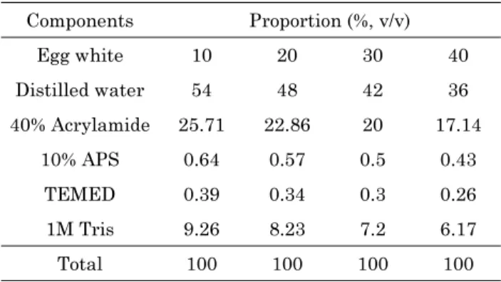
The egg white concentration in the egg white phantom was adjusted from 10-40% in 10% intervals by mixing the egg white with polyacrylamide solution.
The phantom was fabricated as previously described by Takegami et al [6]. Distilled water 1 M Tris, egg white, acrylamide/bis-acrylamide (19:1 ratio), 10%
ammonium persulfate (APS), and 10% N,N,N',N'- Tetramethylethane-1,2-diamine (TEMED) were mixed sequentially. The mixture was hardened by poly- acrylamide polymerization. The polyacrylamide intro- duced fiber crosslinking by copolymerization with N,
N'-Methylenebisacrylamide; polymerization was initi- ated by the addition of APS, which generates the required free radicals in the presence of TEMED.
Table 1 summarizes the phantom composition with increasing egg white.
2. Acoustic properties
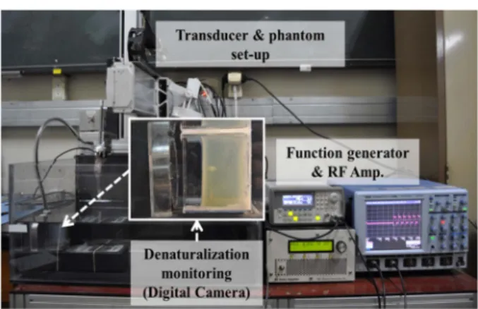
The sound velocity, attenuation coefficient, density, and acoustic impedance were measured for each phantom, which is illustrated in Fig. 1. A water tank was filled with degassed water maintained at 37
oC, similar to the human body temperature. A single 3.5 MHz transducer was used to send and receive the ultrasonic signals (Technisonic Research, Inc., US). A function generator was used as an ultrasonic pulser/
receiver (MKPR-1030, M.K.C Korea Inc., Korea). The received signal was stored in a digital oscilloscope (Waverunner 6100A, Lecroy, USA) and analyzed by Acqknowledge 3.7.5 (BIOPAC Systems, Inc., USA).
The sound velocity in the fabricated phantom was calculated from the echo range by measuring ∆t during a transmitted round trip; the attenuation coefficient was calculated using from the reflected signal as follows (1):
α( f ) = −20/(2d)log
10(A( f )/A
0( f )) (1)
where α, in dB/cm is the attenuation coefficient of the phantom, d is the sample thickness, A( f ) is the reflected signal amplitude at the frequency f with the phantom, and A
0( f ) is the reflected signal amplitude at the frequency f without the phantom. The density of the phantom was calculated by measuring the volume and weight of specimen sized 5 × 5 × 5 cm
3Fig. 1. The experimental setup for evaluating the acoustic properties of the phantom.
Table 1. The phantom composition with increasing egg white.
Components Proportion (%, v/v)
Egg white 10 20 30 40
Distilled water 54 48 42 36
40% Acrylamide 25.71 22.86 20 17.14
10% APS 0.64 0.57 0.5 0.43
TEMED 0.39 0.34 0.3 0.26
1M Tris 9.26 8.23 7.2 6.17
Total 100 100 100 100
9 fabricated phantom. The acoustic impedance was mea-
sured by multiplying the sound velocity and density of each phantom. Ten samples per each egg white concentration were used for measuring the acoustic properties, and they were measured repeatedly 5 times.
After measuring the acoustic properties of egg white phantom, the most similar tissue with the egg white phantom was confirmed by comparing the liver, breast, and soft tissue of human.
3. Thermal properties
To assess the thermal properties, the specific heat was measured in each phantom, and the thermal lesion was characterized by focused ultrasonication.
The temperature change of the water and within the specimen was measured. The specific heat was then calculated based on the law of heat conservation as follows (2):
c
1= (c
2m
2∆T
2)/(m
1∆T
1) (2)
where c
1and c
2are the specific heat of the phantom and water; m
1and m
2are the weights of the phantom and water; and ∆T
1and ∆T
2are the temperature changes of the phantom and water, respectively. The specific heat of the water, c
2is generally 1 at 20
oC.
To determine the lesion generated by a therapeutic ultrasound, a focused ultrasound was performed on the phantom specimen. A concave type single trans- ducer (H-101, Sonic Concepts Co., USA) was used at a 1.1 MHz center frequency, a 100 μs period, 3.5 V
p-pburst wave, and 10 cycles. The electrical power was varied between 40 W, 60 W, and 80 W and lasted 60 s. The focal pressure obtained from each electrical power using a PVDF needle hydrophone in degassed water at 25
oC. For the electrical powers of 40 W, 60 W, and 80 W, the focal pressures were 3.9, 4.70, and 6.2 MPa (acoustic intensity of 523.3, 737.6, and 1,298.8 W/cm
2), respectively. Fig. 2 shows the experi- mental setup for evaluating the thermal lesion by focused ultrasonication. After ultrasonication, the length of the resulting lesion was measured for each phantom according to the egg white concentration.
Fig. 3 shows the mimetic diagram of thermal lesion after ultrasonication. (a) is the width of lesion that
was measured parallel to the lateral direction of ultrasound transducer. (b) is the length of head in a tadpole-shaped lesion, (c) is the distance between the front surface of the phantom to the lesion boundary, (d) is the full-length of lesion. (b), (c), and (d) were measured parallel to the axial direction of ultrasound transducer.
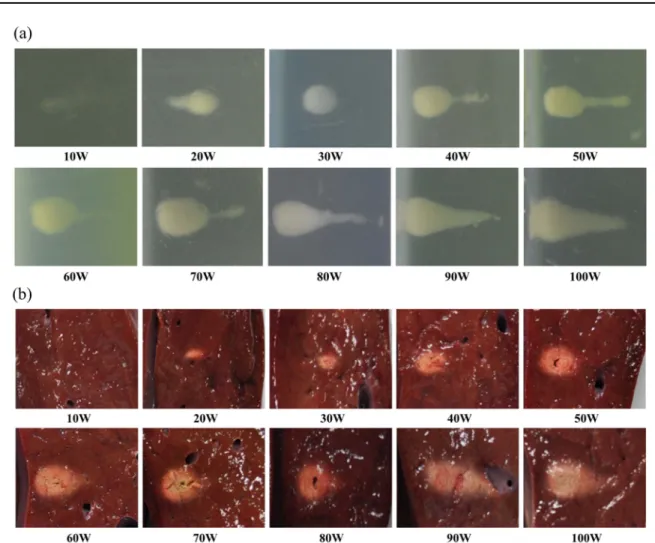
4. Tissue thermal lesion
The tissue morphology was compared between the bovine liver and the egg white phantom. The liver specimens were excised within 24 hours of slaughter to ensure freshness, cut to the size of 8.2 × 8.2 × 4 cm
3, and then fixed into an acrylic frame. A focused ultrasound was performed at conditions identical to those used to evaluate the phantom except the electrical power, which was varied at 10 W intervals between 10-100 W; For each electrical powers, the focal pressures were 2.1, 2.7, 3.4, 3.9, 4.3, 4.7, 5.3, 5.5, 6.2, and 6.4 MPa (acoustic intensity of 146.8, 242.2, Fig. 2. The experimental setup for evaluating the thermal lesion by focused ultrasonication.
Fig. 3. The mimetic diagram of thermal lesion after ultra-
sonication. (a) width, (b) head-length, (c) distance from the
surface, and (d) full-length.
10
385.7, 523.3, 618.6, 737.6, 942.9, 1,002.9, 1,298.8, and 1,353.8 W/cm
2), respectively. To confirm the lesion formation and shape, the lesion in the egg white phantom was monitored in real-time continuously by digital camera (the maximum resolution is 1,230 megapixel). In the liver tissue, the middle of lesion was incised by monitoring with B-mode ultrasonic image to confirm the lesion formation and shape. Its image recorded using the same digital camera. The images of the lesions were traced by the same operator in ImageJ, and the lesion areas were calculated using a MATLAB program.
III. Results
1. Acoustic evaluation
Fig. 4 summarizes the acoustic property changes of the phantom according to the egg white concentration.
The sound velocity decreased as the percentage of egg white increased between 10-30% (10% intervals);
at a 40% concentration, the sound velocity was nearly identical to the velocity at 30% (logarithmic fit, coefficient of determination = 0.9526). The attenuation
coefficient increased linearly (linear fit, coefficient of determination = 0.9057). Both the density and acoustic impedance increased as the egg white concentration increased, but the density plateaued at a 40% con- centration (logarithmic and linear fit, coefficient of determination = 0.9764 and 0.9077, respectively).
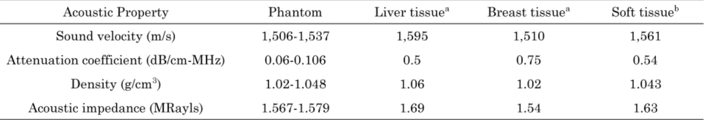
Table 2 summarizes the acoustic property com- parisons between the egg white phantom (10-40% egg white), liver tissue, and breast tissue. The sound velocity of the egg white phantom was 1,506-1,537 m/s, which differed from the liver tissues, but was similar to breast tissue. The acoustic impedance and density were 1.02-1.048 g/cm
3and 1.567-1.579 MRayls, respectively, and were similar among all three tissues.
The attenuation coefficient of the phantom was 0.06- 0.106 dB/cm-MHz), which was 1/5 of the liver tissue coefficient. Overall, the acoustic properties of the phantom were most similar to those of breast tissue, except the attenuation coefficient.
2. Thermal property evaluation
As the egg white concentration increased (10%, 20%, and 30%), the specific heat of the phantoms de-
Fig. 4. The acoustic property changes of the phantom according to the egg white concentration; (a) sound velocity, (b)
attenuation coefficient, (c) density, (d) acoustic impedance.
11 creased to 1.79, 1.532, 1.505, and 1.448 cal/g
oC, re-
spectively (logarithmic fit, coefficient of determination
= 0.9246). On visual examination, the lesion was tadpole-shaped at all concentrations performed at the same electrical power (80 W); the shape was most dis- tinct in the 30% egg white phantom. Fig. 5 illustrates the lesion shapes identified by the focused ultrasound (80 W) at the varying egg white concentrations.
For quantitative analysis, the lesion length was measured parallel to the axial direction of ultrasound transducer, and the perpendicular dimension was designated as the width; the area was calculated at each point along the entire length and width. As the egg white concentration increased, the lesion length also increased until plateauing at a 40% concentration.
However, the distance between the front surface of the phantom to the lesion boundary, directed towards the transducer, decreased as the egg white concen-
tration increased; this confirmed that the lesion formed gradually directed towards the transducer.
These dimensions in each phantom are illustrated in Fig. 6. In addition, the visual transparency of the phantom decreased as the egg white concentration increased. As a result, it was difficult to discern the lesion in the 40% egg white phantom, which had the lowest visual transparency. The color of the formed lesion was unclear due to the low concentration of thermal-sensitive indicator in the 10% egg white phantom; the lesion was easier to distinguish in the 30% egg white phantom compared to the 20% egg white phantom.
3. Morphologic comparison between the egg white phantom and tissue
The length, area, and shape of the lesions in the egg white phantom and bovine liver tissue identified Table 2. The acoustic property comparisons between the egg white phantom (10-40% egg white), liver tissue, and breast tissue.
Acoustic Property Phantom Liver tissue
aBreast tissue
aSoft tissue
bSound velocity (m/s) 1,506-1,537 1,595 1,510 1,561
Attenuation coefficient (dB/cm-MHz) 0.06-0.106 0.5 0.75 0.54
Density (g/cm
3) 1.02-1.048 1.06 1.02 1.043
Acoustic impedance (MRayls) 1.567-1.579 1.69 1.54 1.63
aICRU 1998, bMast et al. 2000.