머루 과피와 종자 추출물의 식품 위해성 세균에 대한 항균성 및 인체 암세포주에 대한 cytotoxicity 분석
원지혜 ・ 김미라1†
경북대학교 식품영양학과, 1경북대학교 식품영양학과 ․ 장수생활과학연구소
Analysis of Antibacterial Activity against Food Spoilage and Food-borne Pathogens and Cytotoxicity on Human Cancer Cell Lines of Extracts from Pericarp and Seed of Vitis coignetiea
Ji Hye Won and Meera Kim1†
Department of Food Science and Nutrition, Kyungpook National University,
1Department of Food Science and Nutrition, Center for Beautiful Aging, Kyungpook National University
Abstract
In this study, antibacterial activity and cytotoxicity of the extracts from pericarp and seed of Vitis coignetiea, which were extracted with 0.1% HCl-60% ethanol, were analyzed. The antibacterial activity of the extracts was determined by paper disc diffusion method against food spoilage and food-borne pathogens. The pericarp extract showed high antibacterial activity against Bacillus cereus, Escherichia coli O157:H7, and Pseudomonas aeruginosa, and the seed extract represented the high antibacterial activity against B. cereus, E. coli O157:H7, and Staphylococcus aureus. The cytotoxicity of the Vitis coignetiea extract against human cancer cells was determined using the MTT assay and SRB assay. The pericarp extract represented strong growth-inhibition activity against G361 and Hep3B cells and the seed extract greatly inhibited the growth of HeLa and G361 cells in the MTT assay. In addition, the pericarp extract displayed a high inhibition activity against the growth of AGS cells and the seed extract greatly inhibited the growth of HeLa, Hep3B, and MCF7 cells in the SRB assay. Especially, the cytotoxicities of the seed extract against HeLa were significantly higher than those of the extract against other cancer cells at all test concentrations. This study demonstrates that the extract from pericarp and seed of Vitis coignetiea possess high antibacterial activity and cytotoxicity.
Key words : antibacterial activity, cytotoxicity, Vitis coignetiea
I. 서 론1)
최근 경제성장과 생활수준의 향상 등으로 삶의 질이 향상 되면서 건강에 대한 관심도 높아지고 있다. 식생활 개선을 통해 건강한 삶을 누리고자 하는 노력이 증가하고 있으며 이
†Corresponding author : Meera Kim, Department of Food Science and Nutrition, Kyungpook National University, Daegu, 702-701, South Korea Tel: +82-53-950-6233
Fax: +82-53-950-6229 E-mail: [email protected]
에 따라 식품이 가지고 있는 항암, 항산화, 항균 같은 생리활 성에 대한 관심도 증가하고 있다. 암, 비만, 심혈관계 질환 및 노화의 원인은 활성산소에 의한 세포손상이 주된 원인으 로 여겨지고 있다. 활성산소종은 우리 몸에서 세포내 신호전 달 물질로서 중요한 역할을 하고 있으나 지나치게 증가되면 조직을 공격하게 되어 생체기능 저하 및 암 유발과 같이 정 상적인 대사를 방해한다(Ludvission J 1993, MeCord JM. 2000,
Park CS 2005). 따라서 항산화작용의 중요성이 부각되고 있으
며 항산화, 항암성을 가지고 있는 식품에 대한 연구들이 수행 되었다(Hwang JH 2002, Jeong SW 2005, Kang HH 2009, Kim YH 등 2008, Lee HJ 2009, Lee HS와 Son JY 2002, Park CS
2005). 이와 더불어 항균작용을 통해 식품의 저장기간을 연장 시키기 위하여 사용되는 식품보존료 중 합성 보존료의 위해
(危害) 가능성이 대두되면서 천연 항균물질에 대한 관심도 증가
하고 있으며(Lee DH 등 2004), 천연 물질인 폴리페놀과 플라보 노이드의 항균성에 대한 연구들이 많이 수행되었다(Han YA 등 2009, Lee DW 2003, Oh SH 등 2006a, Oh SH 등 2006b).
머루(Vitis coignetiea, wild grape)는 쌍떡잎식물강의 갈매나 무목 포도과에 속하는 낙엽성 덩굴식물로 중국, 일본 우리나 라에서 야생으로 자생하고 내한성과 병충해 저항성이 강하다. 우리나라에서는 예로부터 열매는 술을 만들어 먹었으며 잎 추출물은 구토와 설사, 동상, 빈혈 완화를 위하여 민간요법에 이용하였다(Kim NY 등 2006). 머루에는 수용성 비타민 등 필 수 영양소가 풍부하여 두뇌발달과 소화촉진의 기능을 하며 (Cheon KB 1999, Choi SY 등 2006) 동일 vitis 속인 포도보다 10배 이상의 인, 철분, 칼륨, 칼슘을 함유하고 있다(Cheon KB 1999). 또한 머루 뿌리에는 resveratrol이 중심구조인 viniferin, amurensin 등이 함유되어 있는 것으로 나타났으며(Huang KS
등 2000), 이들은 염증치료에 효과적인 것으로 보고되었다
(Choi SY 등 2006). 머루는 술로 많이 섭취하고 있어 이에 대 한 품질 특성과 발효조건 최적화에 대한 연구가 수행되었다 (Kim SH 2008, Kim YH 2009, Lee DH 등 2004, Suh JS
2010,). 머루의 생리활성에 대한 연구로는 머루 종자 추출물
의 항산화물질 탐색(Kim NY 등 2005, Kim NY 등 2006)과 머 루 과피 추출물의 항산화활성 탐색(Choi SY 등 2006), 머루씨 분말첨가가 흰쥐에 대한 항산화활성과 혈액지질 조성에 미치 는 효과(Won HR 2007) 등이 보고되었다. 그러나 동종인 포 도에 비해 머루의 항암 및 항균 활성 등의 생리활성에 대한 연구는 아직까지 미비한 실정으로 본 연구에서는 머루의 생 리활성과 머루를 이용한 기능성 식품 소재에 대한 기초자료 를 얻고자 머루과피와 종자로부터 추출물을 얻고 이들의 식 품위해성 세균에 대한 항균성 및 인체 암세포에 대한 증식 억제 활성을 분석하였다.
II. 재료 및 방법
1. 재료 및 시약
1) 실험재료 및 시약
본 실험에 사용한 머루는 경북 봉화에서 재배하여 수확한 것 을 사용하였다. 3-(4,5-dimethylthiazol-2-yl)-2,5-diphenyltetrazolium (MTT), dimethylsulfoxide(DMSO), sulforhodamineB(SRB)는 Sigma Chemical Co.(St. Louis, MO, USA)로부터 구입하였으며 fetal bovine serum(FBS)은 Gibco BRL(USA), minimum essential medium(MEM, with L-glutamin & Earle's Balanced salts)은 Hyclone Co.(USA)에서 구입하였다. 균 배양을 위한 고체배지로 nutrient agar(Acumedia, USA)를 이용하였으며 액 체배지는 nutrient broth(Acumedia, USA)와 tryptic soy broth(Acumedia, USA)를 사용하였다.
2) 사용 균주 및 암세포주
항균실험에 사용된 균주는 Eschericha coli(KTCT 1682), Escherichia coli O157:H7(ATCC 43894), Pseudomonas aeruginosa(KCTC 1750), Salmonella typhimurium(KCTC 2515), Listeria monocytogenes(KCTC 3569), Bacillus cereus(KCTC 1012), Staphylococcus aureus(KCTC 1916)로 이들을 37℃ 배양 기에서 계대배양하여 실험에 이용하였다.
암세포 증식 억제 활성을 검색하기 위하여 인체 암세포주 인 A549(폐암 세포), AGS(위암 세포), G361(피부암 세포), HeLa(자궁경부암 세포), Hep3B(간암 세포), MCF7(유방암 세 포)를 한국세포주은행(KCLB)으로부터 분양받아 사용하였다. 세포 배양액으로는 10% FBS와 1% penicillin-K -streptomycin이
첨가된 RPMI 1640을 사용하였다. 배양액에 분주된 세포주는
37℃, 5% CO2 배양기에서 배양하여 실험에 사용하였다.
2. 실험 방법
1) 머루 추출물 제조
머루는 과피와 종자를 분리하고 종자는 45℃에서 하루 동 안 건조시킨 후 분쇄하였다. 머루 과피 및 종자 100 g에 각 각 0.1% HCl-60% ethanol(1:1, V/V) 2 L를 가하여 12시간 추 출한 후 여과지(Toyo No. 2, Advantec, Japan)로 여과하는 하 는 과정을 4회 반복하였다. 이 여과액을 회전감압농축기 (EYELA, Rikakiki Co., Japan)로 농축하고 동결건조한 후 추출 수율을 구하였으며, 추출물은 -20℃에서냉동 보관하여 실험에 사용하였다.
2) 총 폴리페놀 및 총 플라보노이드 함량 측정 (1) 총 폴리페놀 함량 측정
총 폴리페놀 함량은 Conforti 등(2007)의 방법을 이용하여 측정하였다. Acetone 40 mL, methanol 40 mL, acetic acid 0.1 mL 및 증류수 0.1 mL를 혼합한 용액 25 mL에 시료 50 mg을 넣고 60℃에서 1시간 동안 방치한 후 상온에서 냉각시켰다. 30초간 vortex로 혼합한 다음 30초간 sonication하였다. 이 용 액 200 μL와 Folin-Ciocalteu 시약 1 mL, 7.5% sodium carbonate 1 mL를 넣고 vortex로 혼합하여 2시간 동안 방치한 후 UV/Visible spectrophotometer(Beckman, USA)를 사용하여 726 nm에서 흡광도를 측정하였다. 표준물질로 gallic acid를 사용하여 총 폴리페놀 함량을 계산하였다.
(2) 총 플라보노이드 함량 측정
머루 과피 및 종자 추출물의 플라보노이드 함량은 Moreno MI 등(2000)의 방법을 이용하여 측정하였다. 80% ethanol에 0.5%로 녹인 시료 0.5 mL와 10% aluminum nitrate 0.1 mL, 1 M potassium acetate 0.1 mL, 80% ethanol 4.3 mL를 혼합한 후 UV/Visible spectrophotometer(Beckman, USA)를 사용하여
510 nm에서 흡광도를 측정하였다. 표준물질로 quercetin을 사 용하여 총 플라보노이드 함량을 계산하였다.
3) 항균성 측정
머루 추출액의 항균성은 paper disc assay를 이용하여 측정 하였다(Bauer AW 등 1966, Kwak DJ 등 2002). 세균은 nutrient broth와 tryptic soy broth를 이용해 37℃에서 12시간 배양하고 1×10⁸ CFU/mL로 희석하였다. 희석된 균 배양액 100 μL를 취해 nutrient agar로 만든 평판배지에 도말하고 균 이 고착되도록 4℃에서 2시간 동안 방치하였다. 균이 도말된 plate 위에 멸균된 직경 6 mm의 paper disc(Advantec, Japan) 를 올리고 syringe filter (pore size 0.2 μm, Advantec, Japan) 를 통과시켜 제균한 시료를 농도별로 각각 20 μL씩 흡수시 킨 후 37℃의 incubator에서 24시간 배양하였다. 배양 후 disc 주위에 생성된 clear zone의 크기를 측정하였다.
4) 인체 암세포주에 대한 cytotoxicity 측정 (1) MTT assay
Carmichael J 등(1987)의 방법을 이용하여 머루 추출물의 MTT assay를 수행하였다. 96 well plate의 각 well에 1×10⁴ cells/mL의 농도로 희석한 암세포 180 µL를 분주하고 37℃의 5% CO2 incubator에서 24시간 동안 선 배양하였다. 그 후, 시 료 100 µL를 첨가하여 같은 조건의 CO2 incubator에서 48시간 동안 배양하였다. 배양 후 각 well에 MTT 용액 20 µL를 첨가 하여 4시간 배양한 후 배양액을 모두 제거하였다. 각 well에 DMSO:ethanol(1:1, v/v) 용액 150 µL를 첨가하여 30분간 교반 하고 ELISA reader (Tecan, Switzerland)를 이용하여 550 nm에서 흡광도를 측정하여 암세포 활성 저해 효과를 구하였다.
Cytotoxicity(%)=(1—시료 첨가구의 흡광도/시료 무첨가군의 흡광도)×100
(2) SRB assay
머루 추출물의 SRB assay는 Doll R과 Peto R(1981)의 방법 을 이용하여 측정하였다. 암세포를 5×10⁴cells/mL 농도로 희 석하여 100 µL씩 96 well plate의 각 well에 첨가하여 24시간 동안 37℃, 5% CO2 incubator에서 선 배양한 후, 각 well당 시료 100 µL씩을 첨가하여 48시간 동안 37℃, 5% CO2
incubator에서 재 배양하였다. 상등액을 제거하고 10% TCA를 각 well에 100 µL씩 첨가하여 1시간 동안 4℃에서 냉장 방치
한 후, TCA를 제거하고 증류수로 5번 세척한 다음 실온에서
건조하였다. 각 well에 1% acetic acid에 녹인 0.4% SBR 용액 을 100 µL씩 첨가하여 30분 동안 염색한 후, 1% acetic acid 로 5번 세척한 다음 건조시켰다. 그 후 10 mM Tris buffer(pH 10.5) 100 µL로 염색된 SRB를 녹여낸 후 540 nm에
서 ELISA reader를 이용하여 흡광도를 측정하고 암세포 활성
저해 효과를 구하였다.
Cytotoxicity(%)=(1—시료 첨가구의 흡광도/시료 무첨가군의 흡광도)×100
5) 통계분석
실험결과는 SPSS(v. 18.0) 프로그램을 이용하여 유의차를 검정하였다. 각 실험값의 평균치간 유의성은 분산분석을 실시 하여 Duncan's multiple range test(p<0.05)로 검정하였다.
III. 결과 및 고찰
1. 머루 추출물의 추출 수율 및 총 폴리페놀 함량, 총 플 라보노이드 함량
머루 과피 및 종자 100 g을 0.1% HCl-60% ethanol로 추출 한 추출물의 수율은 각각 17.51%와 8.39%로 나타나 과피 추 출물의 수율이 종자 추출물의 수율보다 1.5배 이상 높은 것 으로 나타났다(Table 1). 또한 머루 과피 및 종자 추출물의 총 폴리페놀 함량은 각각 5.98과 9.94 mg/g이었으며, 총 플라 보노이드 함량은 2.76, 20.06 mg/g으로 나타나 과피 추출물에 비해 종자 추출물에 총 폴리페놀과 총 플라보노이드가 더 많 이 함유되어 있는 것으로 분석되었다. 이는 포도의 종자 에 탄올 추출물이 과피 에탄올 추출물보다 약 7배 이상의 폴리 페놀화합물을 가지고 있다고 보고한 Park SJ 등(2003)의 연구 결과와 유사하였다. 천연물 추출 관련 선행연구들을 보면 추 출 용매에 따라 폴리페놀 함량이 다르게 나타나는데 머루 과 피의 ethyl acetate 분획물의 폴리페놀 함량은 54.4 mg/100 g 으로 보고되어(Choi SY 등 2006) 0.1% HCl-60% ethanol을 용 매로 이용한 본 연구에서 폴리페놀 함량이 더 높게 나타났 다. 또한 Park HS(2011)은 hexane, chloroform, ethyl acetate, butanol, water 등 여러 용매를 이용하여 머루 과피 추출물의 총 폴리페놀 함량을 분석한 결과 용매에 따라 4.7~53.4 mg/100 g으로 나타나, 본 연구에서의 분석된 총 폴리페놀 함 량이 좀 더 높은 것으로 나타났다. 따라서 본 연구에서 사용 한 0.1% HCl-60% ethanol이 폴리페놀 추출에 효율적인 용매 인 것으로 확인되었다.
Extract Yield (%) Total polyphenol content1) (mg/g)
Total flavonoid content2) (mg/g)
Pericarp 17.51 5.98±0.00 2.76±0.03
Seed 8.39 9.94±0.01 20.06±0.0
1) Total polyphenol content was based on gallic acid as a standard
2) Total flavonoid content was based on quercetin as a standard Table 1. Yield, total polyphenol content, and total flavonoid
content of Vitis coignetiea extract
Extract Microorganism Concentration (mg/disc)
1.5 2 2.5 3 3.5 4
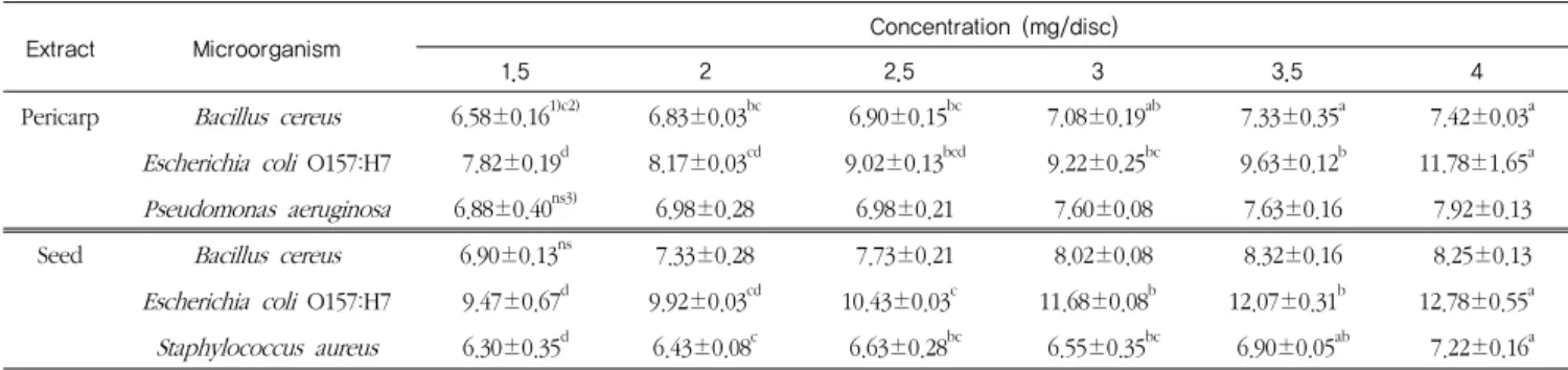
Pericarp Bacillus cereus 6.58±0.161)c2) 6.83±0.03bc 6.90±0.15bc 7.08±0.19ab 7.33±0.35a 7.42±0.03a Escherichia coli O157:H7 7.82±0.19d 8.17±0.03cd 9.02±0.13bcd 9.22±0.25bc 9.63±0.12b 11.78±1.65a Pseudomonas aeruginosa 6.88±0.40ns3) 6.98±0.28 6.98±0.21 7.60±0.08 7.63±0.16 7.92±0.13 Seed Bacillus cereus 6.90±0.13ns 7.33±0.28 7.73±0.21 8.02±0.08 8.32±0.16 8.25±0.13 Escherichia coli O157:H7 9.47±0.67d 9.92±0.03cd 10.43±0.03c 11.68±0.08b 12.07±0.31b 12.78±0.55a Staphylococcus aureus 6.30±0.35d 6.43±0.08c 6.63±0.28bc 6.55±0.35bc 6.90±0.05ab 7.22±0.16a
1) Diameter of clear zone was the mean triplicated experiments.
2) The different superscripts (a-d) are significantly different among various extract concentrations of each part of Vitis coignetiea for each bacterium (p<0.05).
3) ns: not significant
Table 2. Clear inhibition zone for microorganisms by various concentrations of the Vitis coignetiea extract
2. 머루 과피 및 종자 추출물의 항균성
머루 과피 및 종자 추출물의 7가지 식품위해성 세균에 대 한 paper disc assay 결과를 Table 2에 나타내었다. 과피 추출 물에서는 Bacillus cereus, Escherichia coli O157:H7, Pseudomonas aeruginosa에 대해서, 종자 추출물에서는 B.
cereus, E. coli O157:H7, Staphylococcus aureus에 대해서 뚜렷 한 clear zone을 확인할 수 있었다. 또한, 머루 과피 추출물의 농도가 증가할수록 B. cereus와 E. coli O157:H7에 대한 clear zone이 유의적으로 크게 나타났고(p<0.05), 종자 추출물의 농 도가 증가할수록 E. coli O157:H7, S. aureus에 대한 clear zone이 유의적으로 증가하였다(p<0.05). 머루 과피 추출물과 종자 추출물 모두 실험에 사용된 모든 농도에서 E. coli O157:H7에 대해 가장 높은 항균성을 보였다. 머루와 같은 속 (genus)인 포도 추출물의 항균성 연구(Chung HY와 Park DK 2003, Lee MC 등 1997, Nilgun GB 등 2004)에서도 포도 추출 물이 B. cereus, S. aureus, P. aeruginosa에 대해 항균성이 높 다고 보고되었는데 본 실험에서도 이와 유사한 경향을 나타 내었다. 한편 유색 감자에서 추출한 안토시안의 항균성 연구 (Jeon TW 등 2005)와 오배자와 포도 껍질 추출물의 항균성 연구(Lee MC 등 1997)에서는 안토시아닌이 P. aeruginosa에 대해 강력한 항균성을 가지고 있다고 보고하였다. 머루에도 다량의 안토시아닌과 폴리페놀 화합물이 함유되어 있는데
(Park HS 2010) 이러한 안토시아닌과 폴리페놀 화합물은 항균
성을 가지고 있는 것으로 나타나고 있다. 특히 폴리페놀 화 합물의 일종인 resveratrol은 높은 항균성을 가지고 있는데 (Chan MM 2002, Filipa V 등 2003) 머루에는 항균성이 높은 resveratrol이 포도보다 9.6배에서 68.5배까지 많이 함유되어 있다고 보고되었다(Youn JH 등 2003). 또한 Son RH 등(2010) 은 머루 줄기와 자소자로부터 추출한 resveratrol이 S. aureus 에 대해 강력한 항균작용을 가지고 있다고 보고하였고, Toda M 등(1990)과 Ikigai H 등(1993)도 머루 종자 에탄올 추출물 에서 항균작용이 있는 catechin을 분리하였다고 보고한 바 있 다. 따라서 본 연구에서도 머루에 함유되어 있는 안토시아닌
과 폴리페놀 화합물이 식품위해 세균에 대한 항균성에 영향 을 미친 것으로 사료되었다.
3. 머루 과피 및 종자 추출물의 인체 암세포주에 대한 cytotoxicity
머루 과피 및 종자 추출물을 가지고 A549(폐암 세포), AGS(위암 세포), G361(피부암 세포), HeLa(자궁경부암 세포), Hep3B(간암 세포) 및 MCF7(유방암 세포)에 대한 MTT assay 와 SRB assay를 수행한 결과는 다음과 같다.
1) MTT assay
MTT assay는 MTT가 생존 암세포와 효소작용을 하여
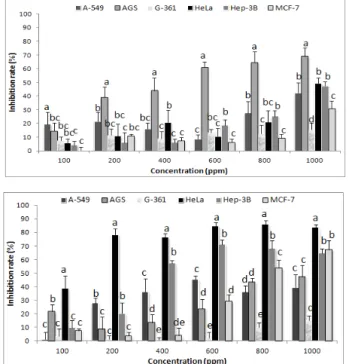
formazan crystal로 침전되는 정도를 흡광도로 측정하여 함암 활성물질에 의한 암세포 사멸정도를 측정하는 방법으로 대량 검색이나 항암효과의 1차적 검색에 적합한 방법이다(Ko SH 1998). 본 실험에서는 MTT assay를 이용하여 각 암세포에 대 한 머루 과피 및 종자 추출물의 농도별 암세포 증식 억제정 도를 측정하였고 그 결과를 Figure 1에 나타내었다. 머루 과 피 및 종자 추출물은 대체로 추출물의 농도가 증가할수록 암 세포 증식 억제 활성도 증가하는 경향을 나타내었다.
머루 과피 추출물은 100, 200 ppm에서 MCF7에 대해 유의 적으로 높은 cytotoxicity를 나타내었고(p<0.05), 600~1,000 ppm에서는 G361에 대해 높은 cytotoxicity를 나타내었다. 머 루 과피 추출물의 경우 800 ppm에서 G361에 대해 cytotoxicity가 80% 이상으로 높게 나왔으며, Hep3B에 대한 cytotoxicity도 70% 이상을 보여주었다. 본 연구에서는 머루 과피 추출물 1,000 ppm 농도에서 MCF7에 대한 cytotoxicity가
54.38%로 나타났는데 이 결과를 안토시아닌이 풍부한 식품인
김의 에탄올 추출물 1,000 µg/assay(약 2,941 ppm) 농도에서 유방암 세포의 증식이 50.49% 억제되었다는 선행연구(Kim SA 등 2005) 결과와 비교할 때, 머루 과피 추출물의 유방암 세포
Figure 1. Inhibitory effects of extract from pericarp and seed of Vitis coignetiea on the human cancer cells by MTT assay (A: pericarp extract, B: seed extract).
Data were the means of triplicate determinations.
The different superscripts (a~e) are significantly different among various cell lines at each concentration of Vitis coignetiea (p<0.05).
억제에 대한 효과가 좀 더 높은 것으로 보였다. Kim YH
(2009)은 머루 과피에서 추출한 안토시아닌으로 항암활성을
측정한 결과, 안토시아닌 농도에 의존적으로 간암세포주인
Hep3B와 HepG2의 생존율이 감소하여 안토시아닌이 항암활
성을 가지고 있음을 확인하였다. 따라서 본 연구에서도 머루 과피에 함유된 안토시아닌이 암세포 증식 억제에 기여했을 것으로 생각되었다. 또한 Jang M 등(1997)은 18주 동안 resveratrol을 투여한 쥐에서 피부암 세포가 68~98% 감소하였 다고 보고하였는데 본 연구에서도 800 ppm과 1,000 ppm 농 도에서 머루 과피 추출물이 다른 암세포주에 비해 피부암 세 포인 G361에 대해 유의적으로 높은 cytotoxicity를 나타내어 (p<0.05) 머루에 함유된 resveratrol 등이 피부암 세포 증식 억 제에 효과를 주었을 것으로 추측되었다.
머루 종자 추출물은 100~400 ppm으로 처리한 MCF7를 제 외하고는 과피 추출물보다 대체로 암세포 증식 억제율이 큰 것으로 나타났다. 머루 종자 추출물은 400 ppm에서 A549, AGS, G361에 대한 cytotoxicity가 60% 이상으로 높았고, HeLa 에 대해서는 600, 800, 1,000 ppm에서 85% 이상의 높은 cytotoxicity를 보여주었다. 머루 종자 추출물은 600~1,000 ppm에서 G361에 대해서도 80% 이상의 cytotoxicity를 나타내 었다. 이러한 높은 암세포 증식 억제율을 선행연구와 비교해
Figure 2. Inhibitory effects of extract from pericarp and seed of Vitis coignetiea on the human cancer cells by SRB assay (A: pericarp extract, B: seed extract).
Data were the means of triplicate determinations.
The different superscripts (a~e) are significantly different among various cell lines at each concentration of Vitis coignetiea(p<0.05).
보면 산수유(Jeon YH 등 2008) 에탄올 추출물의 경우 700 µg/plate(약 3,181 ppm)에서 HeLa에 대해 77.8%의 암세포 증 식 억제율을 나타내었다고 보고되어, 본 연구의 머루 종자 추
출물의 HeLa 세포에 대한 암세포 증식 억제활성이 더 높은
것을 알 수 있었다.
2) SRB assay
SRB assay는 핵 내 sulforhodamin B 단백질량을 측정해 항 암활성을 알아보는 방법으로 항암효과의 1차 검색방법 중 매 우 안정적인 방법으로 알려져 있다(Skehan P 등 1988; Seo DH와 Han DS 2003). 본 연구에서는 머루 과피 및 종자 추출 물의 농도별 암세포 증식 억제 활성을 SRB assay로 측정하여 그 결과를 Figure 2에 나타내었다. 머루 과피 추출물은 200~1,000 ppm의 농도에서 AGS에 대해 유의적으로 높은 세 포증식 억제율을 나타내었으며(p<0.05), 1,000 ppm 농도에서 AGS에 대하여 69.10%의 cytotoxicity를 보였다. AGS에 대한 세포증식 억제를 살펴본 Ham YA 등(2009)의 연구에서 잔대 에탄올 추출물은 1,000 ppm에서 AGS에 대해 64.90%의 항암 활성을 나타내었다고 보고하여 머루 과피 추출물이 AGS에 대 하여 비교적 높은 세포 증식 억제 활성을 가지는 것으로 보 였다.
머루 종자 추출물은 HeLa에 대해 가장 현저한 세포 증식
억제율을 보였고, Hep3B와 MCF7에 대해서도 비교적 높은 억 제율을 나타내었다. HeLa에 대해서는 100~1,000 ppm의 모든 농도에서 다른 세포에 비해 유의적으로 높은 cytotoxicity를 보였고(p<0.05), 600 ppm 이상에서는 80% 이상의 높은 cytotoxicity를 나타내었다. Jeon YH(2008)의 연구에서는 복분 자 에탄올 추출물이 HeLa에 대해 59.5%의 cytotoxicity를 나타 내었다고 보고하여 본 머루 종자 추출물이 HeLa에 대하여 더 높은 암세포 증식 억제 활성을 가지는 것으로 나타났다. 선 행연구에 의하면 폴리페놀 성분 중 catechin이 HeLa에 대해
높은 항암활성을 가지고 있으며 그 중
(-)-epigallocatechin-3-gallate(EGCG)의 세포 증식 억제 활성이 가장 높게 나타났다고 보고되었다(Jin YM 등 2002). 이로 미 루어 볼 때 본 연구에서 머루 종자 추출물의 HeLa에 대한 높 은 억제 활성에도 catechin과 같은 폴리페놀 화합물이 연관될 것으로 사료되며, 향후 이에 대한 후속연구가 더 필요할 것으 로 생각된다.
IV. 요약 및 결론
본 연구에서는 머루 종자와 과피를 0.1% HCl-60% ethanol 로 추출하여 추출물의 식품위해성 세균에 대한 항균성 및 인 체 암세포에 대한 세포 증식 억제 활성을 측정하였으며 주요 결과는 다음과 같다. 식품위해성 세균에 대한 paper disc assay 실시 결과, 머루 과피 추출물은 B. cereus, E. coli O157:H7, P. aeruginosa에 대해서, 종자 추출물은 B. cereus, E. coli O157:H7, S. aureus에 대하여 높은 항균성을 나타내었 다. 또한 MTT와 SRB assay를 이용하여 머루 과피와 종자 추 출물의 인체 암세포 증식 억제 활성을 조사한 결과 MTT assay에서는 머루 과피 추출물이 G361와 Hep3B에 대한 높은 cytotoxicity를 보였으며, 머루 종자 추출물은 600 ppm~1,000 ppm 농도에서 HeLa와 G361 세포에 대해 80% 이상의 높은 cytotoxicity를 나타내었다. SRB assay에서는 머루 과피 추출물 이 AGS에 대해 높은 cytotoxicity를 나타내었고, 머루 종자 추 출물은 HeLa에 대해 현저히 높은 cytotoxicity를 보였으며 Hep3B와 MCF7에 대해서도 비교적 높은 cytotoxicity를 나타내 었다. 전체적으로 볼 때 머루 종자 추출물은 여러 인체 암세 포에 대해 우수한 세포 증식 억제 활성을 가지고 있는 것으 로 나타나 이들 암세포 증식 억제를 위한 새로운 기능성 물 질로서의 개발 가능성을 가지고 있는 것으로 보였다.
참고문헌
Carmichael J, DeGraff WG, Gazdar AF, Minna JD, Mitchell JB. 1987.
Evaluation of a tetrazolium-based semiautomated colorimetric assay: Assessment of chemosensitivity testing. Cancer Res
47:936-942
Chan MY. 2002. Antimicrobial effect of resveratrol on dermatophytes and bacterial pathogens of the skin. Biochem Pharmacol 63(2):99-104
Cheon KB. 1999. Screening of antioxidant from Vitis coignetae, Vitis vinifera L. and comparition of its antioxidant activity. M.A Thesis. Konkuk University. Korea
Choi SY, Cho HS, Sung NJ. 2006. The antioxidative and nitrite scavenging ability of solvent extracts from wild grape (Vitis coignetiea) skin. Korean J Soc Food Sci Nutr 35(8): 61-966 Chung HY, Park DK. 2003. Antimicrobial activity of grape seed
ethanol extract. Korean J Soc Food Sci Nutr 32(1):109-114 Conforti F, Statti GA, Menichini F. 2007. Chemical and biological
variability of hot pepper fruits (Capsicum annuum var.
acuminatum L.) in relation to maturity stage. Food Chem 102:1096-1104
Doll R, Peto R. 1981. The causes of cancer: quantitative estimates of avoidable risks of cancer in the United States today. J Natl Cancer Inst 66(6):1191-1308
Filipa V, Plockováa, M, Šmidrkala, J, Špičkováa, Z, Melzochb, K, Schmidtc Š. 2003. Resveratrol and its antioxidant and antimicrobial effectiveness. Food Chem 83(4):585-593
Ham YA, Choi HJ, Kim SH, Chung MJ, Ham SS. 2009.
Antimutagenic and antitumor effect of Adenophora triphylla extracts. J Korean Soc Food Sci Nut 38(1):25-31
Huang KS, Lin M, Yu LN, Kong M. 2000. Four novel oligostilbenes from the roots of Vitis amurensis. Tetrahedron 56:1321-1329 Hwang JH. 2002. Antioxidative activities of anthocyanins from red
leaf mustard (Brassica juncea Coss. var. integrifolia). Ph.D Thesis. Pusan National University. Korea
Ikigai H, Nakae T, Hara Y, Shimamura T. 1993. Bactericidal catechins damage the lipid bilayer. Biochim Biophys Acta 1147(1):132-136
Jang M, Cai L, Udeani G, Slowing KV, Thomas CF, Beecher CWW, Fong SSD, Farnsworth NR, Douglas Kinghorn A, Mehta RG, Moon RC, Pezzuto JM. 1997. Cancer chemopreventive activity of resveratrol, a natural product derived from grapes. Science 275:218-220
Jeon TW, Cho YS, Lee SH, Cho SM, Cho HM, Jang KS, Park HJ.
2005. Studies on the biological activities and physicochemical characteristics of pigments extracted from Korean purple-fleshed potato. Korean J Food Sci Technol 37(2):247-254
Jeon YH. 2008. Physiological activity of extracts of Rubus coreanum,
and isolation and identification of its flavonoid. M.A. Thesis.
Kyungpook National University. Korea
Jeon YH, Kim MH, Kim MR. 2008. Antioxidative, antimutagenic, and cytotoxic activities of ethanol extracts from Cornus officianalis. J Korean Soc Food Sci Nut 37(1):1-7
Jeong SW. 2005. Antitumor and antioxidant activities of anthocyanins isolated from the seeds of Liriope platyphylla. M.A. Thesis.
Catholic University of Daegu. Korea
Jin YM, Nam SL, Ahn WS. 2002. Growth inhibition and induction of apoptosis in cervical cancer cell lines by green tea polyphenols.
Korean J Soc Obs Gyn 45(4):560-568
Kang HH. 2009. Determination of biological activities of Korean berries and their anthocyanin identification. Ph.D Thesis.
Gyeongsang National University. Korea
Kim NY, Choi JH, Kim YG, Jang MY, Moon JH, Park GH, OH DH.
2006. Isolation and identification of an antioxidant substance from ethanol extract of wild grape (Vitis coignetiea) seed).
Korean J Food Sci Technol 38(1):109-113
Kim NY, Kim YK, Bae KJ, Choi JH, Moon JH, Park GH, Oh DH.
2005. Free radical scavenging effect and extraction condition of ethanol extracts and fractions of wild grape weed (Vitis coignetiea). Korean J Soc Food Sci Nutr 34(6):755-758
Kim SA, Kim J, Woo MK, Kwak CS, MS Lee. 2005. Antimutagenic and cytotoxic effects of ethanol extracts from five kinds of seaweed. J Korean Soc Food Sci Nut 34(4):451-459
Kim SH. 2008. Optimal condition for deacidification fermentation of wild grape wine by mixed culture. Korean J Soc Appl Biol Chem 51(1):17-23
Kim YH. 2009. Optimum fermentation condition and functional quality characteristics of Meoru wine. Ph.D Thesis. Gyeongsang National University. Korea
Kim YH, Kim DS, Woo JS, Kim HH, Lee YS, Kim HS, Ko KH, Lee SK. 2008. Antioxidant activity and cytotoxicity on human cancer cells of anthocyanin extracted from black soybean. Korean J Crop Sci 54(4):407-412
Ko HS. 1998. Characteristics, and antimutagenic and in vitro anticancer effects of chungkookjang manufactured by various variety of soybean. M.A Thesis. Konkuk University. Korea Lee DH, Yu HE, Lee JS. 2004. Quality characteristics and
physiological functionality of wild grape wine. Korean J Natural Sci 15(1):69-78
Lee DW. 2003. Antioxidant and antimicrobial effects of citrus flavonoids and limonoids. Ph. D. Thesis. Yonsei University.
Korea
Lee HJ. 2009. Isolation and identification of anthocyanin in edible rose and determination of their biological activity. M.A. Thesis.
Kangwon National University. Korea
Lee HS, Son JY. 2002. Antioxidant and synergist effect of extract isolated from commercial green, oolong and black tea. Korean J Food Nutr 15(4):377-381
Lee MC, Kim KP, Kim SH, Choung NH, Yim MH. 1997.
Antimicrobial activity of extract from gall-nut and red-grape husk. Korean J Food Nutr 10(2):174-179
Ludvisson J. 1993. Intervention at diagnosis of type Ⅰ diabetes using either antioxidants or photopheresis. Diabetes Metab Rev 9(4):329-336
MeCord JM. 2000. The evolution of free radicals and oxidative stress.
Am J Med 108:652-662
Moreno MI, Isla MI, Sampietro AR, Vattuone MA. 2000. Comparison of the free radical-scavenging activity of propolis from several regions of Argentina. J Ethnopharmacol 71:109-114
Nilgun GB, Gulcan O, Osman S. 2004. Total phenolic contents and antibacterial activity of grape extract. Food Control 15:335-339 Oh SH, Chung JH, Kim YH, Cho JH. 2006a. Study on antioxidative
effects of polyphenols extracted from chestnut inner shell.
Korean J Soc Medi Cor Sci 14(10):536-537
Oh SH, Chung JH, Kim YH, Cho JH. 2006b. Study on antioxidative effects of polyphenols extracted from hop. Korean J Soc Cro Sci 51(1):534-535
Park CS. 2005. Antioxidative and nitrite scavenging abilities of medicinal plant extracts. Korean J Food Preserv 12(6):631-636 Park HS. 2010. Physicochemical property and antioxidant activity of
wild grape (Vitis coignetiea) juice. Korean J Culinary Res 16(4):297-304
Park HS. 2011. Antioxidant activity of solvent extracts from Vitis coignetiea skins. Korean J Culinary Res 17(1):208-217
Park SJ. Lee HY, Oh DH. 2003 Free radical scavenging of seed and skin extracts from Campbell early grape (Vitis labruscana B.). J Korean Soc Food Sci Nutr 32:115-118
Seo DH, Han DS. 2003. Anticancer effects of hexane soluble extract of Houttuynia cordata against cancer cell lines. J Wonkwang dental Research institute 12(1):263-285
Skehan P, Storeng R, Scudiero D, Monks A, McMahon J, Visteca D, Warren JT, Bokesch H, Kenney S, Boyd MR. 1988. New colorimetric cytotoxicity assay for anticancer-drug screening. J Nat Cancer Inst 82(13):1107-1112
Son RH, Chin HS, Ham AR, Mar WC, Nam KW. 2010. Antimicrobial activity of resveratrol oilgomaers and flavonoids from stems of Vitis coignetiae Pulliat and the seeds of Perilla rutescens (L.) Britton. Yakhak Hoeji. 54(1):22-26
Suh JS. 2010. The study of Vitis coignetiae rice wine characteristics manufactured by traditional method. Ph.D Thesis. Yeungnam University. Korea
Toda M, Okubo S, lkigai H, Shimamura T. 1990. Antibacterial and anti-hemolysin activities of tea catechins and their structural relatives. Nihon Saikingaku Zasshi. 45(2):561-566
Won HR. 2007. Antioxidative activity and serum lipid composition of wild grape seed powder in rats fed hypercholesterolemic diet.
Korean J Comm Living Sci 18(3):363-368
Youn JH, Sang HY, Jeon SH, Park HS. 2003. Comparison of resveratrol contents between ‘Gailiangmeru’(Vitis spp.) and
‘Campbell Early’ grape. Kor J Hort Sci Technol 21(Suppl.
II):74