337
Copyright © 2014 The Korean Society of Fisheries and Aquatic Science pISSN:0374-8111, eISSN:2287-8815
서 론
여윔증은
1990
년일본의자주복(Takifugu rubripes)
양식장에 서처음발견되었으나,
최근에는해산양식어류인참돔(Pagrus major),
돌돔(Oplegnathus fasciatus),
넙치(Paralichthys oliva-
ceus)
등에서도여윔증상을보이는질병이확인되어,
양식어민들에게많은피해가발생하고있다
.
이질병에감염된어류 는안구함몰,
두부돌출및어체중감소등의증상을나타내다 가폐사에이르게된다고보고하였다(Tun et al., 2000). Tun et al. (2002)
의연구에따르면,
이러한여윔증상을나타내는어류 의장에서3
종의점액포자충이발견되었으며, Enteromyxum fugu, Enteromyxum leei
및Leptotheca fugu
인 것으로보고하였다
.
여윔질병의원인은명확하게밝혀지지 않았지만,
앞 서동정된점액포자충들이한종또는두종이상이감염되었 을때동일한증상이나타나는것으로확인되었다(Ogawa and Yokoyama, 2001). Yasuda et al. (2005)
은조직병리학적분석 을통해장상피세포에서다량의포자를관찰하였고,
상피의탈 락및 파괴를초래한다고조사하였으며, 3
종의점액포자충에 대한primers
를제작하여PCR
방법을사용하여원인체를진 단하였다.
제주양식산업의대표적품종인넙치는전국양식넙치생산량 의
50%
이상을차지하고국내전체넙치수출량의95%
를점 유하고있는실정이다.
그러나제주양식넙치의질병에의한 연간피해액이700
억원대에이르며해마다급증하고있다.
대제주도 여윔증상 넙치(Paralichthys olivaceus)로부터 분리한 점액포자충의 특성 분석
김승민·전려진
1·박명애
2·정현도
3·정준범*
제주대학교 해양의생명과학부, 1제주대학교 수산백신연구센터, 2국립수산과학원 수산생물방역과, 3부경대학교 수산생명의학과
Characterization of the Myxosporean Parasite Isolated from Emaciated Olive Flounders Paralichthys olivaceus on Jeju Island
Seung Min Kim, Lyu Jin Jun
1
, Myoung Ae Park2
, Hyun Do Jeong3
and Joon Bum Jeong*Faculty of Marine Biomedical Science, Jeju National University, Jeju 690-756, Korea
1
Fish Vaccine Resarch Center, Jeju National University, Jeju 695-814, Korea
2
Aquatic Life Disease Control Division, National Fisheries Research and Development Institute, Busan 619-705, Korea
3
Department of Aquatic Life Medicine, Pukyong National University, Busan 608-737, Korea
To investigate the causes of emaciation in cultured olive flounder Paralichthys olivaceus in Korea. We performed histological examinations and polymerase chain reaction (PCR) with a new primer set. In most cases, the most severe emaciation was observed in the abdominal area Using PCR on extracted livers, kidneys, spleens, gills, brains, and intestines, we found that areas around the kidneys and intestines were as almost always positive. In significantly ema- ciated fish, PCR was positive in all internal organs except the gills. In addition, the homology of 812-bp nucleotide sequences of the 28S rRNA gene was more than 99% in emaciated fish. Partial homology with Myxobolus spp. and Cystodiscus axonis, whose data were obtained from GenBank was 86% and 88%, respectively. Histological examina- tions detected spores in kidneys and intestines but not in other organs. We also performed cohabitation experiments to determine whether infections could be exchanged among species or only within species. Uninfected olive flounder and red sea bream, Pagrus major, cohabitating with emaciated olive flounder showed 100% and 0% cumulative mor- tality, respectively. Thus the cause of emaciation in cultured olive flounder of Korea is likely due to a new parasite.
Key words: Emaciation disease, Olive flounder, PCR, Spore, Cohabitation experiment
This is an Open Access article distributed under the terms of the Creative Commons Attribution Non-Commercial Licens (http://creativecommons.org/licenses/by-nc/3.0/)which permits unrestricted non-commercial use, distribution, and reproduction in any medium, provided the original work is properly cited.
http://dx.doi.org/10.5657/KFAS.2015.0337 Korean J Fish Aquat Sci 48(3) 337-345, June 2015
Received 23 February 2015; Revised 1 June 2015; Accepted 8 June 2015
*Corresponding author: Tel: +82. 64. 754. 3426 Fax: +82. 64. 756. 3493
E-mail address: [email protected]
표적인폐사원인으로는바이러스성질병인
viral hemorrhagic septicemia (VHS)
와기생충성질병인Scuticocilate
가높은비 율을차지하고있으며,
세균성질병인연쇄구균병과에드워드 병외에도,
원인불명으로인한폐사가심각한수준인것으로밝 혀졌다.
국내의여윔증은
2007
년부터제주도넙치양식장에서약20
cm
전후크기의넙치에서발생하고있으며, 1-3
주동안발병하여폐사를야기시키고있다
.
일본의자주복과는달리,
국내넙치 의여윔증발병에대한원인은아직보고되어있지않은상황이 며,
원인불명질병에의한피해를최소화하기위해신속한진단 법을마련하여질병이확산되는것을미연에방지하는것은매 우중요한점이라할수있다.
본연구에서는제주지역에서여윔증상을보이는넙치를대상 으로
,
여윔증의원인을밝혀내기위해분자생물학적분석및조 직병리학적분석을실시하였으며,
또한cohabitation test
를통 하여동일한어종및타어종으로의전이가발생하는지를확인 하고자하였다.
재료 및 방법
실험어
여윔증연구를위한시료는
2010
년(E03), 2011
년(E06), 2012
년(E03)
및2013
년(E12)
에제주지역의넙치양식장으로부터 수집하였다.
채집된 넙치는 광학현미경을 이용하여 기생충 검경을실시하였고,
세균을분리하기위하여tryptic soy agar (Difco Co., USA), thiosulfate citrate bile salts sucrose agar (Difco), Salmonella-Shigella (Difco)
배지에각각병어의간,
비장,
신장조직을도말한후, 25℃
에서배양하였다. Cho et al. (2007)
의방법에따라viral hemorrhagic septicemia virus (VHSV), viral nervous necrosis virus (VNNV), hirame rhab- dovirus (HRV)
및red seabream iridovirus (RSIV)
등4
종의바이러스에대하여
PCR
방법으로감염여부를확인하였다.
DNA추출
여윔증상을보이는넙치로부터간
,
비장,
신장,
장,
뇌및아 가미조직을적출하였고, DNA
를분리하기위하여DNeasy
ⓇBlood & Tissue Kit (Qiagen Hilden, Germany)
를사용하였다.
먼저각조직의
10 mg
에ATL buffer 180 μL
와proteinase K 20 μL
를첨가하여56℃
에서조직이녹을때까지반응시켰다.
반 응후, AL buffer 200 μL
를넣어섞은다음ethanol 200 μL
를더 하여spin column
에옮겨6,000 g
에서1
분간원심분리하였다. Column
을새로운tube
로옮긴후AW1 buffer 500 μL
와AW2 buffer 500 μL
를이용하여세척과정을거친후, AE buffer 50 μL
를첨가하여최종적으로DNA
를분리하였다.
분리된DNA
는실험전까지-20℃
에보관하였다.
Primer 제작
여윔증상을보이는국내의넙치를대상으로여윔증의원인을
분석하기위하여
,
일본에서여윔증의원인체로보고된E. leei
의
small subunit ribosomal DNA gene (SSU rDNA)
으로부터 제작된MM18Sf/MM18Sr primer set (1,589 bp)
를동일하게 제작하였고(Table 1)
국내의여윔증상넙치의장조직으로부터DNA
를분리하여PCR
실험을실시한결과,
모두PCR
음성반 응을나타내었다.
본연구진은GenBank (NCBI, USA)
에등록 된점액포자충(Myxidium sp.)
의염기서열로부터degenerated primers
를제작하였고,
여윔증상넙치로부터DNA
를분리하여PCR
을실시한결과양성반응을확인하였으며, DNA sequenc-
ing
을통한염기서열분석후국내넙치의여윔증진단을위한새로운
EM-F/EM-R primer set
를제작하였다(Table 1).
PCR 및 DNA sequencing
PCR
은microtube
에1 μM
의각primer, 2.5 mM
의각dNTP, 10×G-Taq Buffer, 2.5 U G-Taq DNA polymerase (Gene Pro Themal Cycler Cosmo, Korea)
및template DNA
로서추출된 핵산을첨가한후distilled water
로PCR
혼합물의최종volume
이20 μL
가되게하였다. PCR
조건은95℃
에서3
분간pre- denaturation
시킨후, 95℃
에서30
초denaturation, 55℃
에서30
초annealing, 72℃
에서30
초extension
의반응을1 cycle
로 하여, 35 cycles
를반응시켰다.
그리고, 72℃
에서7
분간post- extension
시켰다. PCR
후증폭산물은1×TAE buffer (40 Mm Tris-acetate, 1 mM EDTA)
를전기영동을위한완충액으로하 여, 0.5 μg/μL ethidium bromide
가첨가된1% agarose gel
상 에서전기영동한후, UV
검출기에서band
를관찰하였다. PCR
증폭산물은gel purification kit (Bioneer, Korea)
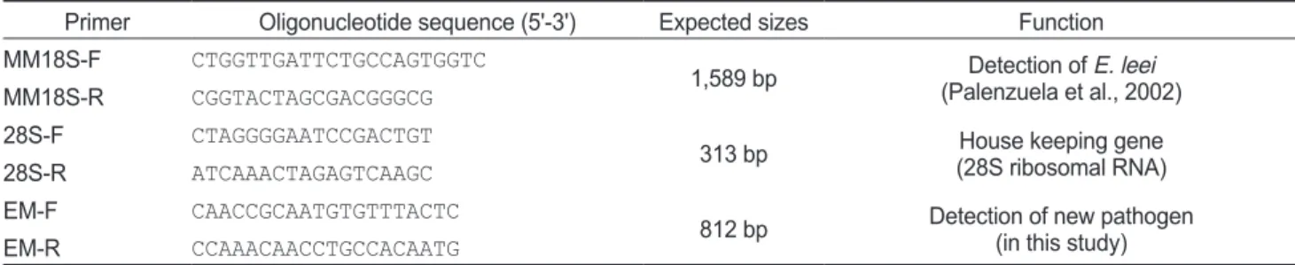
를이용하여Table 1. PCR primers used for the detection of new pathogen
Primer Oligonucleotide sequence (5'-3') Expected sizes Function
MM18S-F CTGGTTGATTCTGCCAGTGGTC
1,589 bp Detection of E. leei
(Palenzuela et al., 2002)
MM18S-R CGGTACTAGCGACGGGCG
28S-F CTAGGGGAATCCGACTGT
313 bp House keeping gene
(28S ribosomal RNA)
28S-R ATCAAACTAGAGTCAAGC
EM-F CAACCGCAATGTGTTTACTC
812 bp Detection of new pathogen (in this study)
EM-R CCAAACAACCTGCCACAATG
회수한후
, ToPo TA cloning
®kit (Invitrogen, USA)
로cloning
하여염기서열분석을의뢰하였다(Solgent, Korea).
조직병리학적 분석
조직병리학적분석을위해여윔 증상을보이는넙치로부터 간
,
비장,
신장,
장,
뇌,
아가미조직을적출하여Bouin's solution
에24
시간동안고정한후70% EtOH
를사용하여탈수하였다.
이후파라핀침투를시키고(Leica EG 1150HC, Germany)
포 매기(Leica Jung 820, Germany)
를사용하여포매를실시하였 다.
이후마이크로톰으로4-5 μm
두께의절편을잘라유리슬 라이드에부착시켜건조시켰다.
제작된조직표본은haematox- ylin
과eosin (H&E)
으로염색을실시한후광학현미경(Zeiss LT60, Germany)
으로검경하였다.
어류 간의 전이실험
감염어로부터물을통한감염이가능한지를확인하기위하여
Ishimatsu et al., (2007)
의방법을사용하여cohabitation
실험 을실시하였다.
먼저PCR
에의해여윔증감염이확인된넙치 를donor group
으로하였고,
음성으로판정된넙치및참돔을recipient group
으로지정하여두가지실험을실시하였다.
첫 번째실험에서는여윔증에걸린넙치(16.3±0.4 cm, 40±5.23
g) 15
마리와 여윔증이 감염되지 않은 넙치(16.7±0.9 cm,
53.4±8.47 g) 15
마리를이용하여cohabitation
실험을실시하 였다.
실험에사용된넙치는100 L
플라스틱수조에recipient
group
의꼬리지느러미를조금잘라내어구별하였다.
두번째실험에서는넙치외타어종인참돔으로여윔증의전이가이루 어지는지를조사하고자하였다
.
참돔이잡아먹히는것을방지하기위하여
100 L
수조내에플라스틱그물망을사용하여구역을나눈후
,
여윔증감염이확인된넙치10
마리와여윔증음 성판정이확인된참돔(6.5±0.5 cm, 3.5±0.7 g) 10
마리를이 용하여cohabitation
실험을실시하였다.
실험기간동안의사육 수온은19±1℃
를유지시켜주었으며1
일1
회씩상업용사료 를공급한후,
사육수를환수시켰다.
매일폐사정도를확인하 였고,
폐사어는조직병리학적관찰및PCR
방법을사용하여여 윔증감염여부를확인하였다.
결 과
여윔증의 원인체 검출
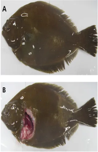
여윔증에감염된넙치의대부분은복부부위가심각하게여 위는것으로관찰되었고
,
일부체색흑화및해부시간출혈등 의증상이관찰되었으나,
일본의자주복에서보고되었던(Tun et al., 2000)
안구함몰이나두부돌출과같은증상은보이지않 았다(Fig. 1A, 1B).
현미경을통해기생충검경을실시한결과,
신장및장에서길이5-8 μm,
폭7-9 μm
의둥근형태의포자가 관찰되었고(Fig. 2),
심각한폐사를보이는일부넙치양식장의경우
, Edwardsiella tarda, Vibrio harveyi
및Streptococcus sp.
등과같은세균성질병과의복합감염증상이나타나기도하였 다
. Viral hemorrhagic septicemia virus (VHSV), viral nervous
Fig. 1. The external (A) and internal (B) signs of the emaciated olive flounder Paralichthys olivaceus.
Fig. 2. The spore from the intestine of emaciated olive flounder Paralichthys olivaceus. Bar=10 μm.
necrosis virus (VNNV), hirame rhabdovirus (HRV)
및red seabream iridovirus (RSIV)
등4
종의viruses
는모든조사대상 양식장에서검출되지않았다(data not shown).
제주도넙치양식장에서여윔증상을보이는넙치를대상으로 새롭게제작한
EM-F/EM-R primer set
를이용하여PCR
을실시한결과
(Table 1),
일본의여윔증에대한연구에서는원인체가주로장에서검출되는것으로보고하였으나
,
본연구에서는 넙치의장에서뿐만아니라신장에서도모두양성의결과를나 타낸다는것을확인하였다(Fig. 3).
여윔증에감염된다양한넙 치를대상으로PCR
을수행한결과,
여윔증원인체의검출정 도가신장,
장,
비장의순서로높은것을확인하였으며,
병원체 의주요감염표적장기는신장및장부위인것으로추정되었 다.
또한,
심각한여윔증상을보이는넙치에서는아가미를제외한모든검사대상조직에서
PCR
양성의결과를확인할수있었다
(Fig. 3).
Fig. 3. PCR amplification from nucleic acids of internal organs from the emaciated olive flounder Paralichthys olivaceus in the level stages of infection. Lanes 1, 7, 13 and 19, Kidney; lanes 2, 8, 14 and 20, Intestine; lanes 3, 9, 15 and 21, Spleen; lanes 4, 10, 16 and 22, Liver; lanes 5, 11, 17 and 23, Brain ; lanes 6, 12, 18 and 24, Gill; M, 1 kb DNA ladder.
검출된
PCR products
에대한유전자염기서열을분석한결과
,
다른시기에제주도의 다양한양식장의 여윔증상넙치로부터검출된병원체들간의유전자상동성은서로
99%
이상인것으로나타났고
,
제주도의넙치양식장에서발생하는여윔 증의원인체는대부분동일한기생충종인것으로추정되었다(Fig. 4). GenBank database
를활용한유전자비교분석을실 시한결과,
일부염기서열인150 bp
가Myxobolus sp. (Acces- sion No. JN616264.1)
및Cystodiscus axonis (Accession No.
JN977605.1)
와부분적으로86%
및88%
의유사성을각각나 타내었으며,
아직GenBank
에등록되지않은미등록종인것으 로조사되었다.
조직병리학적 검사
각장기의조직절편을광학현미경으로관찰한결과
,
여윔증 에감염된넙치의신장과장부위에서원형또는난원형의포자 가다수관찰되었고,
장부위에서보다신장부위에서더욱많은Fig. 4. Comparison analysis of the DNA nucleotide sequences of pathogens detected from the emaciated olive flounder Paralichthys oliva- ceus. Primers used in PCR are in boxes.
수의포자가관찰되었으며
,
기부골절,
사구체,
핵등의파괴와 변형체(plasmodium)
가일부관찰되었다(Fig. 5A and 5B).
감 염된장상피부위에서는포자가관찰되었지만변형체는관찰 되지않았으며,
심한경우조직의탈락및파괴가나타났다(Fig.
5C and 5D).
신장및장을제외한다른조직장기부위에서는포 자가관찰되지않았으며,
특이적인조직학적이상소견도관찰 되지않았다(data not shown).
어류 간의 전이실험
여윔증에대한
PCR
음성결과가확인된넙치및참돔을이용 하여병원체의전이에대한실험을실시한결과,
실험기간중2 groups
의donor
넙치는3
일째에100%
폐사하였고, recipient
넙치에서는12
일째에100%
누적 폐사율이 관찰되었다(Fig.
6A).
감염어로부터 유출된 병원체에 의해 감염된recipient
group
에서의폐사는donor group
에서보다7
일정도후에나 타나기시작하였으며,
폐사가일어난recipient group
의넙치를PCR
법으로확인한결과, 73.3%
의감염률이관찰되었고(Table 2),
조직학적검사결과신장,
장부위에서donor fish
와동일 한 형태의포자가관찰되었다(Fig. 7).
참돔으로의병원체전 이에대한실험결과,
실험기간중3
일째에donor group
에서100%
누적폐사율이관찰되었고, recipient group
에서는실험 종료일까지폐사가발생하지않았으며(Fig. 6B), PCR
검사결 과에서모두음성인것으로판명되었다(Table 2).
조직학적검 사결과에서도모든참돔의조직부위에서포자가관찰되지않 았다(data not shown).
고 찰
본연구에서는일본의여윔증에대한연구방법및결과들을 국내의여윔증연구를위하여적용해보았고
,
우리나라의여윔 증원인체는일본에서보고된여윔증원인체와형태학적으로 는유사하지만,
분자유전학적으로는전혀다른종이라는것을Fig. 5. Histological findings of kidney (A, ×200 and B, ×400) and intestine (C, ×200 and D, ×400) from the emaciated olive flounder Para- lichthys olivaceus. H & E stain. Bar = 20 μm.
원인불명 여윔증 진단
343
0
Day
Cumulative mortality (%)
80 60 40 20
0 2 4 6 8 10 12 14
0
(B)
Day
Cumulative mortality (%)
100 80 60 40 20
0 2 4 6 8 10 12 14
0
(A)
Day
Cumulative mortality (%)
100 80 60 40 20
0 2 4 6 8 10 12 14
0
(B)
Day
Cumulative mortality (%)
100 80 60 40 20
0 2 4 6 8 10 12 14
Fig. 6. Cumulative mortality (%) of olive flounder Paralichthys olivaceus (A) and red seabream Pagrus major (B) after cohabitated with the emaciated olive flounder. ◆, emaciated olive flounder; ○ and ◇, cohabitated olive flounder and red sea bream, respectively.
Fig. 7. Histological findings of kidney (A, ×200 and B, ×400) and intestine (C, ×200 and D, ×400) from the recipient olive flounder Parali- chthys olivaceus cohabitated with the emaciated olive flounder. H & E stain. Bar=20 μm.
Table 2. Infection rate (%) of olive flounder Paralichthys olivaceus and red seabream Pagrus major after cohabitated with the emaciated olive flounder
Donor fish (olive flounder) Recipient fish (olive flounder) Donor fish (olive flounder) Recipient fish (red seabream)
PositivePCR 100%
(15/15) 73.3%
(11/15) 100%
(10/10) 0%
(0/10)
밝혀내었다
.
감염넙치에대한분석결과,
대부분은복부부위 에심각한여윔이관찰되었으며,
감염넙치가감염되지않은넙 치보다체중이30-40%
정도낮은것으로관찰되었고(data not
shown),
간혹육안상복부부위가여위지않은넙치에서도감염이발생하여 감염후여위는것으로판단된다
. Choi et al.
(2012)
은제주도넙치양식장에서발병하는여윔증넙치에서E. tarda, V. harveyi
와같은세균성질병이복합감염되는경우 에대하여보고하였으나,
감염넙치에서공통적으로분리되지 않아여윔증의원인체는아닌것으로추정하였다.
현미경을통 해기생충검경을실시한결과,
길이5-8 μm,
폭7-9 μm
의둥근 형태의포자가관찰되었고(Fig. 2),
이것은일본의자주복에서 발생한여윔증원인체의포자와유사한형태인것으로조사되 었다(Tetsuza et al., 2004).
일본의자주복에서처음발견된여윔증은
Tun et al. (2002)
의연구에서E. fugu, E. leei
와L. fugu
가원인체인것으로의심 되었다.
여윔증에대한분자생물학적진단방법을적용하고자SSU rDNA gene
의MM18Sf/MM18Sr primer set (Palenzuela et al., 2002; Yasuda et al., 2005)
를제작한후여윔증상넙치의 신장과장을이용하여PCR
을실시하였지만음성반응을나타 내었다.
이러한결과는Choi et al. (2012)
의보고와일치하였으 며,
우리나라에서발병하는넙치의여윔증원인체는일본에서 보고된primer set
를사용한PCR
에서는검출되지않는다는것 을확인하였다.
본연구에서는국내양식넙치의여윔증진단을 위한primer set (EM-F/EM-R)
를처음으로개발하였고(Table 1),
여윔증상을나타내는넙치를대상으로한PCR
실험에서명 확한band
가나타나는것을확인하였으며(Fig. 3),
향후국내의 넙치에서여윔증의감염진단이필요한경우,
이와같은방법이 유용하게활용될것으로판단된다.
병원체의염기서열을분석 한결과,
국내넙치의여윔증원인체들간의DNA nucleotide sequences
의상동성은99%
이상인것으로나타났다(Fig. 4).
그리고
, GenBank
에등록된Myxobolus sp.
및Cystodiscus ax- onis
의일부염기서열과80%
정도의상동성이있는것으로확 인되어아직GenBank database
에등록되지않은미등록기생 충종인것으로판명되었다.
향후,
이병원체에대해서는DNA
walking
과같은분자생물학적분석을통하여더욱세부적인연구가뒤따라야할것이다
.
이전연구들에서는
sharpsnout sea bream Puntazzo puntazzo, red sea bream Pagrus major, red drum Sciaenops ocellatus
이Myxidium leei
에감염되었을때공통적으로장조직에서포자를관찰할수있었다고보고하였다
(Le Breton and Marques, 1995; Diamant, 1998; Athanassopoulou et al., 1999).
또한, Myxosporea
에감염된스페인의turbot, Scophthalmus maxi- mus
에서도포자를소화관이나장에서확인할수있었으며이러한결과는
turbot
의성장에영향을미쳤을것이라제시하였다
(Beaman et al., 1999). Bartholomew et al. (1989)
은Cera-
tomyxa shasta
에감염된연어과어류에서도포자형태의기생충을장조직에서 관찰할수있었다고보고하였는데이와같 이유럽의여러연구결과에서점액포자충에감염된어류는주 로장에서감염이이루어지는것으로보고되었다
.
또한,
일본에 서발생하는여윔증의원인기생충도장과담낭에서확인되었 다고보고하였다(Tun et al., 2002).
이러한결과는우리나라여 윔증과조직학적으로유사한경향을보여주고있으며,
여윔증 에심하게감염된넙치의경우,
전장관의상피,
점막고유층,
근 층등에서포자가관찰되는부분과도일치하였다.
그러나,
감염 부위에있어서유럽및일본에서는장조직을주요한감염조직 으로보고하였지만,
본연구결과에서국내넙치의여윔증원인 체는장조직보다신장조직에서더욱높게감염되는것으로조 사되어,
주요감염표적장기는외국에서의보고와차이가있는 것으로판단된다.
일본에서는여윔증의원인체로알려진
E. leei
에감염된어류 를donor fish
로사용하여같은어종및다른어종을대상으로cohabitaiton
실험을실시하였으며그결과,
같은어종및다른 어종으로E. leei
의전이가이루어졌다고보고하였다(Diamant, 1997; Yasuda et al., 2002).
본연구에서는여윔증의원인체를 동일한어종인넙치로전이시켰을때여윔증상이나타났으나 다른어종인참돔으로의전이는일어나지않아일본의연구결 과와는대조적인것으로나타났으며(Table 2),
감염여부를확인하기위해실시한조직학적검사및
PCR
검사에서도모두동일한결과가확인되었다
.
본연구에서는국내의양식넙치에서발생하는여윔증에대하 여분석하였으며
,
기존에일본에서보고된결과와비교했을때,
장조직에서형태학적으로유사한포자가관찰된것은동일하 지만,
병원체의분자생물학적인분석및cohabitation
실험에서 는다른결과를보였고,
아직GenBank
에미등록된새로운기 생충종인것으로조사되었다.
그리고,
국내의넙치양식장에서 많은문제를야기시키는여윔증에대한진단법을처음개발하 여제시하였고,
이것은여윔증에대한다양한연구에있어서중 요한기반이될수있을것이며,
향후에는원인기생충의동정에 대한연구가이루어져야할것이다.
사 사
이논문은해양수산부재원으로 한국해양과학기술진흥원의 지원을받아수행된연구임
(
수산백신연구센터).
References
Athanassopoulou F, Prapas T and Rodger H. 1999. Diseases of
Puntazzo puntazzo cuvier in marine aquaculture systems in
Greece. J Fish Dis 22, 215-218.Bartholomew JL, Smith CH, Rohovec JS and Fryer JL. 1989.
Characterization of a host response to the myxosporean parasite, Ceratomyxa shasta (Noble), by histology, scan- ning electron microscopy and immunological techniques.
J Fish Dis 12, 509-522. http://dx.doi.org/j1365-2761.1989.
tb00561.x.
Beaman HJ, Speare DJ, Brimacombe M and Daley J. 1999.
Evaluating protection against Loma salmonae generated from primary exposure of rainbow trout, Oncorhynchus
mykiss (Walbaum), outside of the xenoma-expression tem-
perature boundaries. J Fish Dis 22, 445,–450. http://dx.doi.org/10.1046/j.1365-2761.1999.00194.x.
Cho MY, Kim MS, Kwon MG, Jee BY, Choi HS, Choi DL, Park GH, Lee CH, Kim JD, Lee JS, Oh YK, Lee DC, Park SH and Park MA. 2007. Epidemiological study of bacterial diseases of cultured olive flounder, Paralichthys olivaceus from 2005 to 2006 in Korea. J Fish Pathol 20, 61-70.
Choi HS, Jun LJ, Kim SM, Jeong HD, Kim YK, Lim H, Yeo Ik and Jeong JB. 2012. Clinical features of fish with patho- gens isolated from emaciated olive flounder Paralichthys
oliveaceus. J Fish Pathol 25, 67-76.
Diamant A. 1997. Fish-to-fish transrmission of a marine myxo- sporean. Dis Aquat Org 30, 99-105.
Diamant A. 1998. Red drum Sciaenops ocellatus (Sciaenidae), a recent introduction to Mediterranean mariculture, is sus- ceptible to Myxidium leei (Myxosporea). Aquaculture 162, 33-39. http://dx.doi.org/10.1016/S0044-8486(97)00307-4.
Ishimatsu A, Hayashi M, Nakane M and Sameshima M. 2007.
Pathophysiology of cultrured tiger puffer Takifugu rubripes suffering from the myxosporean emaciation disease. Fish Pathol 42, 211-217.
Le Breton A and Marques A. 1995. Occurrence of a histozoic
Myxidium infection in two marine cultured species: Pun- tazzo puntazzo C and Pagrus major. Bull Fish Pathol 15,
210-212.Ogawa K and Yokoyama H. 2001. Emaciation disease of cul- tured tiger puffer Takifugu rubripes. Bull Aquacult Suppl 5, 65-70.
Palenzuela O, Redondo MJ and Alvarez-Pellitero P. 2002. De- scription of Enteromyxum scophthalmi gen. nov., sp. nov.
(Myxozoa), an intestinal patasite of turbot (Scophthalmus
maximus L.) using morphological and ribosomal RNA
sequence data. Parasitology 124, 369-370. http://dx.doi.org/10.1017/S0031182001001354.
Tetsuya Y, Yoshinori N, Takeshi K, Yutaka F, Hiroshi Y and Ka- zuo O. 2004. Molecular and Morphological Redescriptions of Enteric Myxozoans, Enteromyxum leei (formerly Myxid-
ium sp. TP) and Enteromyxum fugu comb.n.(syn, Myxidium fugu) from cultrured Tiger puffer. J Fish Pathol 39, 137-143.
Tun T, Yokoyama H, Ogawa K and Wakabayashi H. 2000.
Myxosporeans and their hyperparasitic microsporeans in the intestine of emaciated tiger puffer. J Fish Pathol 35, 145- 156. http://dx.doi.org/10.3147/jsfp.35.145.
Tun T, Ogawa K and Wakabayashi H. 2002. Pathological changes induced by three myxosporeans in the intestine of cultured tiger puffer, Takifugu rubripes (Temminck and
Schlegel). J Fish Pathol 25, 63-72. http://dx.doi.org/10.1046/
j.1365-2761.2002.00333.x.
Yasuda H, Ooyama T, lwata K, Tun T, Yokoyama H and Ogawa K. 2002. Fish-to-fish transmission of Myxidium sp. (Myxo- zoa) in cultured tiger puffer suffering from emaciation dis- ease. J Fish Pathol 37, 29-33. http://dx.doi.org/10.3147/
jsfp.37.29.
Yasuda H, Ooyama T, Nakamura A, lwata K, Palenzuela O and Yokoyama H. 2005. Occurrence of the myxosporean emaciation disease caused by Enteromyxum leei in cultured Japanese flounder Paralichthys olivaceus. Fish Pathol, 40, 175-180.