719
Copyright © 2015 The Korean Society of Fisheries and Aquatic Science pISSN:0374-8111, eISSN:2287-8815
서 론
1990
년대부터각종해수어의양식이활발하게이루어져각국에서종묘수입이성해지자방역체계가미비한상태에서수입 종묘와같이질병도함께도입되어우리나라에없었던질병들 이유행하기시작하였으며
(Chun, 2006),
질병의발병양상또 한,
양식초창기에는고수온기에기생충및세균에의한단독감염이주를이루었으나최근에는수온과상관없이연중다양한 병원체가혼합감염의형태로질병을일으키고있어수산생물 의대량폐사를유발시키기도한다
(Kim et al., 2006).
최근들어 외래질병의유입가능성이증가하고양식어류의질병연관성 에대한관심이증가하면서질병을전반적으로모니터링하거나 질병과의상관관계를구명하고자하는연구가점차증가하고 있는추세이다(Cho et al., 2009; Jung et al., 2012; Song et al.,
제주의 양식 넙치(Paralichthys olivaceus)를 대상으로 한 여윔증 모니터링(2010-2013)
김승민ㆍ전려진
1ㆍ박명애
2ㆍ정승희
3ㆍ정현도
4ㆍ정준범*
제주대학교 해양의생명과학부, 1제주대학교 수산백신연구센터, 2국립수산과학원 수산생물방역과, 3국립수산과학원 병리연구과,
4부경대학교 수산생명의학과
Monitoring of Emaciation Disease in Cultured Olive Flounder Paralichthys olivaceus in Jeju (2010-2013), Korea
Seung Min Kim, Lyu Jin Jun
1
, Myoung Ae Park2
, Sung Hee Jung3
, Hyun Do Jeong4
and Joon Bum Jeong*Faculty of Marine Biomedical Science, Jeju National University, Jeju 63243, Korea
1
Fish Vaccine Resarch Center, Jeju National University, Jeju 63243, Korea
2
Aquatic Life Disease Control Division, National Fisheries Research and Development Institute, Busan 46083, Korea
3
Pathology Division, National Fisheries Research and Development Institute, Busan 46083, Korea
4
Department of Aquatic Life Medicine, Pukyong National University, Busan 48513, Korea
In this study aiming at the cultured olive flounders in Jeju island of Korea, has performed a total of 216 cases of pathogen tests for the olive flounders which seemingly had the symptom of emaciation in 24 farms on the suspicion of its occurrence from June 2010 to December 2013 and intended to get the basic information about the pathogen.
According to the survey results of the emaciation infection status of 24 farms in Jeju over the period of this survey, it was confirmed that 18 (75%) of 24 farms in Jeju are positive in the emaciation infection. Among them, as for the rate of infection per year, it was observed that they are 38% in 2010, 48% in 2011, 50% in 2012 and 60% in 2013, and over the period of this survey the infection rate in accordance with the temperature of water has observed a variety of incidence rates from the summer to the winter season. In addition, according to the results of emaciation infection status for each size, the equivalent showed a detection rate, 54% in 11-20 cm, 43.9% in 21-30 cm and 25.4% in over 31 cm. This result accounts for an important portion among the diseases of farmed olive flounders in Jeju, is consid- ered to be one of the diseases which cause troubles in the farms for olive flounders on land and it is thought that it can be utilized as basic data in order to estimate emaciation which may occur in the similar size of the cultured olive flounders in Jeju island.
Key words: Emaciation, Olive flounder, Monitoring
This is an Open Access article distributed under the terms of the Creative Commons Attribution Non-Commercial Licens (http://creativecommons.org/licenses/by-nc/3.0/) which permits unrestricted non-commercial use, distribution, and reproduction in any medium, provided the original work is properly cited.
http://dx.doi.org/10.5657/KFAS.2015.0719 Korean J Fish Aquat Sci 48(5) 719-724, October 2015
Received 25 August 2015; Revised 12 October 2015; Accepted 29 October 2015
*Corresponding author: Tel: +82. 64. 754. 3426 Fax: +82. 64. 756. 3493
E-mail address: [email protected]
김승민
ㆍ
전려진ㆍ
박명애ㆍ
정승희ㆍ
정현도ㆍ
정준범720
2013).
그러나현재까지국내양식어류에대한질병조사는대부분특정질병을중심으로조사되어왔으며
(Kim et al., 2006;
Cho et al., 2008; Kim et al., 2010),
원인불명의질병에대해서 는연구가미미한실정이다.
국내양식산업의대표적품종인넙치
(Paralichthys olivaceus)
는연간전체양식어류생산량인4
만톤중약50%
를차지하고 있으며,
이중제주지역에서양식넙치의생산량은2
만톤이상 으로전체생산량의절반이상을보이며주요양식어종으로자 리잡고있다(NFRDI, 2013).
하지만,
최근제주도내양식장의약
20 cm
전후크기의넙치에서여위어가는원인불명의질병이발생하여
,
최초발병증상을보인이후1-3
주동안폐사가되 는사례가증가하고있으며,
이질병에감염된넙치는체색흑 화,
어체중감소및간출혈등의증상을나타낸다(Kim et al., 2015). Kim et al. (2015)
의연구에따르면,
주변국가인일본의 터봇여윔증의원인체로보고된Enteromyxum leei
의primer set (1,589 bp)
를제작하여(Palenzuela et al., 2002)
국내여윔 증넙치를대상으로PCR
실험을실시한결과,
음성반응이관 찰되어동일한원인체가 아닌것으로확인되었다.
또한, Kim et al. (2015)
은GenBank (NCBI, USA)
에등록된점액포자충(Myxidium sp.)
의염기서열로부터degenerated primers
를제 작하여,
여윔증상넙치로부터DNA
를분리한후PCR
을실시 한결과양성반응이나타났고, DNA sequencing
을통해염기서 열을분석한결과일부염기서열이점액포자충과일치하였으나아직
GenBank
에등록되지않은미등록종인것으로보고하였다
(Kim et al., 2015).
하지만,
국내에서발생하는여윔증원인 체의발병동향에대한연구는아직보고되어있지않은실정이 며,
양식어류에대한질병모니터링은시기별질병관리에유 용한자료를제공하여,
자국내특정질병에대한기초자료로 서매우중요한역할을하며(OIE, 2008),
양식생물에서발생하 는질병에대하여효과적인방제대책을수립하기위해서는양 식장별질병발생현황에대한정확한모니터링을수집하는것 이선행되어야한다.
본연구에서는
2010
년부터2013
년까지국내양식산업의대 표적품종인넙치에서발생하는여윔증원인체의감염현황을 파악하기위하여Kim et al. (2015)
의방법에따라PCR
법과조 직학적검사를통해여윔증원인체의감염률을조사하고자한 다.
재료 및 방법
실험어
제주도양식넙치를대상으로여윔증원인체의감염현황을 조사하기위해
, 2010
년부터2013
년까지4
년동안양식넙치를 대상으로2010
년에21
회(6, 7, 8, 11, 12
월), 2011
년25
회(3, 7, 8, 10, 11
월), 2012
년4
회(7, 10, 11
월), 2013
년16
회(1, 3, 7, 8, 11, 12
월)
에걸쳐동.하절기및중복된양식장을포함하여24
개소양식장에서
66
회의샘플링을실시하여총216
마리를대상으 로실험을수행하였고,
확보된샘플은육안상여윔증상을보이 며폐사가발생하여의뢰를맡긴양식장과일반적인질병증상 을보이는양식장을대상으로감염조사를실시하였다.
실험전 수질측정기YSI Model 650XL (YSI,USA)
을사용하여수온측 정을실시하였고,
실험실로운반된어류의외부및내부증상을 확인하고,
어체의전장및무게를측정하였다.
DNA 추출
DNA
를분리하기 위하여Kim et al. (2015)
에 보고된방법을참고하여실험어의신장을해부용칼로절개한후
DNeasy
ⓇBlood & Tissue Kit (Qiagen Hilden, Germany)
을 사용하여DNA
를분리하였다.
먼저ATL buffer 180 µL
와proteinase K
20 µL
를첨가하여56℃
에서조직이녹을때까지반응시켰다.
반응후
, AL buffer 200 µL
를섞은다음ethanol 200 uL
를더 하여spin column
에옮겨6,000 g (gravity)
로1
분간원심분리 하였다. Column
을새로운tube
로옮긴후AW1 buffer
와AW2 buffer 500 µL
를이용하여세척과정을거친후, AE buffer 50 µL
를첨가하여DNA
를분리하였다.
분리된DNA
는실험전까 지-20℃
에보관하여사용하였다.
PCR 분석
PCR
분석을위하여Kim et al. (2015)
이여윔증진단을위하여 제시한primer set
를사용하였으며(Table 1), PCR
은microtube
에1 μM
의각primer, 2.5 mM
의각dNTP, 10 x G-Taq Buf- fer, 2.5 U G-Taq DNA polymerase (Gene Pro Themal Cycler Cosmo, Korea)
및template DNA
로서추출된핵산을첨가한 후distilled water
로PCR
혼합물의최종volume
이20 μL
가 되게하였다. PCR
조건은95℃
에서3
분간pre-denaturation
시 킨후, 95℃
에서30
초denaturation, 55℃
에서30
초annealing, 72℃
에서30
초extension
의반응을1
회로하여, 35
회반복하 여반응시켰다.
그리고, 72℃
에서7
분간post-extension
시켰다. PCR
후증폭산물은1×TAE buffer
를전기영동을위한완충 액으로하여, 0.5 μg/μL EtBr
이첨가된1% agarose gel
상에서 전기영동한후, UV
검출기를이용하여, ultraviolet
상에서검출 되는산물의크기를관찰하였다.
조직병리학적 분석
조직병리학적 분석을 위해 넙치의 신장 조직을 적출하여
Bouin’s solution
에24
시간 동안고정한후70% EtOH
탈수Table 1. PCR primers used in this study Primer Oligonucleotide sequence
(5'-3') Expected
sizes Reference EM-F CAACCGCAATGTGTTTACTC
812 bp Kim et al. (2015) EM-R CCAAACAACCTGCCACAATG
many)
포매기(Leica Jung 820, Germany)
를사용하여포매를 실시하였다.
이후마이크로톰으로4-5 µm
두께의절편을잘 라유리슬라이드에부착시켜건조시켰다.
제작된조직표본은haematoxylin
과eosin (H&E)
으로염색을실시한후광학현미 경(Zeiss LT60, Germany)
으로검경하였다.
기타 질병 진단
그 밖에질병은세균성및 바이러스성질병에대해 일반적 인검사방법에따라진단하였다
.
세균성질병인경우는간,
신 장,
비장을Tryptic Soy Agar (Difco Co., USA), Thiosulfate Citrate Bile Salts Sucrose Agar (Difco), Salmonella-Shigella (Difco)
배지에접종하여25℃
에서배양하였고,
필요시API kit (BioMerieux, France)
를사용하여동정하였다.
바이러스질병인경우
Cho et al. (2007)
의방법에따라primer sets
를동일하게제작한후viral hemorrhagic septicemia virus (VHSV), viral nervous necrosis virus (VNNV), hirame rhab- dovirus (HRV)
및red seabream iridovirus (RSIV)
등4
종의바 이러스를대상으로감염여부를확인하였다.
결과 및 고찰
본연구에서는우리나라제주도에서양식되는넙치를대상으 로
2010
년6
월부터2013
년12
월까지총66
회에걸쳐216
마 리의넙치를대상으로여윔증원인체의감염현황을조사하여 향후여윔증연구에대한기초자료로사용하고자하였다.
그결과
, 66
회에걸친조사기간동안의대상양식장중에서50%
(33
회)
에서여윔증양성반응이나타났고,
총216
마리의넙치 중93
마리(43%)
에서양성반응을나타내었다(Fig. 1, Table 2).
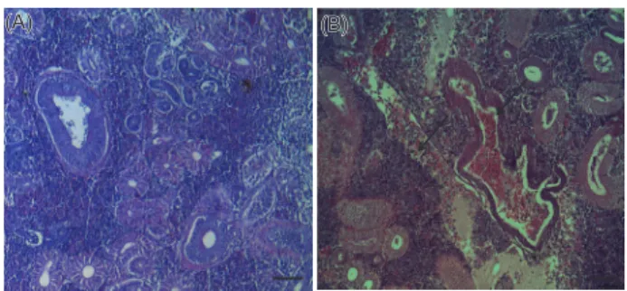
원형또는난원형의포자가다수관찰되었고
,
기부골절,
사구 체,
핵등의파괴와변형된변형체(plasmodium)
가일부관찰되었으며
(Fig. 2B), PCR
음성반응을보인넙치에서는정상적인신장형태가관찰되었다
(Fig. 2A).
이러한결과는Kim et al.
(2015)
의보고와일치하였으나,
기생충성질병으로잘알려진Ichthyobodo sp., cryptocaryon sp., myxobolus sp., miamien- sis sp., myxidium sp.
등과조직학적으로비교했을때,
대부분 다른형태가관찰되었고,
숙주및표적장기에서도다른경향을 나타내어,
새로운기생충종인것으로추정되며,
향후에는여윔 증에대한형태학적동정및real-time PCR
을통한정량적분석 에대한연구가이루어져야할것이다.
양식장연도별감염률은
2010
년도21
개소중8
개소(38%), 2011
년도25
개소중12
개소(48%), 2012
년도4
개소중2
개소(50%), 2013
년도16
개소중11
개소(60%)
에서양성임을확인 하였다(Table 2).
그중양식장에서여윔증발생이의심되어분 석을의뢰해온경우에는분석결과, 59.5% (75
마리/126
마리)
의높은검출율을보였고,
무작위로여윔증모니터링을실시한 경우에는20% (18
마리/90
마리)
의낮은검출율을나타내었다(data not shown).
또한,
검출된양식장은이후의감염발생율이 높은것으로확인되었고,
향후에는발병경로,
기회감염성,
숙주 의감수성등질병에대한전반적인연구가뒤따라야할것이다.
여윔증원인체에의한연도별감염률은
2010
년도60
마리중17
마리(28.3%), 2011
년도75
마리 중32
마리(42.6%), 2012
년도24
마리 중8
마리(33.3%), 2013
년도57
마리 중36
마리(63.1%)
등으로나타났고,
조사기간중수온에따른감염률은 여름철부터겨울철까지다양하게나타나는것으로조사되었다(Table 2).
제주도의양식장은대부분연중17℃
내외의지하해 수를혼합사용하여여름철에도사육수온을23℃
내외,
겨울철 에는14℃
이상을유지하고있어(Oh et al., 1998),
여윔증이연 중발생하는원인으로서제주도의시기적인수온변동이크지 않은것도연관성이있을것으로추정된다.
현재까지어류에서보고되는여윔증원인체에대한생활사가 밝혀진종은없으며
,
감염되는어류의크기및감염경로등에대Fig. 1. PCR results of emaciation disease positive and negative samples. Lane 1 and 9 show emaciation disease positive and nega- tive results. Lane1, Farm-A (June, 2010); lane 2, Farm-B (Octo- ber, 2011); lane 3, Farm-C (October, 2011); lane 4, Farm-D (No- vember, 2011); lane 5, Farm-E (October, 2012); lane 6, Farm-F (November, 2012); lane 7, Farm-G (July, 2013); lane 8, Farm-N (December, 2010); lane 9, Farm-O (December, 2010); 1 kb DNA ladder.
800 600 400 200
0 11-20cm 21-30cm 31-40cm
Fish weight (g)
M N P 1 2 3 4 5 6 7 8 9
500bp 1,000bp
(A) (B)
(A) (B)
Fig. 2. Histological changes in the kidney of the olive flounder Paralichthys olivaceus. A: control olive flounder (×100); B: emaci- ated olive flounder (×100). H. E. stain Bar = 20 µm.
800 600 400 200
0 11-20cm 21-30cm 31-40cm
Fish weight (g)
M N P 1 2 3 4 5 6 7 8 9
500bp 1,000bp
(A) (B)
(A) (B)
김승민
ㆍ
전려진ㆍ
박명애ㆍ
정승희ㆍ
정현도ㆍ
정준범722
해서도조사가부족한실정이다
.
본연구에서넙치의크기별로 감염현황을조사한결과, 11-20 cm 74
마리중40
마리(54%),
21-30 cm 91
마리중40
마리(43.9%), 31-40 cm 51
마리중13
마리(25.4%)
의검출률을나타내었다(Table 3).
Table 2. Prevalence of emaciation disease in cultured olive flounder Paralichthys olivaceus of Jeju from 2010 to 2013
Year Month Water temp(℃)
Farm &
No. of fish
Infection rate (%) (positive no. / total no.)
Fish Farm
PCR test Total Histological examination PCR test Total
2010
June 18.5 A(6)*, B(3) 33.3%
(3/9)
28.3%
(17/60)
44.4%
(4/9) 50%
(1/2)
(8/21)38%
July 19.5 C(3), D(3), E(3),
F(3), G(3) 0%
(0/15) 0%
(0/15) 0%
(0/5)
August 20 E(3)*, H(3)* 66.6%
(4/6) 66.6%
(4/6) 100%
(2/2)
November 18.1 I(6)* 83.3%
(5/6) NT 100%
(1/1) December 15 A(1)*,D(1), J(1),
K(3), L(3)*M(3)*, N(2), O(3), P(3), Q(1)*, R(3)
20.8%
(5/24) NT 36.3%
(4/11)
2011
May 15 S(6)* 100%
(6/6)
42.6%
(32/75)
NT 100%
(1/1)
(12/25)48%
July 19.5 A(3)*,M(3),T(3)
,U(3)*,V(3)*,W(3), X(3) 28.5%
(6/21) 33.3%
(7/21) 28.5%
(3/7) August 21.5 A(3), B(3)*,M(3), T(3)*,
V(3)*,W(3), X(3), 33.3%
(7/21) NT 28.5%
(3/7) October 20 A(3), B(3)*,C(1)*, M(3)*,V(3),
X(3), W(3),Y(2) 33.3%
(7/21) 42.8%
(9/21) 37.5%
(3/8)
November 18.5 T(3)*, D(3)* 100%
(6/6) NT 100%
(2/2)
2012
July 20 D(9), F(3) 0%
(0/12)
33.3%
(8/24)
NT 0%
(0/2)
50%(2/4)
October 21 E(6)* 33.3%
(2/6) 33.3%
(2/6) 100%
(1/1)
November 18 F(6)* 100%
(6/6) 100%
(6/6) 100%
(1/1)
2013
January 14.8 J(6) 0%
(0/6)
63.1%
(36/57)
NT 0%
(0/1)
(11/16)60%
May 16 Q(4)* 100%
(4/4) 100%(4/4) 100%
(1/1)
July 19.7 G(4)*, Q(4)* 87.5%
(7/8) 100%(8/8) 100%
(2/2)
August 22 E(5)*, K(5)* 80%
(8/10) NT 100%
(2/2)
September 21 S(2)* 100%
(2/2) 100%(2/2) 100%
(1/1)
November 18.8 K(3)*, J(4)*,U(4)* 63.6%
(7/11) NT 100%
(3/3) December 15.6 C(1), J(3)*, Q(3), R(3), V(3)*, Y(3) 50%(8/16) 62.5%(10/16) 33.3%
(2/6)
Total 43%
(93/216) 50%
(33/66)
* : Positive NT: not tested
넙치의 여윔증 감염 조사
723
본연구조사에서여윔증감염으로판정된양식넙치의외부임 상증상을관찰해본결과
,
전체적으로는체색흑화및복부여윔 증상이주로관찰되었고,
내부임상증상관찰에서는간출혈및 신장부위가하얗게변색되는증상을보였으며,
그외에특별한 증상은관찰되지않았다(data not shown).
하지만,
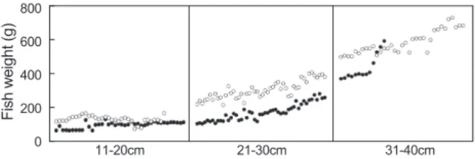
여윔증에감염된넙치가여윔증이발병하지않는넙치보다
30-40%
낮은체중을나타내었고
(Fig. 3),
이러한결과는Kim et al. (2015)
이 보고한결과와일치하였다.
이는병원성에따른병리학적특성 을평가하는데에있어유용한기초자료로활용될것으로기대 되며,
여윔증에의한어체중감소와여윔증병원체의병원성과 의상관관계에대해서는앞으로좀더체계적인연구가이루어 져야될것이다.
기생충은넙치의종묘생산시기부터출하전까지넙치의성장
과폐사에밀접한영향을미치는것으로알려져있으며
(Chun,
2006),
세균이나바이러스등에의한2
차감염을유발하는것으 로알려져있다(Kim et al., 2010).
또한, Cho et al. (2007)
은넙 치로부터분리된병원체의감염상황을비교분석한결과, 2
종 이상병원체의혼합감염이46%
를차지하였으며,
세균끼리의 혼합감염보다는세균과기생충의혼합감염,
세균과바이러스 의혼합감염또는세균,
바이러스및기생충이모두혼합되는경 우가많은것으로나타났다.
본연구의병원체감염현황결과
,
여윔증과주요세균성질 병의혼합감염은Vibrio sp.
와의혼합감염이36
건(38.7%)
으 로가장많았으며,
다음으로는Streptococcus sp. 27
건(29%), Edwardsiella sp. 9
건(9.6%)
순으로여윔증과혼합감염되는세 균성질병은주로Vibrio sp.
인것으로나타났다(Table 4).
하지 만,
여윔증과주요바이러스성질병이혼합감염되는경우는없 었으며,
여윔증단독으로발생하는경우가21
건(22%)
으로나 타났다(data not shown).
이러한결과는Chun (2005)
이보고한Vibrio sp.
는기생충감염이나스트레스를받게되면2
차감염 으로발병할수있다는사실을뒷받침할수있으나,
다른세균 성질병과의혼합감염에대한연구는아직밝혀진사례가없으 며,
향후여윔증과세균성질병과의혼합감염에대해서는연구 가필요할것으로판단된다.
본연구에서는
2010
년부터2013
년까지4
년간제주도양식넙 치에서발생하는여윔증원인병원체의발병현황을조사하기위하여
, PCR
방법및병리조직학적관찰등의방법을사용하여모니터링하였으며
,
매년넙치양식장에서38-60%
의매우 높은감염률을나타낸다는것을확인하였다.
제주도넙치양식장에서발생하는아직밝혀지지않은대량폐사의원인을규명 하는데있어서도본연구결과는중요한자료가될수있을것이 며
,
향후국내넙치에서발생하는여윔증연구에서도좋은기초 자료로활용가능할것이다.
사 사
이논문은국립수산과학원수산과학연구사업의지원으로수 행된연구이며
,
연구비지원에감사드립니다.
References
Cho MY, Kim MS, Kwon MG, Jee BY, Choi HS, Choi DL, Park GH, Lee CH, Kim JD, Lee JS, Oh YK, Lee DC, Park SH and Park MA. 2007. Epidemiological study of bacterial diseases of cultured olive flounder, Paralichthys olivaceus from 2005 to 2006 in Korea. J Fish Pathol 20, 61-70.
Cho MY, Kim MS, Choi HS, Park GH, Kim JW, Park MS and Park MA. 2008. A statistical study on infectious diseases of cultured olive flounder, Paralichthys olivaceus in Korea. J Fish Pathol 21, 271-278.
Cho MY, Jee BY, Park GH, Lee CH, Lee DC, Kim JW, Park MS and Park MA. 2009. Monitoring of fish pathogens in wild marine fish of Korean coastal offshore water in 2008. J Fish Pathol 22, 75-83.
Chun SK. 2005. Paralichthys olivaceus: disease and treatment.
Fig. 3. Monitoring of the index content in the emaciated olive flounder Paralichthys olivaceus.○: Uninfected olive flounder, ●:
infected olive flounder.
800 600 400 200
0 11-20cm 21-30cm 31-40cm
Fish weight (g)
500bp 1,000bp
(A) (B)
(A) (B)
Table 3. Size distribution of fish isolated with emaciation disease Fish species Fish size
(cm) No. of fish
Distribution Infection rate
Olive flounder
11-20 74 54%(40/74)
21-30 91 43.9%(40/91)
31-40 51 25.4%(13/51)
Table 4. Occurrence of mixed infection by emaciation agent and main bacteria in cultured olive flounder Paralichthys olivaceus of Jeju from 2010 to 2013
Year E+E E+V E+S
2010 11.7% (2/17) 47% (8/17) 23.5% (4/17) 2011 9.3% (3/32) 37.5% (12/32) 34.3% (11/32) 2012 12.5% (1/8) 50% (4/8) 25% (2/8) 2013 8.3% (3/36) 33.3% (12/36) 27.7% (10/36) Total 9.6% (9/93) 38.7% (36/93) 29% (27/93) E+E, Emaciation+Edwardsiella sp.; E+V, Emaciation +Vibrio sp.;
E+S, Emaciation+Streptococcus sp.
김승민
ㆍ
전려진ㆍ
박명애ㆍ
정승희ㆍ
정현도ㆍ
정준범724
Kor susantimes 112-118.
Chun SK. 2006. Fish parasitology. Kor susantimes 11-68.
Jung SH, Choi HS, Jeung WD, Kim MS, Kwon MG, Seo JS, Hwang JY, Kim SR, Cho YR, Kim JD, Park MA, Jee BY, Cho MY and Kim JW. 2012. Monitoring of bacteria and parasites in cultured olive flounder, black rockfish, red sea bream and shrimp during summer period in Korea from 2007 to 2011. J Fish Pathol 25, 231-241. http://dx.doi.
org/10.7847/jfp.2012.25.3.231.
Kim JW, Jung SH, Park MA, Do JW, Choi DL, Jee BY, Cho MY, Kim MS, Choi HS, Kim YC and Lee JS. 2006. Moni- toring of pathogens in cultured fish of Korea for the summer period from 2000 to 2006. J Fish Pathol 19, 207-214.
Kim JW, Cho MY, Park GH, Won KM, Choi HS, Kim MS and Park MA. 2010. Statistical data on infectious diseases of cultured olive flounder Paralichthys olivaceus form 2005 to 2007. J Fish Pathol 23, 369-377.
Kim SM, Jun LJ, Park MA, Jeong HD and Jeong JB. 2015.
Characterization of the myxosporean parasite isolated from emaciated olive flounders Paralichthys oliveceus on Jeju Island. J Fish Aquat 48, 337-345. http://dx.doi.org/10.5657/
KFAS.2015.0337.
NFRDI. 2013. Statistical yearbook of marine fisheries.
Oh SP, Kim DH, Lee JJ and Lee CH. 1998. Bacterial diseases in flounder farms of Cheju Island. J Fish Pathol 11, 23-27.
OIE. 2008. Guidelines for aquatic animal health surveillance. In Aquatic animal health code (eleventh ed,). World organiza- tion for animal health Paris France, 245.
Palenzuela O, Redondo MJ and Alvarez-Pellitero P. 2002. De- scription of Enteromyxum scophthalmi gen. nov., sp. nov.
(Myxozoa), an intestinal patasite of turbot(Scophthalmus
maximus L.) using morphological and ribosomal RNA
sequence data. Parasitology 124, 369-370. http://dx.doi.org/10.1017/S0031182001001354.
Song JY, Choi JH, Choi HS, Jung SH and Park MA. 2013. Mon- itoring of Kudoa septempunctata in cultured olive flounder and wild fish in Jeju Isiand during 2012. J Fish Pathol 26, 129-137.