97 97
�Corresponding Author :
스쿠티카충은 Oligohymenophorea강 Scutic- ociliatia아강에 속하는 기생성 원충류로 크기는 약 31.5(21-37) nm × 18.5(11-28) nm (Jung et al., 2007)이며 대부분의 섬모충과는 달리 신장 그리 고 혈관 및 뇌조직까지 감염을 유발하여 해산어 인 넙치에 폐사를 일으킴으로써 양식장에 경제 적인 손실을 입히고 있다 (Lee et al., 1994; Jin et
al., 2007a). 특히 넙치 치어에 기생하면 매우 침입성이 강하며 숙주 조직을 파괴하고 숙주세포
들을 섭식하여 폐사를 일으킨다 (Yoshinaga and Nakazoe, 1993; Dkoval and Figueras, 1994; Mun- day et al., 1997; Song et al., 2009). 넙치 Par-
alichthys olivaceus에서 스쿠티카증을 일으키는원인종으로 우리나라에서는 Uronema marinum (Jee et al., 2001), Philasterides dicentrarchi (Kim et
al., 2004a), Pseudocohnilemdus persalinus (Kim et al., 2004b)가 보고 된 적이 있다. 스쿠티카섬모충은 크기와 형태가 유사하여 종을 분류에 있어
온도가 스쿠티카충 Miamiensis avidus의 증식과 넙치에 감염시 폐사에 미치는 영향
배민지∙임은영∙김흥윤∙정성주
�전남대학교 수산생명의학과
The effect of temperature to scuticociliatida Miamiensis avidus proliferation, and to mortality of infected olive flounder
Paralichthys olivaceus
Min-Ji Bae, Eun -Young Im, Heung-Yun Kim and Sung-Ju Jung� Department of Aqualife Medicine, Chonmam National University, Chunnam 550-749, Korea
Scuticociliates Miamiensis avidus (syn. Philasterides dicentrarchi) causes high mortality and bad growth in olive flounder Paralichthys olivaceus. Temperature is an important factor not only for growth of pathogens but also for host immune system in poikilothermal animal. In this study, temperature affecting ciliate growth and pathogenicity against olive flounder were examined. Doubling time for the ciliate growth was 61.82 hours at 5℃, 26.32 hours at 10℃, 21.14 hours at 15℃, 16.86 hours 20℃ and 16.21 hours at 25
℃. Maximum ciliate numbers were similar at 10~20℃ at the range of 1.54~1.75×10⁵/ ml. Duplicated intraperitoneal injections were conducted with the ciliates by the concentrations of 1×10², 1×10³, 1×10⁴and 1×10⁵/ fish (average 8.34 cm, 4.33 g) then kept at 10℃, 15℃ and 20℃. Cumulative mortality was low at 10℃ and the mortality was increasing at higher water temperatures. In addition, cumulative mortality was higher at higher dose of infections. In conclusion, Scuticocilite M. avidus grew well at higher temperature (at 5℃, 10℃, 15℃ and 25℃) in vitro, and olive flounder mortality due to M. avidus was high- ly water temperature and dose dependent. The results of this study suggest that water temperature control may one of the essential factor to reduce mortality due to M. avidus infection.
Key words: Scuticociliatida, Miamiensis avidus, Philasterides dicentrarchi, Paralichthys olivaceus, Tem- perature
�Corresponding Author : Sung-Ju Jung, Tel : 061-659-3175 Fax : 061-659-3175, E-mail : [email protected]
어려움이 있는데 선행연구에서 형태적 관찰과 small subunit rRNA분석을 통해서 Philasterides
dicentrarchi와 Miamiensis avidus가 동일종인 것으로 보고한 바 있다 (Jung et al., 2007). 또한 M.
avidus, Pseudocohnilembus persalinus, Pseudo- cohnilembus hargisi와 U. marinum을 복강주사하
여 병원성을 확인해 본 결과 M. avidus만이 넙치 치어에 강한 병원성을 가지고 있음을 보고하였 다(Song et al., 2009). 그러므로 본 연구에서는 넙 치에 질병을 일으키는 중요한 스쿠티카종인 M.
avidus를 대상으로 연구하였다.
우리나라 제주도의 스쿠티카증은 연중 발생 하기는 하지만 대체로 5월에서 9월까지 많이 발병하며 수온이 23℃내외인 7월과 8월에 많이 발병한다고 보고되고 있다 (Jin et al., 2007b). 양 식어류에서 질병의 발생은 계절적 유행의 시기 가 있는데 이는 숙주와 병원체, 그리고 이를 둘 러싼 수온, 용존산소와 사육밀도 등의 환경인자 의 영향에 의한다 (Bowden, 2008). 특히 넙치의 질병은 고수온기에 많이 발생하는데 이는 병원 체가 고온에서 잘 증식하거나 고수온에 따른 넙 치의 스트레스가 원인으로 생각할 수 있을 것이 다. 또한, 수온을 조절함으로써 폐사를 줄일 수 있는 어류질병으로 Infectious hematopoietic necrosis virus (IHNV) (Smail and Munro, 2001) Viral hemorrhagic septicemia (VHSV) (Sano et al., 2009)등을 들 수 있으며 이들 질병은 수온을 각 각 15℃와 20℃ 이상으로 높여 질병 제어를 할 수 있다. 또한 섬모충인 백점충Ichtyophthirius
multifilis은 수온을 올리면서 충의 생활사의 회전을 빨리하면서 약욕 처리를 함으로써(Lom and Dykova, 1992) 충의 제어를 하는 방법이 사 용되고 있어 수온의 조절은 질병에 걸린 어류의 폐사를 경감시키기 위한 방법으로 사용되고 있 다. 본 연구에서는 병원체가 되는 스쿠티카 M.
avidus의 증식에 적합한 온도범위를 in vitro에서
알아보았으며, 10℃, 15℃와 20℃에서 동일한 농 도의 충을 넙치에 감염시켰을 때 수온에 따른 넙치의 폐사율의 차이를 확인하였고, 수온을 조
절함으로써 폐사를 경감시킬 수 있는 지 알아보 고자 하였다.
재료 및 방법
Miamiensis avidus의 분리와 배양
실험에 사용한 스쿠티카충은 2005년 7월 21 일 여수에서 사육하던 넙치의 뇌에서 분리된 것 으로 Jung et al. (2005)의 방법에 따라 분리, 배양, 클로닝한 후 strain YS2로 명명하였다. 이후 클로 닝 된 한 충체를 대량배양하여 형태학적 관찰과 small subunit rRNA분석 (GenBnak accession EU831200)을 통하여 M. avidus로 동정하였다.
YS2 strain은 먹이원이 되는 chinook salmon On-
corhynchus tshawytscha의 embryo 유래의 상피성세포(CHSE-214 cell line)에 접종하여 10℃에서 지속적으로 배양하였으며 평균 한달에 한번 계 대하였다.
Miamiensis avidus 증식에 미치는 배양온도의 영향
CHSE-214 cell line를 24 well plate에 1 ml씩 분
주하고 분주한 CHSE-214 세포의 수를 혈구계산
판을 이용해 2회 측정하였다. CHSE-214 세포는
1.125 ~1.675 × 10
6/ ml / well의 농도로 24 well
plate에 분주하였고, 분주 후 3~4시간이 경과하
여 CHSE-214세포가 well 바닥에 부착되었을 때
충을 접종하였다. 충은 25 cm
2세포배양용 플라스
크에 미리 배양해 둔 것을 10 ul 취해 에펜돌프
튜브에 넣고 10% 포르말린에 1:9로 고정하여 충
체가 움직이지 않도록 한 후 혈구 계산판을 이
용해 계수하였으며 오차를 줄이기 위해 반복 실
시하였다. 각 well 당 스쿠티카충을 20 cell로 접
종하여 plate는 지퍼백에 넣어 5℃, 10℃, 15℃,
20℃와 25℃의 BOD 배양기에서 각각 배양하였
다. 배양한 충의 계수를 위하여 접종 후 1, 2, 3,
5, 7, 14, 21일과 37일째 각각 2 well의 배양액 전
체를 회수하여 10% 포르말린에 고정하였고 혈
구 계산판으로 스쿠티카충을 계수하였다. 이분
열을 위해 걸리는 시간을 계산하기 위하여 2일, 3일, 5일과 7일의 충수를 이용하여 나온 계산값 의 평균을 산출하였고, 5℃에서는 증식이 늦어 5 일, 7일, 14일, 21일의 충수로 계산하였다.
감염실험어
넙치는 고흥의 양식장에서 운반해 온 것으로 평균 전장은 8.34 (7.4-9.3) cm, 평균 전중은 4.33 (4.0-6.0) g을 사용하였으며 실험실로 옮겨오기 이전 현미경관찰에 의한 기생충검사, BHI (brain heart infusion)배지 배양에 의한 세균검사 및 PCR 법에 의한 VHSV검사 (primers VG1: 5'- ATGGAATGGAACACTTTTTTC-3', VD3: 5'- TGTGATCATGGGTCCTGGTG-3') (Miller et al., 1998)를 실시하여 모두 음성임을 확인하였다. 양 식장에서의 사육은 20℃ 전후에서 이루어졌으 며 실험실로 옮겨온 후 냉각기와 여과기가 설치 된 200 L 수조에 200마리씩 넣고 매일 2도씩 온 도를 낮추어 10℃와 15℃로 맞추고 20℃ 그룹 도 동일한 셋트의 수조에서 10일간 순치한 것을 1차 감염실험에 사용하였다. 2차 감염실험은 동 일한 넙치를 32일간 순치시켜 사육하던 것을 사 용하였다.
수온에 따른 Miamiensis avidus의 병원성
감염실험은 10℃로 설정한 저온실에서 하였 다. 50 L의 수조에 20 L의 해수를 넣은 후, 10℃
는 히터를 넣지 않고 15℃와 20℃의 실험구에는 히터를 넣어 수온을 맞춘 후 각 온도에서 순치 된 넙치치어를 각 수조에 20마리씩 수용하고 산 소 공급을 하였다. 감염에 사용한 YS2 strain은 75 ㎠의 세포배양용 플라스크에서 배양한 CHSE-214 cell line에서 배양하였고, 배양 7일 후 충을 2000 rpm에서 5분간 원심 분리한 후 혈구 계산판으로 계수하여 넙치 한 마리당 1×10
2, 1×10
3, 1×10
4과 1×10
5/ 20 ul씩 31게이지의 인 슈린주사기 (Becton, Dickinson, USA)를 사용하 여 복강에 접종하였다. 각 수온에서의 대조구는 실험구와 같은 방법으로 FBS를 함유하지 않은
DMEM(Dulbecco's modified eagle medium, GIB- CO, USA)을 20 ul씩 접종하였으며, 11일간 폐사 를 관찰하였다. 2차 실험은 1차 실험과 동일한 방법으로 하였고 24일간 폐사를 관찰하였다.
결 과
Miamiensis avidus 증식에 미치는 배양온도의 영향
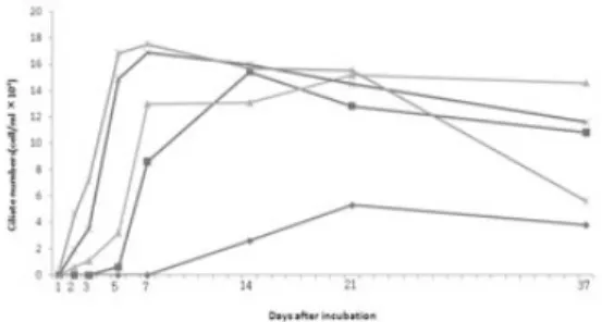
배양온도에 따른 시간 경과별 스쿠티카충 수 의 변화를 관찰하였다(Fig. 1). 스쿠티카충은 10
℃, 15℃, 20℃와 25℃에서 양호하게 증식하였으 며 고온 일수록 더 빠르게 분열하였으며 최고로 증식 가능한 수는 1.54~1.75× 10
5/ ml의 범위 로 유사했다. 그러나 5℃에서는 분열이 느리고, 5.3× 10
4/ ml가 최고로 배양 가능한 충수였다 (Table1). M. avidus는 이분열로 분열 하는데 이 분열에 걸리는 시간은 Table 1로부터 계산한 결 과 5℃에서 61.82시간, 10℃에서 26.32시간, 15
℃에서 21.14시간, 20℃에서 16.86시간 그리고 25℃에서는 16.21시간이었다. 배양시간이 경과 할수록 충체의 크기는 작아졌으며 14일을 기점 으로 사멸하는 세포들이 보였다. 25℃에서는 다 른 온도구에 비하여 37일 이후 사멸하는 충이 많았다.
감염실험
어체당 1×10
5/ 20 ul 농도를 감염시켰을 때 11 일간의 누적폐사율은 10℃에서는 65%, 15℃에
Fig. 1. Effect of temperature concentration on the growth of Miamiensis avidus
at 5℃(-◆-), 10℃(-■-), 15℃(-▲-) 20℃(-×-), 25℃(-*-)
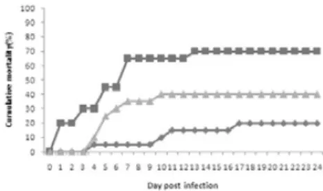
Fig. 3. Accumulated mortality of olive flounder by intraperitoneal injection (1×10⁴ciliates/fish) under various temperature at 10℃(-◆-), 15℃(-■-), 20℃(-▲-)
Fig. 5. Accumulated mortality of olive flounder by intraperitoneal injection (1×10²ciliates/fish) under various temperature at 10℃(-◆-), 15℃(-■-), 20℃(-▲-) Fig. 4. Accumulated mortality of olive flounder by
intraperitoneal injection (1×10³ciliates/fish) under various temperature at 10℃(-◆-), 15℃(-■-), 20℃(-▲-) Fig. 2. Accumulated mortality of olive flounder by intraperitoneal injection (1×10⁵ciliates/fish) under various temperature at 10℃(-◆-), 15℃(-■-), 20℃(-▲-)
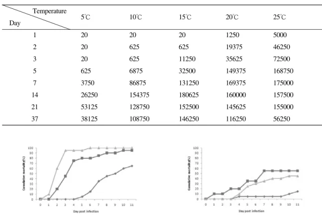
Table 1. Influence of temperature on in vitro growth of Miamiensis avidus in CHSE-214 cell line. Data are shown as cell numbers / ml.
Temperature
Day 5℃ 10℃ 15℃ 20℃ 25℃
1 20 20 20 1250 5000
2 20 625 625 19375 46250
3 20 625 11250 35625 72500
5 625 6875 32500 149375 168750
7 3750 86875 131250 169375 175000
14 26250 154375 180625 160000 157500
21 53125 128750 152500 145625 155000
37 38125 108750 146250 116250 56250
서 95%, 20℃에서 100%였다(Fig. 2). 어체당 1×
10
4을 감염시켰을 때 누적폐사율은 10℃에서는 10%, 15℃에서 25%, 20℃에서 40%였다(Fig. 3).
어체당 1×10
3을 감염시켰을 때 누적폐사율은 10℃에서는 0%, 15℃에서 5%, 20℃에서 40%였
으며(Fig. 4), 어체당 1×10
2을 감염시켰을 때 누 적폐사율은 10℃에서는 5%, 15℃에서 0%, 20℃
에서 20%였다(Fig. 5). 대조구에서는 폐사가 발 생하지 않았다.
두 번째 감염실험은 첫 번째 실험구와 동일한
조건에서 행하였고 24일간 폐사를 관찰하였다.
어체당 1×10
5을 감염시켰을 때 누적폐사율은 10℃에서는 70%, 15℃에서 100%, 20℃에서 100%였다(Fig. 6). 어체당 1×10
4을 감염시켰을 때 누적폐사율은 10℃에서는 20%, 15℃에서 70%, 20℃에서 40%였다(Fig. 7). 어체당 1×10
3을 감염시켰을 때 누적폐사율은 10℃에서는 5%, 15℃에서 15%, 20℃에서 45%였으며(Fig. 8), 어체당 1×10
2/ 20 ul 농도를 감염시켰을 때 누적 폐사율은 10℃에서는 5%, 15℃에서 5%, 20℃에 서 25%였다(Fig. 9). 폐사의 패턴은 첫 번째와 유 사하였으며 두 번째 실험에서는 24일간 폐사의 추이를 관찰하였는데 10일에서 11일 사이에 폐 사는 안정된 이후 실험을 종료하는 시점까지 많 은 폐사는 발생하지 않았다. 각 수온별 대조구에 서는 폐사가 발생하지 않았다.
감염 실험에서 폐사어의 아가미, 피부, 복수, 뇌 를 관찰한 결과, 어체당 1×10
5/ 20 ul 의 농도로
감염시켰을 때 20℃는 감염 1일째 아가미(피부) 에만 충이 관찰되다가 감염 3일째 뇌에서도 관 찰되었으며, 15℃는 감염 2일째 아가미(피부)에 서 충이 보였고 감염 4일째 뇌에서 충이 관찰되 었다. 그리고 10℃는 감염 5일째 아가미(피부)에 서 충이 관찰되었고, 감염 6일째 뇌에서도 보였 다. 어체당 1×10
4을 감염시켰을 때 20℃는 감염 4일째 아가미(피부)에만 충이 관찰되다가 감염 6일째 뇌에서도 보였고, 15℃는 감염 5일째 아 가미(피부)에만 보이다가 감염 7일째 뇌에서도 관찰되었다. 그리고 10℃는 감염 8일째 아가미 (피부)와 뇌에서 충이 관찰되었다. 어체당 1×10
3을 감염시켰을 때 20℃는 감염 7일째 아가미(피 부)와 뇌에서 충이 보였고, 15℃는 감염 8일째 아가미(피부)에서만 충이 관찰되었고, 10℃에서 는 감염 9일째 아가미(피부)에서만 충이 관찰되 었고 감염 17일째에도 뇌에서는 충을 관찰할 수 없었다. 어체당 1×10
2을 감염시켰을 때 20℃는
Fig. 8. Accumulated mortality of olive flounder byintraperitoneal injection (1×10³ciliates/fish) under various temperature at 10℃(-◆-), 15℃(-■-), 20℃(-▲-) Fig. 6. Accumulated mortality of olive flounder by intraperitoneal injection (1×10⁵ciliates/fish) under various temperature at 10℃(-◆-), 15℃(-■-), 20℃(-▲-)
Fig. 9. Accumulated mortality of olive flounder by intraperitoneal injection (1×10²ciliates/fish) under various temperature at 10℃(-◆-), 15℃(-■-), 20℃(-▲-) Fig. 7. Accumulated mortality of olive flounder by intraperitoneal injection(1×10⁴ciliates/fish) under various temperature at 10℃(-◆-), 15℃(-■-), 20℃(-▲-)
감염 7일째 아가미와 뇌에서 충이 관찰되었고, 10℃에서는 감염 8일째 아가미에서만 충이 관 찰 되었다. 모든 폐사어는 붉은색의 복수로 충만 해있었고 복수액을 현미경으로 관찰하면 활동 성이 강한 다수의 스쿠티카충이 적혈구를 세포 질 내에 함유하고 있는 것을 관찰할 수 있었다.
폐사직전의 넙치는 체색흑화와 복수증상을 보 였고 경련과 탈장을 일으키는 개체도 보였다. 감 염 10일 이후부터 경미한 궤양이 형성되는 개체 가 약간 보였다.
고 찰
M. avidus는 양식현장에서 넙치에 감염되는 스
쿠티카충 중에 어체 조직이나 뇌 속에 침투하여 치명적인 피해를 입히는 종이다 (Jung et al., 2007; Jin et al., 2009). M. avidus는 pH 6~9 범위 에서 증식이 이루어지며 온도는 10~30℃ 범위 에서 생존 및 증식이 가능하나 최적 온도 범위 는 10~25℃인데 10℃와 15℃보다 20℃에서 증 식이 더 빨리 일어난다고 보고하고 있다 (Jin et
al., 2007a). 또한, Iglesias et al. (2003a)은 13℃보다는 18℃와 23℃ 사이에서 증식이 잘 되며 최고 배양가능 농도는 표준 Leibovitz's L-15 배지 (Sig- ma-Aldrich Chimie, Germany)에서 1~2×10
5이라 하였다. 본 연구에서는 클로닝을 통하여 분리한
M. avidus를 10% FBS가 함유된 DMEM에서 배양한 CHSE-214 cells을 먹이원으로 배양하였을 때 in vitro상에서 5℃, 10℃, 15℃, 20℃와 25℃에 서 최대 증식가능한 수는 1.54~1.75 × 10
5의 범 위로 다른 보고들과 유사하였고, 5℃에서는 증식 이 느리고 최대성장가능한 수도 5.4 × 10
4으로 다른 배양온도에서 보다 적었다. 이분열에 걸리 는 시간은 5℃에서 61.82시간, 10℃에서 26.32시 간, 15℃에서 21.14시간, 20℃에서 16.86시간 그 리고 25℃에서는 16.21시간으로 배양온도가 높 아질수록 이분열에 걸리는 시간이 짧은 것을 알 수 있었다. 충은 먹이원이 되는 CHSE-214세포 가 완전히 없어진 후에도 모든 실험온도에서 37
일째에도 생존해 있었으나 25℃에서는 크기가 작고 죽은 충이 많이 관찰되었다. 본 실험은 충 을 24 well plate에서 배양하였으나 25 cm
2와 75 cm
2의 세포배양용 플라스크의 큰 배양계를 사용 하는 경우에도 25℃는 다른 온도보다 분열이 빨 리 일어나고 사멸하는 세포도 빨리 생겼다. 먹이 원이 되는 CHSE-214세포는 3일에서 7일 사이 에 모두 없어지게 되는 데 먹이원이 없는 상태 에서는 분열은 하지 않지만 생존은 가능하여 양 식현장에서도 박멸이 어려울 것으로 보인다.
스쿠티카충에 의한 폐사는 연중 발생하지만 고수온기에 피해가 더 심한데 이는 충의 증식이 고수온기에 더 활발하기 때문인 것으로 생각된 다. 그러나 한편으로는 스쿠티카충의 침입에 대 항하는 어체의 면역반응 증강이 보고되고 있으 며, 충체를 이용한 백신의 개발이 기대되고 있다 (Iglesias et al., 2003b; Jung et al., 2006; Sanmartín
et al., 2008). 숙주가 되는 넙치는 광염성어류로서식온도는 10~27℃의 범위이고 최적 사육수 온은 21~24℃이다 (넙치양식표준지침서, 2006).
어류는 최적수온범위에서 면역반응이 가장 강 하고 활발하게 일어나는데 (Manning and Nakan- ishi, 1996; Bowden et al., 2007; Bowden, 2008) 이 온도범위에서 스쿠티카충에 감염이 일어나면 숙주가 충을 이겨낼 수도 있을 것으로 기대되므 로 실제 충을 어체에 감염시켰을 때의 폐사율은
in vitro 배양조건과는 차이가 날 수 있다. 스쿠티카충을 생체에 감염시켰을 때의 수온에 따른 병 원성의 차이는 보고된 바가 없어 본 연구를 실 시하였다. 실험은 2회 실시하였으며 10℃, 15℃, 20℃의 수온에서 어체 마리당 1×10
2, 1×10
3, 1
×10
4, 1×10
5ciliates을 접종시켜 1회째 실험은
11일간, 2회째 실험은 24일간 폐사율을 관찰하
였다. 폐사는 10일 정도까지 발생한 후 안정되어
두 번의 실험에서 비슷한 폐사패턴을 보였다. 이
는 복강으로 감염시킨 충에 의한 초기의 폐사가
주이며, 감염어(폐사어)에서 다른 개체로의 감염
에 의한 폐사는 경미한 것으로 판단할 수 있을
것이다. 온도가 높을수록 폐사율이 높았고 뇌에
서 충이 보이기 시작하는 시점도 짧았다. 또한 감염충의 농도가 높을수록 폐사율이 높았다. 20
℃의 넙치의 면역이 높은 상태에서 충에 의한 폐사가 많이 발생하였고, 10℃의 면역이 낮은 것 으로 생각되는 온도에서 폐사가 적게 발생한 결 과는 충에 의한 폐사율이 스쿠티카충에 대항하 는 어체의 면역반응보다는 충체의 증식속도에 더 영향을 많이 받는 것을 나타내고 있는 것으 로 생각된다. 그러므로 수온을 어체의 면역이 높 은 성장적온으로 유지하는 방법은 스쿠티카충 에 대해서는 적절하지 않은 방법으로 생각되며 고수온기에는 차광망을 시설을 하고, 환기를 잘 시키고, 환수율을 증가시키는 등의 수온이 높아 지지 않도록 하는 방법을 모색해야할 것이다.
요 약
본 연구는 양식넙치에 높은 폐사율과 성장저 하를 일으키는 스쿠티카섬모충 Miamiensis
avidus (syn. Philasterides dicentrarchi)의 성장과병원성에 미치는 온도의 영향을 알아보았다. in
vitro 배양조건에서 이분열에 걸리는 시간은 5℃에서 61.82시간, 10℃에서 26.32시간, 15℃에서 21.14시간, 20℃에서 16.86시간 그리고 25℃에서 는 16.21시간이었다. 최고로 배양 가능한 충수는 1.54~1.75 × 10
5/ ml의 범위로 10℃이상의 모든 온도범위에서 유사했다. 넙치(평균 8.34 cm, 4.33 g)마리당 1×10
2/ ul, 1×10
3/ ul, 1×10
4/ ul 과 1×
10
5/ ul 으로 스쿠티카충을 복강에 감염시킨 후 10℃, 15℃와 20℃에서 사육하면서 폐사를 관찰 하는 실험을 2회 실시한 결과, 수온 10℃에서는 폐사가 현저히 낮았고 온도가 높아질수록 폐사 가 증가하였다. 또한 감염농도가 높아질수록 폐 사는 증가하였다. 그러므로 스쿠티카섬모충 M.
avidus는 온도가 높을수록 증식이 잘 되며 또한
수온이 높아질수록 온도의존적으로 폐사가 증 가하므로 폐사를 경감시키기 위해서는 고수온 기의 수온관리가 중요한 것으로 사료된다.
감사의 글
이 논문은 2006년도 전남대학교 연구년교수 연구비 지원에 의하여 연구되었습니다.
참 고 문 헌
Bowden, T.J.: Modulation of the immune system of fish by their environment. Fish Shellfish Im- munol., 25: 373-383, 2008.
Bowden, T.J., Thompson, K.D., Morgan, A.L., Gratacap R.M.L., Nikoskelainen, S.: Sea- sonal variation and the immune response: A fish perspective. Fish Shellfish Immunol., 22: 695-706, 2007.
Dkoval, I. and Figueras, A.: Histopathological changes in turbot Scophthalmus maximus
due to a histophagous ciliate. Dis. Aquat.Org., 18: 5-9, 1994.
Iglesias, R., Parama, A., Alvarez, M.F., Leiro, J., Aja, C. and Sanmartin, M.L.: In vitro growth requirements for the fish pathogen Philas-
terides dicentrarchi (Ciliophora, Scuticocili-atida). Vet. Parasitol., 111: 19-30, 2003a.
Iglesias, R., Parama, A., Alvarez, M. F., Leiro, J., Ubeira, F.M. and Sanmartin M.L.: Philas-
terides dicentrarchi (Ciliophora: Scuticocili-atida) expresses surface immobilization anti- gens that probably induce protective im- mune responses in turbot. Parasitology 126:
125-134, 2003b.
Jee, B.Y., Kim, Y.C. and Park, M.S.: Morphology and biology of parasite responsible for scu- ticociliatosis of cultured olive flounder par-
alichthys olivaceus. Dis. Aquat. Org., 47:49-55, 2001.
Jin, C.N., Harikrishnan, R., Moon, Y.G., Kim,
M.C., Kim, J.S., Balasundaram, C., Azad,
I.S. and Heo, M.S.: Histopathological
changes of Korea cultured olive flounder,
Paralichthys olivaceus due to scuticociliato-sis caused by histophagous scuticociliate,
Philasterides dicentrarachi. Vet Parasitol.161: 292-301, 2009
Jin, C.N., Kang, H.S., Lee, C.H., Lee, Y.D., Lee, J.H. and Heo, M.S.: Biological Characteris- tics of Scuticociliate, Philasterides dicen-
trarchi Isolated from Cultured Olive Floun-der, Paralichthys olivaceus. J. Aquac. 20:
106-113, 2007a.
Jin, C.N., Kang, H.S., Moon, Y.G., Lee, Y.D., Lee, J.H., Song, C.B., and Heo, M.S.: Scuticocil- iatosis in flounder farms of Jeju island. J.
Fish Pathol. 20: 93-98, 2007b.
Jung, S.J., Kitamura S.I., Aoyama, M., Song, J.Y., Kim, B.K. and Oh, M.J.: Immune response of olive flounder Paralichthys olivaceus against Miamiensis avidus (Ciliophora: Scu- ticociliatida). J. Fish Pathol., 19: 171-181, 2006.
Jung, S.J., Kitamura S.I., Song J.Y. and Oh, M.J.:
Miamiensis avidus (Ciliophora: Scuticocili-
atida) causes systemic infection of olive flounder Paralichthys olivaceus and is a se- nior synonym of Philasterides dicentrarchi.
Dis. Aquat. Org., 73: 227-234, 2007.
Jung, S.J., Kitamura, S.I., Song, J.Y., Joung, I.Y.
and Oh, M.J.: Complete small subunit rRNA gene sequence of the scuticociliate Mi-
amiensis avidus pathogenic to olive flounder Paralichthys olivaceus. Dis. Aquat. Org., 64:159-163, 2005.
Kim, S.M., Cho, J.B., Kim, S.K., Nam, Y.K. and Kim, K.H.: Occurrence of Scuticociliatosis in olive flounder Paralichthys olivaceus by
Philasterides dicentrarchi (Ciliophora: Scu-ticociliatida). Dis. Aquat. Org., 62: 233-238, 2004a.
Kim, S.M., Cho, J.B., Lee, E.H., Kwon, S.R., Kim, S.K., Nam, Y.K. and Kim, K.H.: Pseudo-
cohnilemdus persalinus (Ciliophora: Scutic-ociliatida) is an additional species causing Scuticociliatosis in olive flounder Par-
alichthys olivaceus. Dis. Aquat. Org., 62:239-244, 2004b.
Lee, N.S., Park, J.H., Han, K.S. and Huh, M.D.:
Histopathological changes in fingerlings of Japanese flounder, Paralichthys olivaceus, with severe scuticociliatosis. J. Fish Pathol., 7: 151-160, 1994.
Lom, J and Dykova, I.: Protozoan parasites of fish- es. Elsevier, Amsterdam, 315p., 1992.
Manning, J. and Nakanishi, T.: The specific im- mune system: cellular defenses in The fish immune system, pp.183-185, ed., Iwama, G.
and Nakanishi, T. Academic press, San Diego, 1996.
Miller, T.A., Rapp, J., Wastlhuber, U., Hoffmann, R.W. and Enzmann P.-J.: Rapid and sensi- tive reverse transcriptase-polymerase chain reaction based detection and differential di- agnosis of fish pathogenic rhabdoviruses in organ samples and cultured cells. Dis.
Aquat. Org., 34:13-20, 1998.
Munday, B. L., O'Donoghue, P. J., Watts, M., Rough, K. and Hawkesford.: Fetal en- cephalitis due to the scuticociliate Uronema
nigricans in sea-caged, southern bluefin tyna Thunnus maccoyii. Dis. Aquat. Org., 30: 17-25, 1997.
Sanmartín, M.L., Paramá, A.,Castro, R., Cabaleiro, S., Leiro, J., Lamas, J., Barja, J.L.: Vaccina- tion of turbot, Psetta maxima (L.), against the protozoan parasite Philasterides dicen-
trarchi: effects on antibody production andprotection. J. Fish Dis. 31: 135-140, 2008.
Sano, M., Ito, T., Matsuyama, T., Nakayasu, C. and
Kurita, J.: Effect of water temperature shift- ing on mortality of Japanese flounder Par-
alichthys olivaceus experimentally infectedwith viral hemorrhagic septicemia virus.
Aquaculture. 286: 254-258, 2009.
Smail, D.A. and Munro, A.L.S.: The Virology of Teleost. In Fish Pathology. pp.238, 3rd ed., Roberts, R. J.. Harcourt Publisher, 2001.
Song, J.Y., Kitamura, S-I., Oh, M.J., Kang, H.S., Lee, J.H., Tanaka, S.J. and Jung, S.J.: Patho- genicity of Miamiensis avidus (syn. Philas-
terides dicentrarchi), Pseudocohnilembus persalinus, Pseudocohnilembus hargisi and Uronema marinum (Ciliophora, Scuticocili-atida). Dis. Aquat. Org., 83: 133-143, 2009.
Yoshinaga, T. and Nakazoe, J.: Isolation and in vitro cultivation of an unidentified ciliate causing scuticociliatosis in Japanese flounder par-
alichthys olivaceus. Fish Pathol., 28: 131-134, 1993.
손맹현, 박민우, 김응오, 임한규, 김대중, 안철민, 엄기혁, 김성길, 조용철, 이창훙, 황형규, 윤 성종, 한석중, 최낙중, 박영병, 어윤양: 넙치 양식표준지침서, pp.5-6, 도서출판 해인, 2006.
Manuscript Received : July 17, 2009 Revised : August 12, 2009 Accepted : August 22, 2009