Anti-oxidative and Anti-hyperglycemia Effects of Dung Beetle Extracts on the High Fat Diet SD Rats
Ha Jeong Kim, Ban Ji Kim and Mi Young Ahn*
Department of Agricultural Biology, National Academy of Agricultural Science, RDA, 166 Nongsaengmyung-Ro, Iseo-Myun, Wanju-Gun 55365, Korea
Received March 31, 2016 /Revised May 13, 2016 /Accepted May 16, 2016
Dung beetle (Catharsius molossus, CA) is a well-known group of insects thanks to their exploitation of animal feces, a behavioral trait with a global impact on earth’s ecosystems. This study was con- ducted to investigate the effect of CA extract on a high-fat diet in SD rats. Male rats were divided into 5 groups. Animals were fed on a high-fat diet for seven weeks before and dung beetle extract for a month during the administration. Weight gain was decreased in ethanol extract from CA group.
Administration of CA extract reduced the organ weight of testis and kidney, and adipose tissue weight. Lipid oxidative stress was evaluated measuring malondialdehyde level in liver. There were no significant differences in groups. Protein oxidative stress was evaluated measuring protein carbonyl content in blood. The protein carbonyl in blood was significantly decreased in ethanol and acetone extracted dung beetle groups (p<0.05). Meanwhile, the protein carbonyl in hepatocyte was not sig- nificant among the groups. Fibronectin and laminin by using D-HUVEC cell in vitro were measured by ELISA assay. There was significance in CA extract. The level of IL-10, IL-1β, VEGF, eNOS was evaluated by ELISA. There was significance in IL-10 compared to control (p<0.05). SOD and GPx tend- ed to increase by CA extract. Furthermore, CAT was increased significantly by CA extract (p<0.05).
After administration of CA extracts the composition of saturated fatty acid in adipose tissue tend to decrease, while unsaturated fatty acid increases. In conclusion, dung beetle had anti-hyperglycemia ef- fects of oxidative stress and antioxidant activity.
Key words : Cytokine, dung beetle, high-fat diet, oxidative stress
*Corresponding author
*Tel : +82-63-238-2975, Fax : +82-63-238-3833
*E-mail : [email protected]
This is an Open-Access article distributed under the terms of the Creative Commons Attribution Non-Commercial License (http://creativecommons.org/licenses/by-nc/3.0) which permits unrestricted non-commercial use, distribution, and reproduction in any medium, provided the original work is properly cited.
Journal of Life Science 2016 Vol. 26. No. 7. 772~781 DOI : http://dx.doi.org/10.5352/JLS.2016.26.7.772
서 론
당뇨병의 세계적인 추이는 유행 상태에까지 이르렀다고 한 다. 세계보건기구(World Health Organization; WHO)에 의하 면 2011년을 기준으로 하였을 때 세계적으로 약 3억 6,620만 명의 당뇨병 환자가 있으며, 2030년까지는 약 1.51배 증가되어 5억 5,180만 명이 될 것이라고 예측하였다[32]. 당뇨병은 식후 혈중 당과 인슐린 농도가 비정상적으로 급격히 상승하는데, 고혈당의 지속은 망막, 신장, 신경과 같은 기관에 합병증을 초래한다. 현재까지 당뇨의 근원적 치료방법은 개발되어 있지 않은 실정이며, 혈당치를 정상적인 수준으로 유지하는 것이 최선의 치료 방법으로 알려져 있다[9, 36, 53].
다양한 혈당강하제 중 α-glucosidase inhibitor인 acarbose, voglibose 등은 소장에서 탄수화물 분해를 억제시켜 섭취한
탄수화물의 분해를 지연시킴으로써 식후 혈중 당 농도의 급격
한 상승을 막을 수 있다[18]. 하지만 이런 합성 혈당 강하제는
저혈당의 위험이 높고 복부팽창, 부글거림, 설사 등의 부작용
이 나타날 수 있다[47]. 현재 제2형 당뇨 치료제로써 널리 사용
되고 있는 메트포민은 glycogen의 분해율을 감소시켜 혈당치
를 낮추고 췌장으로부터 인슐린 분비도 촉진하지 않으며, 체
중 감소 효과도 나타낸다. 또한 인슐린 유무와 상관없이 지방
산 산화에 영향을 미쳐 혈장 지질 감소에도 좋은 효과를 나타
낸다[26]. 하지만 메트포민 치료요법의 주요 부작용은 구토,
복부 불쾌감이나 복부 팽만감 등 소화기 계통에서 주로 많이
나타나며 이러한 부작용으로 인해 당뇨치료를 중단하는 경우
가 상당히 많다고 보고되고 있다. 당뇨환자가 메트포민을 장
기 복용한 경우 체내 칼슘대사에 영향을 미쳐 비타민 B12의
흡수를 저해함으로써 피로감을 동반한 혀의 통증이나 신경증
을 유발할 수 있다. 메트포민은 소장점막에서 당을 젖산으로
전환시키는데, 일부 신장 기능이 저하된 당뇨환자에게 지속적
으로 메트포민을 투여한 경우 혈액 내 축적된 메트포민이 간
에서 젖산이 당으로 전환되는 것을 저해하여 혈액 내 젖산
농도가 올라가게 되고 호흡곤란이나 복부 통증 등을 일으키는
젖산산증을 유발한다. 이와 같이 신장 기능이 약하거나 심장
질환자 그리고 간 기능이 약한 환자에게 사용 시 심각한 부작
용을 초래할 수도 있다[4, 34, 54]. 따라서 최근 당뇨병 치료약 제의 부작용을 극복하기 위해서 천연물을 소재로 하는 당뇨병 예방 및 치료보조제에 관한 연구가 활발히 이루어지고 있다 [24]. 천연물의 항당뇨 효능연구에서 직접적인 혈당강하작용 외에 확인이 필요한 효능으로 항비만 효과가 있다.
쇠똥구리는 농업적으로 매우 중요한 역할을 하고 있어 그 생태에 관한 연구 또한 중요해서 동물의 분을 지하에 묻거나 소비함으로써 영양분을 재활용하고 토질을 개선시키고[11], 가축의 분을 제거, 결과적으로 병원균의 서식처를 제거함으로 써 소, 양, 말 등 가축 등을 병원균으로부터 보호하는 역할을 한다. American Institute of Biological Science에서는 2006년 에 가축의 분뇨를 제거함으로써 3억 8,000만 달러의 가축이 보호된다고 추산하기도 할 정도로 그 경제적 가치는 매우 크 다[38]. 쇠똥구리의 농업적 중요성이 인식되어 농촌진흥청에 서 한반도에 서식하는 애기뿔소똥구리(Copris tripartitus) 사육 에 성공하고, 쇠똥구리 자체에서 분비되는 항박테리아성 펩타 이드에 대한 연구가 이루어져 펩타이드의 구조를 부분적으로 규명하였다[27]. 쇠똥구리 추출물의 비교군으로 사용된 귀뚜 라미 추출물은 Ahn [3]의 연구에 의하면 귀뚜라미를 사료에 첨가 급여시 계육에서 필수지방산을 비롯한 다가불포화 지방 산이 증가되었고, 소량의 EPA 증가, 육질의 향미와 육색의 명도 및 황색도 증가로 육질이 향상되었다. 또한 고지방식이 를 먹인 흰쥐에서 지방조직의 축적을 방지하는 항비만 효과를 확인한 바가 있다[3]. 양성대조군으로 사용된 Krill oil은 오메 가-3 지방산의 근원으로 혈장의 중성지방을 낮추는 항비만 효 과[48]와 당뇨환자에서 심혈관질환의 위험성을 낮추는 효과가 확인되었다[31]. 따라서 이와 같은 합성 치료제의 부작용을 최 소화하고 혈당강하효능과 함께 항비만 효능은 당뇨의 예방과 치료 및 부작용을 줄일 수 있는 중요한 요소가 되는 천연자원 곤충 소재 개발이 절실하게 요구되고 있다.
본 연구에서는 쇠똥구리 추출물을 이용하여 흰쥐에서 고지 방식이로 고혈당을 유도해 in vivo에서 지질과 단백질의 산화 적 스트레스 방지와 항산화 효과를 구명하고 당뇨 혈관내피세 포(D-HUVEC)를 이용하여 in vitro에서 cytokine 연구를 수행 하였다.
재료 및 방법
실험동물 및 사육조건
SD 4주령 수컷 랫드를 (주) 샘타코바이오코리아(경기도, 오 산)로부터 구입하여 7일간의 순화기간을 거쳐 건강한 동물만 을 골라 실험에 사용하였다. 사육 조건은 온도 21~23℃, 습도 40~60%, 조명 12시간 명암으로 유지시켰으며, 시험동물용 고 형사료((주) 샘타코바이오코리아, 오산)와 여과된 정제수를 자 유 섭취시켰다. SD 수컷 쥐를 5군으로 나뉘어 PBS 대조군, 쇠똥구리로부터 추출한 에탄올 추출물(CA EtOH, 100 mg/
kg/day), 쇠똥구리(100 mg/kg/day)로부터 추출한 아세톤 추 출물 (CA acetone), 귀뚜라미(Gryllus bimaculatus,)로부터 추출 한 아세톤 추출물(Gb acetone, 100 mg/kg/day), 양성대조군 으로 Krill oil (Mercola Health Resources, LLC., Aurora, USA)을 100 mg/kg/day 농도로 사용하였다. 본 실험에 사용 된 쇠똥구리는 강량(생약)의 형태로서 중국 연길 의약품 검사 소장이 감정하고 한국으로 들어온 것으로서, 시료를 동결건조 후 분쇄시켜 70% EtOH 추출여과 후 농축시켜 쇠똥구리 EtOH 추출물을 얻었고, EtOH 추출 후 남은 찌꺼기를 건조시켜 ace- tone 추출물을 여과 후 농축하여 쇠똥구리 acetone 추출물을 획득하였다. 귀뚜라미는 강원도 정선의 아리농장에서 냉동제 품으로 구입하였다. 동결건조 후 분쇄시켜 EtOH 추출 후 남은 찌꺼기를 건조시켜 acetone 추출과 여과 후 농축시켜 귀뚜라 미 acetone 추출물을 획득하였다. 13주령 쥐에 고지방식이로 비만을 7주간 유도하고, 20주령부터 시험물질을 고지방 식이 를 하면서 한 달간 투여하였다. 모든 동물 실험과정은 농촌진 흥청 동물실험윤리 위원회의 승인을 받았다(No. NAAS1501).
현미경을 통한 adipocyte의 수 및 크기 관찰
조직을 4% formaldehyde로 고정시킨 후 4 um로 절편을 만들어 toluidine 염색[23]을 시행한 후 현미경(Leica CTR 6000, microsoft systems)을 통해 adipocyte의 수와 크기를 관 찰하였다. adipocyte의 수는 현미경의 배율을 100배로 관찰하 여 통계 처리했고, 간 조직의 adipocyte의 수와 크기는 확대하 여 400배로 측정하였다.
간조직의 과산화지질 수준측정
채혈 후 간을 적출하여 0.9% 생리식염수에 세척하고 -70℃
에서 24시간 보관한 다음 단백질 추출용액(Intron Biotechnol- ogy, Gyeonggi-do, Korea)으로 분쇄하고, 조직 분쇄물 0.2 ml 에 8.1 % SDS 용액 0.2 ml, 증류수 0.6 ml을 첨가하여 실온에서 5분 정도 방치한 다음, 20% acetic acid (pH 3.5) 1.5 ml, 0.8
% TBA (thiobarbituric acid) 1.5 ml을 넣어 잘 섞은 뒤 95℃에 서 1시간 동안 가열하였다. 가열이 끝난 후 실온에서 식혀 증 류수 1 ml과 n-butanol:pyrimidine (15:1) 용액 5 ml를 첨가하 고, 8,000 rpm에서 20분간 원심분리 하여 상층액을 취하여 532 nm에서 과산화 생성물인 thiobarbituric acid reactive sub- stances (TBARS)의 흡광도를 측정하였으며[46], 지질의 과산 화 정도를 1, 1, 3, 3-tetraethoxypropane을 표준물질로 하여 측정하였다.
Protein carbonyl assay
글로빈과 hepatocyte 샘플당 10 mg protein에 0.2 DNPH와
2N-HCl을 넣고 1시간 동안 암실에서 상온 방치하며 15분마다
vortexing을 해준다. 10% TCA (최종농도) 넣어 단백질 침전시
키고, 5분간 아이스에 방치한 후 원심분리 13,000× g 2분 동안
돌리고 600 ul 6M guanidine-HCl 용액에 약간의 초음파처리 를 가해 펠렛을 녹인다. 원심분리 13,000× g 2분간 실시하고 상층액으로 360-380 nm에서 흡광도를 측정한다. 단백질 양은 bovine serum albumin을 대조로 하여 Bradford 방법으로 595 nm에서 흡광도를 측정하여 결정하였다.
간조직 중의 항산화효소 활성도 측정
간의 catalase 활성은 Abei 방법[1]에 따라, glutathione per- oxidase 활성은 Lawrence와 Burk의 방법[37]에 따라, super- oxide dismutase 활성은 McCord와 Fridovich의 방법[40]에 따라 각각 분광 광도계(Shimadzu, Japan)로 측정하였다.
세포실험
당뇨 혈관내피세포(Human Microvascular Endothelial Cells (CARDIAC) Diabetes type II ; D-HUVEC)를 Clonetics사 (Cambrex Bio Science Rockland, Walkersville, USA)로부터 구입하여 FBS, hydrocortisone, hFGF, VEGF, IGF, ascorbic acid, hEGF, GA-1000이 첨가된 EGM-2 MV SingleQuots (Lonza, MD, USA)를 EBM-2 배지에 넣어 95% 공기, 5% CO2, 습기가 충분한 37℃의 대기로 2~3일에 한 번씩 분주하여 배양 하였다. 곤충추출물 시료는 PBS 대조군, 쇠똥구리로부터 추출 한 에탄올 추출물(1 mg/ml), 쇠똥구리로부터 추출한 아세톤 추출물, 귀뚜라미로부터 추출한 아세톤 추출물(1 mg/ml), 양 성대조군으로 Krill oil (1 mg/ml)을 사용하여 처리하였다.
Adipocytokine과 염증인자 분석
사이토카인 분석을 위해 랫의 serum에서 IL-10과 IL-1ß를 효소면역측정법인ELISA kit를 사용하여 분광 광도계로 흡광 도를 측정하였고, VEGF와 eNOS는 D-HUVEC에서 곤충 시료 를 처리하여 24시간 인큐베이터에서 배양한 후, ELISA kit를 사용하여 분광 광도계로 흡광도를 측정하였다. 단핵구/대식 세포에서 분비되어 adipocyte의 염증반응에서 기여하는 M2 (anti-inflammatory effects) 마커로 IL-10 (Koma biotech Inc., South Korea)을, M1 (pro-inflammatory effect) 마커로 IL-1ß (Koma biotech Inc., South Korea), VEGF (R&D systems, MN, USA), eNOS (R&D systems, MN, USA)을 효소면역측정법인 ELISA kit로 확인하였다.
Laminin과 fibronectin ELISA
24 well plate에 D-HUVEC을 well당 1×10
5을 분주하고 쇠 똥구리 추출물질을 0.2 mg/ml 농도로 처리한 다음 하루 동안 37℃ 인큐베이터에서 배양한 다음 laminin (Millipore, CA, USA), fibronectin (Millipore, CA, USA) ELISA를 각각 시행하 였다. 표준물질과 샘플시료에 rabbit anti-human laminin, rab- bit anti-human fibronectin을 각각 동량으로 섞어 실온에서 10분간 반응시킨 다음, 100 ul를 덜어 well plate에서 1시간
동안 반응시킨 다음, wash buffer로 4회 수세한다. 100 ul goat anti-rabbit IgG-HRP conjugation을 각 well에 분주한 다음, 실온에서 30분간 반응시키고, wash buffer로 4회 수세한다.
100 ul TMB/E substrate를 각 well에 분주해서 실온에서 10분 간 반응시켜 분광 광도계로 450 nm에서 흡광도를 측정하고 표준식을 사용하여 농도를 계산하였다.
GC/Mass에 의한 지방산 분석법
지방조직으로부터 지방산을 추출하기 위해 복부지방과 부 고환지방 각각에서 0.1g 무게를 측량해서, 클로로포름:메탄올 (2:1) 혼합액으로 지방산 추출(24시간)한 후, 여과지로 여과하 고, 인지질의 알칼리 가수분해에 의한 비누화 반응(100℃, 0.5N NaOH in MeOH)과 메틸레이션(100℃, 14% BF
3로 15분 동안) 후, 25가지 지방산을 GC/MS로 분석했다. GC/MS는 (Agilent 6890 GC), Agilent 5973N mass detector, EI mode HP-5 capillary column (Agilent Technologies, Palo Alto, CA, USA)을 사용하였다. 주입구 온도는 250℃, MS 이동선은 23 0℃을 항상 유지하고, 오븐 온도는 180℃에서 20분간 유지된 다음, 10℃/분 증가되어 230℃까지 상승하고 10분간 유지하 고, 정량은 37지방산 standard (Sigma L9405, 10 rg/ml)를 사 용하였다(Sigma-Aldrich Inc.).
통계처리 및 결과 처리
실험결과는 평균±표준편차(Mean±SD)로 계산하였고, 각 시료농도군에 대한 유의성 검정은 대조군과 비교하여 Stu- dent’s t-test 한 후 p<0.05 값을 통계적으로 유의성 있는 결과 로 간주하였다.
결과 및 고찰
Adipocyte의 수 및 크기 관찰
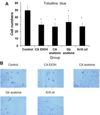
쇠똥구리 추출물을 먹인 랫 간의 adipocyte 수를 현미경을 사용하여 관찰하였다. 쇠똥구리 추출물을 먹인 SD rat의 간조 직을 toluidine blue 염색을 한 후 adipocyte를 관찰한 결과 대조군에 비해 CA EtOH, CA acetone, Gb acetone, Krill oil에 서 유의적으로 감소하였다(Fig. 1A). 이로써 쇠똥구리 추출물 이 adipocyte 수를 감소시키는 것으로 사료된다. 쇠똥구리 추 출물을 먹인 랫 간의 adipocyte 수와 크기를 현미경을 사용하 여 400배 확대하여 관찰하였으나 adipocyte 크기는 군간에 차 이가 없었다(Fig. 1B).
과산화지질 생성억제 효과 측정
쇠똥구리 추출물 투여에 대한 지질과산화의 산물인 mal-
dondialdehyde (MDA) 생성억제작용은 Fig. 2A에 나타나있
다. MDA는 3개의 탄소로 이루어진 저분자알데히드로서 다중
불포화지방산에 대한 유리기의 공격에 의해 생성되는 과산화
Toluidine blue
A
B
Fig. 1. The numbers of adipocyte and liver histology were de- termined according to different liver sites, using tolui- dine blue staining. (A) the numbers of mast cells by tol- uidine blue staining. (B) liver histology of PBS control, ethanol extract derived from dung beetle group, acetone extract from dung beetle, acetone extract from Gryllus bimaculatus, krill oil by toluidine blue staining (original magnification, x400). The data represent the mean±SD of three separate experiments (significant as compared to Control. *p<0.05).
A
B
C
Fig. 2. Oxidative stress levels of lipid and protein in dung beetle extracts. (A) Malondialdehyde (MDA) level in hep- atocyte from SD rats treated with dung beetle extract and other substances over two months. Protein carbonyl lev- els in blood (B) and liver (C) of SD rats treated with CA and other substances over two months. The data rep- resent the mean±SD of three separate experiments (significant as compared to Control. *p<0.05).
물의 분해산물이다. 유리기는 지질 단백질 및 DNA를 손상시 킴으로서 세포손상을 초래하여 노화 및 심혈관질환의 원인이 되며[5, 41] 항산화 효과가 있는 물질을 급여함으로서 유리기 의 활동을 감소시키는데 여러 유리기 중 과산화지질 산물은 유리기에 의한 지질손상의 지표로 가장 많이 이용되고 있다 [12]. 본 실험에서 쇠똥구리 추출물은 군간에 유의성이 없었다 (Fig. 2A).
단백질 산화스트레스의 측정
단백질 산화 스트레스의 지표로 protein carbonyl은 다른 산화 스트레스 지표에 비해 혈액 내에서 비교적 안정적이고, 산화 과정 중에 생기는 공통적인 현상이므로 산화 스트레스의 정도를 유용하게 측정할 수 있는 것으로 알려져 있다[16]. 만성 신부전, 당뇨, Alzeimer’s 질환 등의 질환에서는 protein car- bonyl 부산물이 질병의 병인과 관련이 있는 것으로 연구되고 있고 급성호흡곤란증후군, 기관지폐형성이상(bronchopul- monarydysplasia) 등의 폐질환에서도 증가를 관찰할 수 있다 [17]. Protein carbonyl group은 산화적인 스트레스의 중요하
고 즉각적인 바이오마커이다. Protein carbonyl이 DNPH로 태
깅되면 안정한 DNP hydrazone을 생성하는데 이는 375nm에
서 정량가능하다. 쇠똥구리 추출물을 먹인 SD rat의 혈액과
간에서 단백질의 산화적 스트레스 측정을 위해 단백질 카르보
닐 함량의 수준을 Fig. 2에 나타내었다. 혈액에서 대조군과 비
A
B
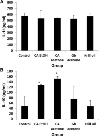
Fig. 3. Serum IL-1β and IL-10 levels of SD rats treated with dung beetle extracts over two months. (A) IL-1β cyto- kine activity, (B) IL-10 cytokine activity. The data repre- sent the mean±SD of three separate experiments (significant as compared to Control. *p<0.05).
교해 CA EtOH와 CA acetone군에서 유의적으로 감소하였고 (Fig. 2B), 간에서는 군간에 유의성이 없었다(Fig. 2C).
효소면역측정법을 이용한 사이토카인 활성의 수준 비만에 의한 지방세포로부터 유리된 사이토카인들은 비만 유래 이차성 질환인 심장질환 및 인슐린 저항성을 유발하는 중요한 요소가 된다[22, 35]. IL-10은 Th1과 Th2의 면역반응을 교차조절(cross regulation)하여 면역반응을 조절하는 cyto- kine이다[50]. 면역 조절작용에 관여하는 것으로 알려져 있다 [47, 49]. 또한 전염증성 cytokine들이 과량 분비되어 IL-10과 균형을 이루지 못할 경우, 숙주의 생존력에 영향을 줄 수 있으 므로[14] 염증 반응에서 IL-10의 분비는 매우 중요하다. IL-1β 는 TNF-α와 함께 대표적인 염증성 cytokine으로, NO를 생성 하게 하는 매개물질이다[7-8, 43]. 급성기 염증반응에서 이들 두 cytokine의 혈액 내 농도는 급격히 증가하며, 상호 상가효 과를 나타낸다[44]. 쇠똥구리 추출물을 먹인 SD rat의 혈청 IL-1beta의 수준은 군간에 유의성이 없었고, 항비만 사이토카 인인 IL-10은 대조군에 비해 CA EtOH와 CA acetone에서 유 의성있게 증가되어 항비만 효과가 있었다(Fig. 3).
혈관내피세포에서의 세포부착단백질 발현량 측정(In vitro) 세포 외막의 세포부착단백질인 laminin과 fibronectin의 발 현량 측정을 위해 D-HUVEC을 이용, 쇠똥구리 추출물을 처리 하여 실험한 결과가 Fig. 4에 나타나있다. 쇠똥구리 추출물을 처리한 세포의 CA EtOH군에서 대조군에 비해 laminin 발현 량이 감소되었고(Fig. 4A), fibronectin은 Gb acetone, Krill oil, Gb oil에서 발현량이 감소되었다(Fig. 4B).
세포에서 사이토카인 활성 측정
혈관내피세포는 신생혈관 생성에 중요한 역할을 한다. 신생 혈관 생성은 기존의 혈관에서 새로운 혈관이 만들어지는 일련 의 과정이며 유도물질과 억제물질간의 균형에 의하여 정교하 게 조절되는 것으로 알려져 있다. 신생혈관은 상처의 치유나 종양의 성장 과정에서 영양분과 산소의 공급 및 노폐물의 제 거를 위한 필수적인 과정이다[37]. VEGF가 강력한 혈관형성 촉진인자로 알려져 있다. 혈관 생성은 VEGF 등 혈관 생성 유 도물질에 의해 시작되어, 혈관을 둘러싼 기저 막이 분해되고, 혈관내피세포가 성장, 이동되어 공간적인 배열이 이루어지면 서 혈관이 생성된다[28].
NO는 혈관유지, 염증조절, 신경전달 등의 기능을 가지는 물질로서 NO의 농도는 뇌 허혈로 인한 손상과 직접적으로 연관이 되어 있을 뿐 아니라, 신경세포 손상에 따른 퇴행성 뇌 질환 발병에도 중요한 역할을 한다[13]. 이러한 NO는 뇌 허혈과 같은 저산소 상태가 되면 뇌세포 내에 칼슘이온(Ca2+) 의 농도가 높아지면서 NOS의 활성이 증가하게 되어 생성이 된다[10, 45]. NOS isoforms에는 칼슘이온의 농도에 의존적인
nNOS, eNOS와 칼슘이온의 농도에 비의존적이며 면역자극이나 염증과 같은 외부자극으로부터 유도되는 iNOS가 있는데 이런 NOS isoform의 종류에 따라 생성되는 NO가 뇌신경에 대해 보호작용을 하거나 독성작성을 하게 된다[29]. 뇌허혈 상 태에서 nNOS와 iNOS로부터 다량 생성되는 NO는 미토콘드 리아의 전자전달계 complex I, II 및 포도당 연소에 필요한 aconitase를 억제하여 에너지 생성을 방해하고 미토콘드리아 의 호흡을 저해함으로써 세포사멸을 유도하게 되지만, eNOS 에서 생성되는 NO는 대뇌 혈류의 흐름을 원활하게 하여 뇌신 경을 보호하는 역할을 한다[6]. 또한, eNOS는 심장근육세포, hippocampus의 pyramidal 세포, 혈관내피세포에 존재하여 [30] 혈관이완, 신생혈관형성, 혈소판응집억제, 발기(corpora cavernosa 이완) 등의 기능을 갖고 있다[42]. D-HUVEC을 이 용하여 쇠똥구리 추출물을 처리하여 사이토카인 활성을 측정 한 것이 Fig. 4에 나타나있다. 쇠똥구리 추출물을 처리한 VEGF (Fig. 4C)와 eNOS (Fig. 4D) 활성은 군간에 유의성이 없었다.
쇠똥구리 추출물의 간 중 항산화활성 효능
쇠똥구리 추출물의 항산화활성이 Fig. 5에 나타나있다. 생
체 내의 반응성 활성 산소종(reactive oxygen species, ROS)들
A
B
C
D
Fig. 4. Laminin, fibronectin, VEGF, eNOS assays in human microvascular en- dothelial cells diabetes type II treat- ed with dung beetle extracts. (A) Laminin activity, (B) Fibronectin ac- tivity, (C) VEGF activity, (D) eNOS activity. The data represent the mean±SD of three separate experi- ments (significant as compared to Control. *p<0.05).
Fig. 5. Antioxidant activities of dung beetle extracts in SD rat liver. (A) Superoxide dismutase (SOD) activity, (B) Catalase activity, (C) Glutathione peroxidase (GPx) activity. The data represent the mean±SD of three separate experiments (significant as compared to Control. *p<0.05).
은 체내의 항산화 물질들, 즉 비타민 C와 E, carotenoids, glu- tathione 및 항산화 효소들에 의해 조절된다[52]. 항산화 효소 들 중에서 superoxide dismutase (SOD)는 superoxide anion 을 hydrogen peroxide로 전환시키며, 생성된 hydrogen per- oxide는 catalase (CAT)와 glutathione peroxidase (GSH-Px) 에 의해 분해되는데, 분해되지 못한 여분의 hydrogen per- oxide는 Fe
2+와 반응하여 세포에 매우 강한 독성을 주는 hy- droxyl radical로 전환되고, 세포 내 단백질, 세포막, DNA 등
에 손상을 주어 세포의 노화와 괴사 등을 초래한다[19-21, 33].
고지방 식이로 인한 지속적인 고혈당은 간 중 항산화 효소 (SOD, CAT, GPx) 활성을 약화시켰으나, 쇠똥구리 추출물 투 여 시, 줄어들었던 항산화 효소 활성이 유의적으로 증가되었 다.
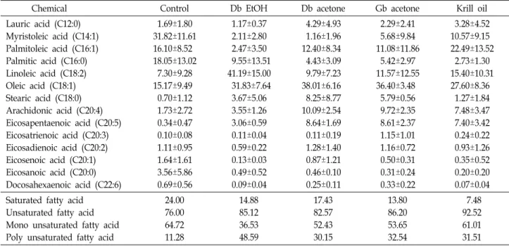
쇠똥구리 추출물을 먹인 랫 지방조직에서의 지방산 조성
지방조직에서의 지방산 조성비는 Table 1-2에 나타내었다.
Table 1. Effect of CA extracts on fatty acid composition ratio of abdominal fat tissues of SD rats over a 5 weeks period
Chemical Control Db EtOH Db acetone Gb acetone Krill oil
Lauric acid (C12:0) Myristoleic acid (C14:1) Palmitoleic acid (C16:1) Palmitic acid (C16:0) Linoleic acid (C18:2) Oleic acid (C18:1) Stearic acid (C18:0) Arachidonic acid (C20:4) Eicosapentaenoic acid (C20:5) Eicosatrienoic acid (C20:3) Eicosadienoic acid (C20:2) Eicosenoic acid (C20:1) Eicosanoic acid (C20:0) Docosahexaenoic acid (C22:6)
1.69±1.58 5.16±8.95 21.71±18.81 44.86±17.37 2.88±4.57 14.26±8.14 0.97±1.02 3.76±5.34 1.47±2.03 0.51±0.30 0.26±0.11 0.58±0.62 0.90±0.81 0.98±1.49
0.36±0.36 N.D 42.01±18.71 14.74±7.60 7.25±10.75 20.01±8.29 6.15±9.39 2.78±0.51 1.29±1.01 0.48±0.44 1.39±1.00 1.63±2.40 1.04±1.53 0.87±1.40
1.27±1.72 4.21±7.20 47.93±12.82 29.17±20.52 1.58±1.78 9.13±8.96 3.31±5.63 1.22±1.16 0.19±0.15 0.17±0.20 0.28±0.32 0.59±0.79 0.80±1.14 0.16±0.05
0.94±0.37 9.35±13.43 9.92±17.18 9.45±13.56 10.70±11.52 12.14±12.83 2.03±2.81 6.04±4.55 3.49±3.38 0.57±0.61 1.28±1.40 0.56±0.40 1.93±2.11 0.17±0.11
0.78±0.74 7.29±8.49 19.19±16.62 12.86±11.26 14.19±5.95 32.57±16.51 7.79±4.00 2.11±1.18 0.49±0.66 0.33±0.30 0.59±0.67 0.88±1.24 0.62±0.51 0.31±0.19 Saturated fatty acid
Unsaturated fatty acid Mono unsaturated fatty acid Poly unsaturated fatty acid
48.42 51.58 41.72 9.86
22.28 77.72 63.65 14.07
34.54 65.46 61.86 3.61
14.36 85.64 63.38 22.26
22.05 77.95 59.94 18.01
Table 2. Effect of CA extracts on fatty acid composition ratio of epididymidal fat tissues of SD rats over a 5 weeks period
Chemical Control Db EtOH Db acetone Gb acetone Krill oil
Lauric acid (C12:0) Myristoleic acid (C14:1) Palmitoleic acid (C16:1) Palmitic acid (C16:0) Linoleic acid (C18:2) Oleic acid (C18:1) Stearic acid (C18:0) Arachidonic acid (C20:4) Eicosapentaenoic acid (C20:5) Eicosatrienoic acid (C20:3) Eicosadienoic acid (C20:2) Eicosenoic acid (C20:1) Eicosanoic acid (C20:0) Docosahexaenoic acid (C22:6)
1.69±1.80 31.82±11.61 16.10±8.52 18.05±13.02 7.30±9.28 15.17±9.49 0.70±1.12 1.73±2.72 0.34±0.47 0.10±0.08 1.11±0.95 1.64±1.61 3.56±5.86 0.69±0.56
1.17±0.37 2.11±2.80 2.47±3.50 9.55±13.51 41.19±15.00 31.83±7.64 3.67±5.06 3.55±1.26 3.06±0.59 0.11±0.04 0.59±0.22 0.13±0.03 0.49±0.52 0.09±0.04
4.29±4.93 1.16±1.96 12.40±8.34 4.43±3.09 9.79±7.23 38.01±6.16 8.25±8.77 10.09±2.54 8.64±1.69 0.11±0.19 1.28±1.40 0.87±1.21 0.46±0.10 0.25±0.11
2.29±2.41 5.68±9.84 11.08±11.86 5.42±2.97 11.57±12.55 36.40±3.48 5.79±0.56 9.72±2.35 8.61±2.37 1.15±1.01 1.16±0.72 0.50±0.31 0.31±0.24 0.33±0.22
3.28±4.52 10.57±9.15 22.49±13.52 2.73±1.30 15.40±10.31 27.60±8.36 1.27±1.84 7.48±3.47 7.40±3.42 0.24±0.22 0.93±1.26 0.35±0.52 0.20±0.20 0.07±0.04 Saturated fatty acid
Unsaturated fatty acid Mono unsaturated fatty acid Poly unsaturated fatty acid
24.00 76.00 64.72 11.28
14.88 85.12 36.53 48.59
17.43 82.57 52.43 30.15
13.80 86.20 53.65 32.54
7.48 92.52 61.01 31.51
n-6계 다중불포화지방산은 linoleic acid (18:2n-6), arach-
idonic acid (20:4n-6) 그리고 n-3계 다중불포화지방산은 LNA (linolenic acid, 18:3n-3), EPA (eicosapentaenoic acid, 20:5n-3), DHA (docosahexaenoic acid, 22:6n-3)를 들 수 있 는데 전자가 결핍 될 경우에 성장지연, 피부손상, 생식장애, 지방간, 조갈증 등이 야기되는 반면에 후자의 경우에는 학습 능력 감퇴, 망막기능 장애, 시각 장애 등의 증상이 나타나, 각 기 다른 계열의 다중불포화지방산은 결핍시에 서로 다른 증상 을 유발시키는 것으로 알려져 있다[15, 25].
쇠똥구리 추출물을 먹인 SD rat의 복부지방조직에서 불포
화지방산인 myristoleic acid은 대조군에 비해 Gb acetone,
Krill oil에서 증가하였고, palmitoleic acid는 CA EtOH, CA
acetone에서 증가하였고, linoleic acid는 CA acetone, Gb ace-
tone, Krill oil에서 증가하였고, oleic acid는 CA EtOH, Krill
oil에서 증가하였고, arachidonic aci와 EPA는 Gb acetone에서
증가하였다. 대조 군에 비해 CA EtOH, CA acetone, Gb ace-
tone, Krill oil의 포화지방산이 감소하고, 불포화지방산이 증
가하였고, 다가불포화지방산은 대조 군에 비해 CA EtOH, Gb
acetone, Krill oil에서 증가하였다(Table 1). 쇠똥구리 추출물 을 먹인 SD rat의 부고환지방조직에서 불포화지방산인 lino- leic acid, oleic acid, arachidonic acid, EPA가 대조 군에 비해 CA EtOH, CA acetone, Gb acetone, Krill oil에서 증가하였다.
대조 군에 비해 CA EtOH, CA acetone, Gb acetone, Krill oil의 포화지방산이 감소하고, 불포화지방산이 증가하였고, 다가불 포화지방산은 대조군에 비해 CA EtOH, CA acetone, Gb ace- tone, Krill oil군에서 증가하였다(Table 2).
감사의 글
본 논문은 농촌진흥청 기본과제(PJ011853)로 수행된 연구 결과의 일부입니다.
References
1. Abei, H. 1974. Catalase. Method. Enzy. Anal. 2, 673-684.
2. Ahn, M. Y., Ryu, K. S., Park, B. Y., Kim, D. W., Kim, I.
and Kim, S. H. 2000. Effect of cricket on the chicken and its egg. Kor. J. Poult. Sci. 27, 197-202.
3. Ahn, M. Y., Kim, M. J., Kwon, R. H., Hwang, J. S. and Park, K. K. 2015. Gene expression profiling and inhibition of adi- pose tissue accumulation of G. bimaculatus extract in rats on high fat diet. Lipids Health Dis. 14, 116.
4. Altun, E., Kaya, B., Paydaş, S., Sarıakçalı, B. and Karayaylalı, I. 2014. Lactic acidosis induced by metformin in a chronic hemodialysis patient with diabetes mellitus type 2. Hemodial.
Int. 18, 529-531.
5. Babiy, A.V., Gebicki, J. M. and Sullivan, D. R. 1990. Vitamin E content and low density oxidizability induced by free radicals. Atherosclerosis 81, 175-182.
6. Beckman, J. S. and Koppenol, W. 1996. Nitric oxide, super- oxide, and peroxynitrite: the good, the bad, and ugly. Am.
J. Physiol. 271, 1424-1437.
7. Benvenistem, E. N. 1997. Immunology of the Nervous System. Oxford University Press. pp. 419-459. New York, USA.
8. Benvenistem, E. N. 1998. Cytokine actions in the central nervous system. Cytokine Growth F. R. 9, 259-275.
9. Boulé, N. G., Haddad, E., Kenny, G. P., Wells, G. A. and Sigal, R. J. 2001. Effects of exercise on glycemic control and body mass in type 2 diabetes mellitus: a meta-analysis of controlled clinical trials. JAMA 286, 1218-1227.
10. Bredt, D. S., Ferris, C. D. and Snyder, S. H. 1992. Nitric oxide synthase regu-latory sites; Phosphorylation by cyclic AMP- dependent proteinkinase, protein kinase,calcium /calmodulin protein kinase; identification of flavin and calmodulin bind- ing sites. J. Biol. Chem. 16, 10976-10982.
11. Brown, J., Scholtz, C. H, Janeau, J. L, Grellier, S. and Podwo- jewski, P. 2010. Dung beetles (Coleoptera: Scarabaeidae) can improve soil hydrological properties. Appl. Soil Ecol. 46, 9- 16.
12. Buege, J. A. and Aust, S. D. 1978. Microsomal lipid perox-
idation. In: Fleischer S, Packer, L. eds. Methods of Enzymelogy (London, Academic Press) 52, 302-309.
13. Choi, J. and Kim, D. 2003. Theraputic angiogenesis for car- diovascular diseases: the present and future. Kor. Circ. J. 33, 739-745.
14. Clerici, M., Ferrario, E., Trabattoni, D., Viviani, S., Bonfganti, V. and Vanzon, D. J. 1994. Multiple defects of T helper cell function in newly diagnosed patients with Hodgkin’s disease. Eur. J. Cancer 30, 1464-1470.
15. Connor, W. E., Neuringer, M. and Reisbick, S. 1996. Essential fatty acids: The importance of n-3 fatty acids in the retina and brain. Nutr. Rev. 50, 21-29.
16. Dalle-Donne, I., Rossi, R., Giustarini, D., Milzani, A. and Colombo, R. 2003. Protein carbonyl groups as biomarkers of oxidative stress. Clin. Chim. Acta. 329, 23-38.
17. Dalle-Donne, I., Giustarini, D., Colombo, R., Rossi, R. and Milzani, A. 2003. Protein carbonylation in human diseases.
Trends Mol. Med. 9, 169-176.
18. Derosa, G. and Maffioli, P. 2012. α-Glucosidase inhibitors and their use in clinical practice. Arch. Med. Sci. 8, 899-906.
19. Emerit, J., Beaumount, C. and Trivin, F. 2001. Iron metabo- lism, free radicals, and oxidative injury. Biomed. Pharmacother.
55, 333-339.
20. Fridovich, I. 1978. The biology of oxygen radicals. Science 201, 875-880.
21. Guemouri, L., Artur, Y. and Herbeth, B. 1991. Biological var- iability of superoxide dismutase, glutathione peroxidase, and catalase in blood. Clin. Chem. 37, 1932-1937.
22. Guzik, T. J., Mangalat, D. and Korbut, R. 2006. Adipocytoki- nesnovel link between inflammation and vascular function.
J. Physiol. Pharmacol. 57, 505-528.
23. Hausma, G. J. 1981. Techniques for studying adipocytes.
Stain Technol. 56, 149-154.
24. Heo, S. J., Hwang, J. Y., Choi, J. I., Han, J. S., Kim, H. J.
and Jeon, Y. J. 2009. Diphlorethohydroxycarmalol isolated from Ishige okamurae, a brown algae, a potent alpha-glucosi- dase and alpha-amylase inhibitor, alleviates postprandial hyperglycemia in diabetic mice. Eur. J. Pharmacol. 615, 252-256.
25. Holman, R. T. 1968. Essential fatty acid deficiency. Prog.
Lipid Res. 9, 275-348.
26. Hundal, R. S., Krssak, M., Dufou,r S., Laurent, D., Lebon, V., Chandramouli, V., Inzucchi, S. E., Schumann, W. C., Petersen, K. F., Landau, B. R. and Shulman, G. I. 2000.
Mechanism by which metformin reduces glucose pro- duction in type 2 diabetes. Diabetes 49, 2063-2069.
27. Hwang, J. S. et al. 2009. Isolation and characterization of a defensin-like peptide (Coprisin) from the dung beetle, Copris tripartitus. Int. J. Pept. Article ID 136284.
28. Hwang, J. S., Sung, D. I., Lee, W. M., Chung, Y. S. and Kim, H. B. 2014. Ethanol extracts of Chungkookjang stimulate the proliferation and migration of human umbilical vascular en- dothelial cells. Kor. J. Microbiol. 50, 223-226.
29. Ignarro, L. and Murad, F. 1995. Nitric oxide: Biochemistry, molecular biology, and therapeutic implications. Adv.
Pharmacol. 34, 511-516.
30. Janssens, S. P., Shimouchi, A., Quertermous, T., Bloch, D.
B. and Bloch, K. D. 1992. Cloning and expression of a cDNA encoding human endothelium-derived relaxing factor/nitric oxide synthase. J. Biol. Chem. 267, 14519-14522.
31. Jessika, M. L., Lauren, C. D., Amber, D. B., Marie, E. C., Joann, R. P. and Ramin, A. 2015. Effects of krill oil on endo- thelial function and other cardiovascular risk factors in par- ticipants with type 2 diabetes, a randomized controlled trial BMJ Open Diabetes Res. Care 3, e000107.
32. Joo, Y. I. 2009. Preventive medicine and public health.
Gyechulk Press. pp. 369, 2nd ed. Seoul, Korea.
33. Kim, J. H., Son, I. S., Kim, J. S., Kim, K. H. and Kwon, C.
S. 2008. Lipase-inhibitory and anti-oxidative activity of the methanol extract and the powder of Phellinus linteus. J. Kor.
Soc. Food Sci. Nutr. 37, 154-161.
34. Kim, O. K., Oak, C. H., Jeong, J. M., Lee, J. W., Shin, M.
H. and Kim, N. H. 2012. A case of metformin-induced lactic acidosis with acute kidney injury misdiagnosed as hep- atorenal syndrome in a cirrhosis patient. Kor. J. Med. 82, 241-246.
35. Kobalava, Z. D., Villevalde, S. V. and Isikova, K. V. 2009.
The role of adiponectin in development and progression of cardiovascular diseases. Kardiologiia 49, 51-58.
36. Koivisto, V. A. 1993. Insulin therapy in type II diabetes.
Diabetes Care 3, 29-39.
37. Lawrence, R. A. and Burk, R. F. 1976. Glutathione perox- idase activity in selenium-deficient rat liver. Biochem. Bioph.
Res. Co. 71, 952-958.
38. Losey, J. E. and Vaughan, M. 2006. The economic value of ecological services provided by insects. BioScience 56, 311- 323.
39. Mathiak, G., Grass, G., Herzmann, T. and Luebke, T. 2000.
Cu-Zetina C, Boehm SA. Capase-1-inhibitor ac-YVAD-cmk reduces LPS-lethality in rats without affecting haematology or cytokine responses. Brit. J. Pharmacol. 131, 383-386.
40. McCord, J. M. and Fridovich, I. 1969. Superoxide dismutase an enzymic function for erythrocuprein (hemocuprein). J.
Biol. Chem. 244, 6049-6055.
41. Miquel, J. and Quintailha, A. T. 1989. Historical introduction to free radical and antioxidant biomedical research. Vol. 1, pp. 3-16. In: CRC Handbook of Free Radical and Antiox- idants in Biomedicine. Weber, H. (ed.). CRC Press, Inc., Boca Raton, FL, USA.
42. Moncada, S. and Higgs, A. 1993. The L-arginine-nitric oxide pathway. N. Engl. J. Med. 329, 2002-2012.
43. Murphy, S., Simmons, M. L., Agullo, L., Garcia, A., Feinstein, D. L., Galea, E., Reis, D. J., Minc-Golomb, D. and Schwartz,
J. P. 1993. Synthesis of nitric oxide in CNS glial cells. Trends Neurosci. 16, 323-328.
44. Murphy, S. 2000. Production of nitric oxide by glial cells:
regulation and potential roles in the CNS. Glia. 29, 1-13.
45. Naka, M., Nanbu, T., Kobayashi, K., Kamanaka, Y., Komeno, M., Yanase, R., Fukutomi, T., Fujimura, S., Seo, H. G., Fujiwara, N., Ohuchida, S., Suzuki, K. and Kondo, K. 2000.
A potent inhibitor of inducible nitric oxide synthase, ONO-1714, a cyclic amidine derivative. Biochem. Bioph. Res.
Co. 270, 663-667.
46. Ohkawa, H., Ohishi, N. and Yagi, K. 1979. Assay for lipid peroxide in animal tissues by thiobarbituric acid reaction.
Anal. Biochem. 95, 351-358.
47. Pender, S.L., Breese, E. J. and Gϋnther, U. 1999. Suppression of T-cell-mediated injury in human gut by interleukin 10:
Role of matrix metalloproteinases. Gastroenterology 115, 573-583.
48. Rolf K. B., Marie, S. R., Pavol, B., Asbjørn, S., Jan, E. N., Espen, R., Inge, B. and Bodil, B. 2015. Krill oil reduces plas- ma triacylglycerol level and improves related lipoprotein particle concentration, fatty acid composition and redox sta- tus in healthy young adults - a pilot study. Lipids Health Dis. 14, 163.
49. Schotte, H. B., Schluter, P., Willeke, E., Mickholz, A., Schora, W. and Domschke, M. G. 2004. Long-term treatment with etanercept significantly reduces the number of proin- flammatory cytokine secteting peripheral blood mono- nuclear cells in patients with rheumatoid arthritis. Rheuma- tology 4, 112-118.
50. Shawn, B., Danuta, M., Skowronski, G., Kent, H., Robert, C. and Brunham, L. 2004. Aggregate content influences the Th1/Th2 immuneresponse to influenza vaccine: Evidence from a mousemodel. J. Med. Virol. 72, 138-142.
51. Standl, E. and Schnell, O. 2012. Alpha-glucosidase inhibitors 2012 - cardiovascular considerations and trial evaluation.
Diab. Vasc. Dis. Res. 9, 163-169.
52. Toda, N., Ayajiki, K. and Okamura, T. 2009. Cerebral blood flow regulation by nitric oxide: recent advances. Pharmacol.
Rev. 61, 62-97.
53. Toeller, M. 1991. Diet therapy of diabetes mellitus. Fortschr Med. 109, 41-42, 45.
54. Zhou, G., Myers, R., Li, Y., Chen, Y., Shen, X., Fenyk-Melody, J., Wu, M., Ventre, J., Doebber, T., Fujii, N., Musi, N., Hirshman, M. F., Goodyear, L. J. and Moller, D. E. 2001.
Role of AMP-activated protein kinase in mechanism of met- formin action. J. Clin. Invest. 108, 1167-1174.
초록:고지방식이로 유발한 흰쥐에서 쇠똥구리 추출물의 항산화 효과 및 혈당강하에 미치는 영향
김하정․김반지․안미영*
(농촌진흥청 국립농업과학원 농업생물부)