Gallic acid가 Lipopolysaccharide로 활성화된 마우스 대식세포의 케모카인과 성장인자 생성에 미치는 영향
박완수*
경원대학교 한의과대학 병리학교실
Inhibitory Effect of Gallic acid on Production of Chemokine and Growth Factor in Mouse Macrophage Stimulated by Lipopolysaccharide
Wan Su Park*
Department of Pathology, College of Oriental Medicine, Kyungwon University
Chemokine and Growth Factor are major mediumtors of immuno-inflammatory pathway. The purpose of this study is to investigate whether productions of Chemokine and Growth Factor in lipopolysaccharide (LPS)-induced mouse macrophage RAW 264.7 cells are modulated by Gallic acid (GA), which is easily founded in tannin-containing natural materials such as red wine, green tea, grape juice, and Corni Fructus. Productions of Chemokine and Growth Factor were analyzed by High-throughput Multiplex Bead based Assay with Bio-plex Suspension Array System based on xMAP Ⓡ (multi-analyte profiling beads) technology. At first, cell culture supernatant was obtained after treatment with LPS and GA for 24 hour. Then, the antibody-conjugated beads were added and incubated for 30 minutes. After incubation, detection antibody was added and incubated for 30 minutes. And Strepavidin-conjugated Phycoerythrin (SAPE) was added. After incubation for 30 minutes, the level of SAPE fluorescence was analyzed on Bio-plex Suspension Array System. Based on fluorescence intensity, concentrations of Chemokine and Growth Factor were determined. The results of the experiment are as follows. GA significantly inhibited the production of interferon-inducible protein (IP)-10, keratinocyte-derived chemokine(KC), and vascular endothelial growth factor (VEGF) in LPS-induced RAW 264.7 cells at the concentration of 25, 50, 100, 200 uM (
p<0.05). GA significantly inhibited the production of monocyte chemoattractant protein-1(MCP-1) and macrophage-colony stimulating factor(M-CSF) in LPS-induced RAW 264.7 cells at the concentration of 50, 100, 200 uM (
p<0.05). GA diminished the production of granulocyte macrophage-colony stimulating factor (GM-CSF) in LPS-induced RAW 264.7 cells. But GA did not show the inhibitory effect on the production of leukemia inhibitory factor (LIP) and macrophage inflammatory protein (MIP)-2 in LPS-induced RAW 264.7 cells. These results suggest that GA has the immuno-modulating activity related with its inhibitory effects on the production of IP-10, KC, MCP-1, VEGF, and M-CSF in LPS-induced macrophages.
Key words : Gallic acid, Macrophage, Chemokine, Growth factor, Inflammation, Lipopolysaccharide
* 교신저자 : 박완수, 성남시 수정구 복정동 경원대학교 한의과대학
․E-mail : hangl98@naver.com, ․Tel : 031-750-8821
․접수 : 2010/06/23 ․수정 : 2010/07/20 ․채택 : 2010/07/29
서 론
갈릭산(gallic acid; GA, 몰식자산)은 녹차, 홍차, 포도주, 커 피, 머루 등에 많이 함유되어 있는 탄닌(tannin)의 주요 구성성분 으로 식료품뿐만 아니라 산수유(山茱萸), 오미자(五味子), 산사자
(山査子), 오배자(五倍子) 등의 한약재에도 많이 함유되어 있는 것으로 알려져 있다 1,2) . 최근 정 등 3) 은 GA가 Hydrogen Peroxide 로 인한 손상으로부터 피부멜라닌세포를 보호하는 효과가 있음 을 보고한 바 있으며, 주 4) 는 발효차중의 미량성분인 gallic acid 산화물 purpurogallin carboxylic acid가 항염효능이 있음을 보고 하였다.
외부의 병원체로부터 인체를 방어하는 대표적인 체계가 인
체 면역염증체계이다. 인체의 면역염증방어체계는 세균, 바이러
스, 진균 혹은 이물질이 인체내부로 침입하였을 때 다양한 방법 을 동원하여 침입한 병원체(pathogen)들을 제거하고 손상된 자 신의 조직을 복구한다. 이에 관여하는 인체의 세포들이 다양한 면역염증세포들(immuno-inflammatory cells)이며 대표적인 면역 염증세포가 대식세포(macrophage)이다. 대식세포는 병원체 침입 의 초기단계에 침입한 항원을 ‘침입자’로 인식하고, 림프구와 같 은 인체 내의 다른 면역세포들에 적극적인 신호전달을 통하여 면역방어기능이 활성화되도록 한다 5,6) . 대식세포의 이러한 병원 체성 항원(pathogenic antigen)인식은 인터루킨(interleukin)뿐만 아니라 interferon-inducible protein (IP)-10, keratinocyte-derived chemokine(KC), monocyte chemoattractant protein-1(MCP-1)과 같은 케모카인(chemokine), vascular endothelial growth factor (VEGF), macrophage-colony stimulating factor(M-CSF), granulocyte macrophage-colony stimulating factor(GM-CSF)와 같은 성장인자(growth factor)의 생성증가로 이어진다. 외부로부 터 침입한 병원체에 의한 대식세포의 케모카인과 성장인자의 생 성증가는 면역염증반응의 필수적 조건이다. 그러나 적절히 제어 되지 못한 각종의 케모카인과 성장인자의 과잉생성은 급성 및 만성의 염증성 질환으로 발전할 수 있다 7-10) . 그러므로 한약재에 포함되어 있는 성분들을 대상으로 염증매개물질로 활성화된 대 식세포의 케모카인 및 성장인자 과잉생성을 조절할 수 활성을 조사함으로써 염증질환에 사용되는 한약재들의 항염효능을 규명 해 나갈 수 있을 것이다.
이에 저자는 식물성 한약재에 많이 포함되어 있는 갈릭산 (GA)이 그람음성균의 세포벽으로부터 유래된 지질다당체 lipopolysaccharide(LPS)로 마우스 대식세포 RAW 264.7의 케모 카인과 성장인자의 생성을 유발시키고 이에 대한 GA의 영향을 알아보기 위하여 in vitro 실험을 수행하고 유의한 결과를 얻었 기에 이에 보고하는 바이다.
재료 및 방법
1. 재료
1) 시약 및 기기
본 실험에 사용된 시약 중 gallic acid, dimethyl sulfoxide (DMSO), lipopolysaccharide(LPS), 1×PBS solution 등은 Sigma사 (MO, USA)로부터 구입하여 사용하였으며, 케모카인과 성장인자 분석을 위한 multiplex cytokine assay kit는 Bio-rad사(CA, USA) 와 Panomics사(CA, USA) 제품을 사용하였다. 각 시약의 품질은 분석용 등급 이상의 것으로 하여 사용하였으며 본 실험에 사용된 기기는 Bio-plex 200(Bio-Rad, USA), CO 2 incubator(NUAIRE, USA), rotary vacuum evaporator(Eyela, Japan), air compressor(Tamiya, Japan), homogenizer(Omni, USA), research microscope(Becton dickinson, USA), centrifuge(Hanil, Korea), fume hood(Hanil), clean bench(Jeio thec, Korea), ultrasonic cleaner(Branson, USA), thermo aluminum bath(Fine PCR, USA), vortex mixer(Vision Scientific Co, Korea), water bath(iNtRON biotech., Korea), ice-maker(Vision Scientific Co), deep
freezer(Revco, USA) 등이다.
2. 방법 1) Cell line
실험에 사용된 대식세포는 mouse macrophage(RAW 264.7 cell line)이며, 한국세포주은행(KCLB, Korea)에서 구입하였다.
2) 세포 배양
RAW 264.7 cells는 37℃, 5% CO 2 조건에서 10% FBS, penicillin(100 U/mL), streptomycin(100 ug/mL)이 첨가된 DMEM 배지로 배양되었다. RAW 264.7 cells는 75 cm 2 flask (Falcon, USA)에서 충분히 증식된 후 배양 3일 간격으로 배양세 포 표면을 phosphate buffered saline(Sigma, USA) 용액으로 씻 어준 후, 50 mL flask 당 1 mL의 0.25% trypsin-EDTA용액을 넣 고 실온에서 1분간 처리한 다음 trypsin용액을 버리고 37℃에서 5분간 보관하고 세포를 탈착하여 계대 배양하였다. 탈착된 세포 는 10% FBS가 첨가된 DMEM 배양액 10 mL에 부유시킨 다음, 새로운 배양용기에 옮겨 1 : 2의 split ratio로 CO 2 배양기(37℃, 5% CO 2 )에서 배양하였다.
3) 멀티플렉스 싸이토카인 분석(multiplex cytokine assay) LPS로 활성화된 RAW 264.7 cells의 각종 케모카인과 성장 인자 생성과 관련된 시료의 영향을 알아보기 위해 Yoon 등 11) 의 방법을 참고하여 다음과 같이 실험을 시행하였다.
96 well plate에 1×10 4 cells/well의 농도로 분주되도록 1×10 5
cells/mL의 cell을 100 μL씩 넣고 37℃, 5% CO 2 incubator에서
24시간동안 배양한 후, 배지를 버리고 배양세포 표면을
phosphate buffered saline (PBS) 용액으로 씻어주었다. LPS(1
ug/mL) 단독 혹은 배지에 녹인 시료(50, 100, 200, 400 uM)와 함
께 각 well에 처리하고 24시간 동안 배양하였다. 배양이 끝나면
상층액을 채취하여 다음의 멀티플렉스 어세이를 실시한다. 세포
배양 상층액을 분주하기 전에 96 well plate 타입의 Filter plate를
wash buffer로 세척 후, 특정항체가 결합되어 있는 beads를 분주
후 다시한번 wash buffer로 세척한다. 세척이 끝나면 세척액을
모두 제거한 후 준비된 세포배양상층액과 표준물질(standard
antibody)을 각 well에 50 μL씩 분주한다. 분주가 끝나면 실온에
서 30분간 500 rpm의 속도로 shaking한다. 30분간의 shaking
incubation이 끝나면 wash buffer를 이용, 3회의 세척을 실시한
다. 세척이 끝나면 세척액을 모두 제거한 후 미리 혼합된
Detection Antibody를 각 well에 25 μL씩 분주하고 실온에서 30
분간 500 rpm의 속도로 shaking한다. 30분간의 shaking
incubation이 끝나면 wash buffer를 이용, 3회의 세척을 실시한
다. 세척이 끝나면 세척액을 모두 제거한 후 미리 잘 섞인
Streptavidin-PE를 각 well에 50 μL씩 분주하고 실온에서 30분간
500 rpm의 속도로 shaking한다. 30분간의 shaking incubation이
끝나면 wash buffer를 이용, 3회의 세척을 실시한다. 세척이 끝
나면 세척액을 모두 제거한 후 각 well에 Reading buffer를 120
μ L씩 분주하고 실온에서 5분간 500rpm의 속도로 shaking한 후
Bioplex-200을 이용, 각 케모카인과 성장인자들의 생성량을 측정,
비교한다.
3. 통계처리
실험성적은 평균치 ± 표준오차 (Mean ± SEM)로 나타내었 으며, 대조군과 각 실험군과의 평균 차이는 Student t-test로 분석 하여 p <0.05일 때 통계적으로 유의한 것으로 판정하였다.
결 과
1. GA가 LPS로 유발된 mouse macrophage RAW 264.7의 IP-10 생성증가에 미치는 영향
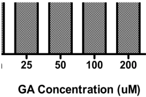
LPS(1 ug/mL)와 함께 GA(0, 25, 50, 100, 200 uM)가 포함된 배지에 RAW 264.7 cells을 24시간 동안 배양한 결과 LPS로 인한 IP-10 생성증가(4639.9 pg/mL)를 GA가 25, 50, 100, 200 uM의 농도에서 각각 1988.9, 2039.3, 1948.5, 2008.4 pg/mL로 모두 유의 ( p <0.05)하게 감소시켰다(Fig. 1).
Nor Con 25 50 100 200
GA Concentration (uM)
Fig. 1. Effect of GA on producton of IP-10 in RAW 264.7 cells using Bioplex cytokine assay.
Cells were incubated with LPS (1 ug/mL) and GA (0, 25, 50, 100, 200 uM) for 24 h. Results are represented as mean ± SEM. Nor : Normal group treated with culture medium only. Con : Control group treated with LPS only. * represents p < 0.05 compared to the control.2. GA가 LPS로 유발된 mouse macrophage RAW 264.7의 KC 생 성증가에 미치는 영향
LPS(1 ug/mL)와 함께 GA(0, 25, 50, 100, 200 uM)가 포함된 배지에 RAW 264.7 cells을 24시간 동안 배양한 결과 LPS로 인한 KC 생성증가(300.6 pg/mL)를 GA가 25, 50, 100, 200 uM의 농도 에서 각각 28, 29.8, 25.8, 27.3 pg/mL로 모두 유의( p <0.05)하게 감소시켰다(Fig. 2).
Nor Con 25 50 100 200
*
* * *
GA Concentration (uM)
Fig. 2. Effect of GA on producton of KC in RAW 264.7 cells using Bioplex cytokine assay.
Cells were incubated with LPS (1 ug/mL) and GA (0, 25, 50, 100, 200 uM) for 24 h. Results are represented as mean ± SEM. Nor : Normal group treated with culture medium only. Con : Control group treated with LPS only. * represents p < 0.05 compared to the control.3. GA가 LPS로 유발된 mouse macrophage RAW 264.7의 VEGF 생성증가에 미치는 영향
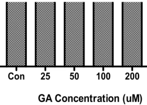
LPS(1 ug/mL)와 함께 GA(0, 25, 50, 100, 200 uM)가 포함된 배지에 RAW 264.7 cells을 24시간 동안 배양한 결과 LPS로 인한 VEGF 생성증가(2206.4 pg/mL)를 GA가 25, 50, 100, 200 uM의 농도에서 각각 1641.6, 1784, 1428.9, 1701.6 pg/mL로 모두 유의 ( p <0.05)하게 감소시켰다(Fig. 3).
Nor Con 25 50 100 200
GA Concentration (uM)
Fig. 3. Effect of GA on producton of VEGF in RAW 264.7 cells using Bioplex cytokine assay.
Cells were incubated with LPS (1 ug/mL) and GA (0, 25, 50, 100, 200 uM) for 24 h. Results are represented as mean ± SEM.Nor : Normal group treated with culture medium only. Con : Control group treated with LPS only. * representsp < 0.05 compared to the control.
4. GA가 LPS로 유발된 mouse macrophage RAW 264.7의 MCP-1 생성증가에 미치는 영향
LPS(1 ug/mL)와 함께 GA(0, 25, 50, 100, 200 uM)가 포함된 배지에 RAW 264.7 cells을 24시간 동안 배양한 결과 LPS로 인한 MCP-1 생성증가(17115.5 pg/mL)를 GA가 50, 100, 200 uM의 농 도에서 각각 14710.2, 15116.8, 14666.5 pg/mL로 유의( p <0.05)하 게 감소시켰다(Fig. 4).
Nor Con 25 50 100 200
GA Concentration (uM)
Fig. 4. Effect of GA on producton of MCP-1 in RAW 264.7 cells using Bioplex cytokine assay.
Cells were incubated with LPS (1 ug/mL) and GA (0, 25, 50, 100, 200 uM) for 24 h. Results are represented as mean ± SEM.Nor : Normal group treated with culture medium only. Con : Control group treated with LPS only. * representsp < 0.05 compared to the control.
5. GA가 LPS로 유발된 mouse macrophage RAW 264.7의 M-CSF 생성증가에 미치는 영향
LPS(1 ug/mL)와 함께 GA(0, 25, 50, 100, 200 uM)가 포함된
배지에 RAW 264.7 cells을 24시간 동안 배양한 결과 LPS로 인한
M-CSF 생성증가(62.6 pg/mL)를 GA가 50, 100, 200 uM의 농도
에서 각각 55.5, 55.8, 58 pg/mL로 유의( p <0.05)하게 감소시켰다 (Fig. 5).
Nor Con 25 50 100 200
GA Concentration (uM)
Fig. 5. Effect of GA on producton of M-CSF in RAW 264.7 cells using Bioplex cytokine assay.
Cells were incubated with LPS (1 ug/mL) and GA (0, 25, 50, 100, 200 uM) for 24 h. Results are represented as mean ± SEM.Nor : Normal group treated with culture medium only. Con : Control group treated with LPS only. * representsp < 0.05 compared to the control.
6. GA가 LPS로 유발된 mouse macrophage RAW 264.7의 GM-CSF 생성증가에 미치는 영향
LPS(1 ug/mL)와 함께 GA(0, 25, 50, 100, 200 uM)가 포함된 배지에 RAW 264.7 cells을 24시간 동안 배양한 결과 LPS로 인한 GM-CSF 생성증가(2448 pg/mL)를 GA가 50, 200 uM의 농도에서 각각 1915.5, 1954 pg/mL로 유의( p <0.05)하게 감소시켰다(Fig. 6).
Nor Con 25 50 100 200
GA Concentration (uM)
Fig. 6. Effect of GA on producton of GM-CSF in RAW 264.7 cells using Bioplex cytokine assay.
Cells were incubated with LPS (1 ug/mL) and GA (0, 25, 50, 100, 200 uM) for 24 h. Results are represented as mean ± SEM.Nor : Normal group treated with culture medium only. Con : Control group treated with LPS only. * representsp < 0.05 compared to the control.