Cerebellar Artery
Spectrum of Audiovestibular Loss
Hyung Lee, MD; Ji Soo Kim, MD; Eun-Ji Chung, MD; Hyon-Ah Yi, MD; In-Sung Chung, MD;
Seong-Ryong Lee, MD; Je-Young Shin, MD
Background and Purpose—To define the detailed spectrum of audiovestibular dysfunction in anterior inferior cerebellar artery territory infarction.
Methods—Over 8.5 years, we prospectively identified 82 consecutive patients with anterior inferior cerebellar artery territory infarction diagnosed by MRI. Each patient completed a standardized audiovestibular questionnaire and underwent a neuro-otologic evaluation, including bithermal caloric tests and pure tone audiogram.
Results—All but 2 (80 of 82 [98%]) patients had acute prolonged vertigo and vestibular dysfunction of peripheral, central, or combined origin. The most common pattern of audiovestibular dysfunction was the combined loss of auditory and vestibular function (n ⫽49 [60%]). A selective loss of vestibular (n⫽4 [5%]) or cochlear (n⫽3 [4%]) function was rarely observed. We could classify anterior inferior cerebellar artery territory infarction into 7 subgroups according to the patterns of neuro-otological presentations: (1) acute prolonged vertigo with audiovestibular loss (n ⫽35); (2) acute prolonged vertigo with audiovestibular loss preceded by an episode(s) of transient vertigo/auditory disturbance within 1 month before the infarction (n ⫽13); (3) acute prolonged vertigo and isolated auditory loss without vestibular loss (n ⫽3); (4) acute prolonged vertigo and isolated vestibular loss without auditory loss (n⫽4); (5) acute prolonged vertigo but without documented audiovestibular loss (n ⫽24); (6) acute prolonged vertigo and isolated audiovestibular loss without any other neurological symptoms/signs (n ⫽1); and (7) nonvestibular symptoms with normal audiovestibular function (n ⫽2).
Conclusions—Infarction in the anterior inferior cerebellar artery territory can present with a broad spectrum of audiovestibular dysfunctions. Unlike a viral cause, labyrinthine dysfunction of a vascular cause usually leads to combined loss of both auditory and vestibular functions. (Stroke. 2009;40:3745-3751.)
Key Words: anterior inferior cerebellar artery 䡲 audiovestibular loss 䡲 infarction
I n infarction involving the distribution of the anterior inferior cerebellar artery (AICA), vertigo is usually asso- ciated with other neurological symptoms or signs such as hearing loss, facial weakness, facial sensory loss, crossed sensory loss, Horner syndrome, gait ataxia, and limb ataxia.1–3
Adams
1was the first who completely described the syndrome associated with AICA occlusion. In his patient, neuro- otologic symptoms such as vertigo, tinnitus, and bilateral hearing loss were early symptoms. Subsequent reports on AICA infarction have focused on the brainstem and cerebel- lar findings with little attention to the associated neuro- otological dysfunction.
2,4 –7 Before 2000, few studies
8,9 had carefully investigated the audiovestibular disturbances that occur with AICA infarction.
AICA usually arises from the caudal third of the basilar artery and supplies the inner ear, lateral pons, middle cere- bellar peduncle, and anterior inferior cerebellum, including the flocculus.
2,5Because AICA is an important artery for vascular supply to the peripheral and central vestibular structures and its occlusion commonly causes vertigo of either peripheral or central origin, we considered that analyz- ing the patterns of vestibular loss may shed light on the mechanism of vertigo occurring in vascular compromise within the posterior circulation.
Although recent studies
10 –17have emphasized that audio- vestibular loss is an important sign for the diagnosis of AICA infarction, detailed spectrum of audiovestibular dysfunctions has not been systematically studied in AICA territory infarc-
Received August 6, 2009; accepted August 25, 2009.
From the Department of Neurology (H.L., E.-J.C., H.-A.Y.) and the Brain Research Institute (H.L., H.-A.Y., I.-S.C., S.-R.L.), Keimyung University School of Medicine, Daegu, South Korea; and the Department of Neurology (J.S.K., J.-Y.S.), Seoul National University College of Medicine, Seoul National University Bundang Hospital, Seongnam, South Korea.
H.L. and J.S.K. had cocorresponding author positions.
Correspondence to Hyung Lee, MD, Department of Neurology, Keimyung University School of Medicine, 194 Dongsan dong, Daegu, 700-712 South Korea. E-mail [email protected]
© 2009 American Heart Association, Inc.
Stroke is available at http://stroke.ahajournals.org DOI: 10.1161/STROKEAHA.109.564682
3745
by guest on February 2, 2017http://stroke.ahajournals.org/Downloaded from
tion. Thus, we sought to investigate the patterns of the audiovestibular dysfunction in a large series of consecutive patients with AICA territory infarction and to consider its clinical implication.
Patients and Methods
Between January 2000 and July 2008, we identified 90 consecutive patients from the acute stroke registry at 2 university hospitals in Korea, who had an acute infarction in the distribution of the AICA.
We determined the AICA territory using the anatomic diagrams of Amarenco and Hauw3and the diagnosis of the AICA infarction was made when the MRI lesions involved at least one of the following anatomic structures: middle cerebellar peduncle, lateral inferior pontine area, or anterior cerebellar hemisphere. The typical AICA territory pontine lesion is like a triangle with an anterolateral base and an apex directed toward the fourth ventricle between the middle cerebellar peduncle and anterolateral pontine region. After excluding 8 patients with incomplete audiovestibular evaluation, 82 patients were finally selected for this study. In every patient, diagnostic tests were performed to determine risk factors of stroke. The average age of the patients was 64.9⫾15.3 years with a range from 23 to 93 years. Each patient completed a standardized dizziness questionnaire that included a detailed description on acute audiovestibular distur- bances and underwent a neuro-otologic evaluation performed by the authors (H.L. and J.S.K.).
Vertigo was defined as a spinning illusion of the environment or the patients themselves. Caloric testing was performed with 30°C cold and 44°C warm irrigations of each ear for 20 seconds. Canal paresis (CP) was calculated using the Jongkees formula. Details regarding the audiovestibular tests have been previously pub- lished.3,18 We defined the auditory loss of a vascular cause as follows: (1) the patients noted a definite decline of hearing during the attack of AICA infarction; and (2) pure tone audiogram also documented the sensorineural hearing loss (SNHL). The diagnosis of vestibular loss of a vascular cause was based on the following criteria: (1) the patient had acute prolonged (⬎24 hours) vertigo at the time of AICA infarction; and (2) standardized caloric stimulation showed a reduced response ⬎25% in the lesion side. Central vestibular dysfunction was defined when: (1) the patient had acute prolonged (⬎24 hours) vertigo at the time of AICA infarction; (2) standardized caloric stimulation showed normal caloric response;
and (3) patients showed at least one of the following signs, including asymmetrical abnormalities of pursuit or optokinetic nystagmus, gaze-evoked bidirectional nystagmus, or impaired modulation of the vestibular response using visual input.
Neuro-otological evaluations were mostly performed during the acute period (0 to 43 days; mean interval of 6.7 days for vestibular evaluation and 5.3 days for auditory evaluation). In most patients, the MRI, including diffusion-weighted imaging and MR angiography (MRA), was performed during the acute period (0 to 30 days; mean intervals, 4.1 days).
Statistical analyses were performed by using SPSS program (Version 12.0; SPSS, Chicago, Ill).2test was used to compare the
result of MRA findings in patients with and without prodromal audiovestibular loss. The MRA findings in patients with and without multiple posterior circulation infarcts were also compared using the
2test. Significance was assumed at a value of P⬍0.05.
All experiments followed the tenets of the Declaration of Helsinki and informed consents were obtained after the nature and possible consequences of the study had been explained to the participants.
Because the present study included all consecutive patients during the research period, 23 patients previously reported3,10 –12,16,17were included, but new information is added in this report.
Results Clinical Features
All were alert and oriented on admission. In most patients (80 of 82 [98%]), acute spontaneous prolonged vertigo ( ⬎24 hours) with nausea/vomiting was the presenting or main symptom. The spontaneous nystagmus was predominantly horizontal and 55 (67%) patients showed spontaneous nys- tagmus beating away from (84% [46 of 55]) or toward (11%
[6 of 55]) the side of the lesion. The other 3 showed seesaw, upbeat, or pure torsional nystagmus. Asymmetrical bidirec- tional gaze-evoked nystagmus was also found in 35 patients.
Other findings included limb dysmetria (n ⫽55 [67%]), gait ataxia (n ⫽52 [63%]), facial sensory loss (n⫽23 [28%]), facial weakness (n ⫽23 [21%]), body sensory loss (n⫽5 [6%]), Horner syndrome (n ⫽3 [4%]), dysarthria (n⫽3 [4%]), eye motion limitation (n ⫽2 [2%]), and limb weakness (n⫽2 [2%]). The complete AICA syndrome described by Adams was found in only 2 (2%) patients in whom the classic pontine symptoms such as facial sensory loss and weakness, crossed sensory sign, and Horner syndrome developed in addition to prolonged vertigo, hearing loss, and cerebellar signs. The most common risk factor was hypertension (66%) followed by diabetes mellitus (40%), current smoking (27%), a history of stroke (23%), atrial fibrillation (7%), and cardiac disease (6%). At least 2 vascular risk factors were found in 40 (49%), whereas no identifiable risk factor was present in 8 (10%) patients.
Characteristics of Audiovestibular Loss
There were 7 subgroups of AICA territory infarction accord- ing to the patterns of neuro-otological presentations (Table 1):
(1) 35 patients presented with prolonged vertigo and had acute audiovestibular loss (ie, CP and SNHL); (2) 13 patients had an episode(s) of transient vertigo, hearing loss, and/or tinnitus within 1 month before the infarction in addition to
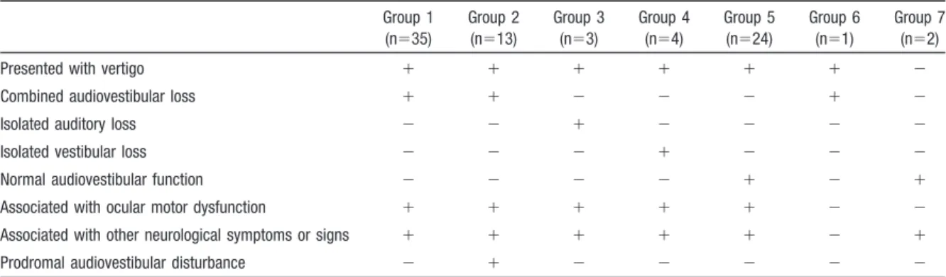
Table 1. Patterns of Audiovestibular Loss in 82 Patients With AICA Territory Infarction Group 1
(n⫽35)
Group 2 (n⫽13)
Group 3 (n⫽3)
Group 4 (n⫽4)
Group 5 (n⫽24)
Group 6 (n⫽1)
Group 7 (n⫽2)
Presented with vertigo ⫹ ⫹ ⫹ ⫹ ⫹ ⫹ ⫺
Combined audiovestibular loss ⫹ ⫹ ⫺ ⫺ ⫺ ⫹ ⫺
Isolated auditory loss ⫺ ⫺ ⫹ ⫺ ⫺ ⫺ ⫺
Isolated vestibular loss ⫺ ⫺ ⫺ ⫹ ⫺ ⫺ ⫺
Normal audiovestibular function ⫺ ⫺ ⫺ ⫺ ⫹ ⫺ ⫹
Associated with ocular motor dysfunction ⫹ ⫹ ⫹ ⫹ ⫹ ⫺ ⫺
Associated with other neurological symptoms or signs ⫹ ⫹ ⫹ ⫹ ⫹ ⫺ ⫹
Prodromal audiovestibular disturbance ⫺ ⫹ ⫺ ⫺ ⫺ ⫺ ⫺
by guest on February 2, 2017http://stroke.ahajournals.org/Downloaded from
prolonged vertigo and acute audiovestibular loss as in Group 1; (3) 3 patients presented with prolonged vertigo and had acute onset of isolated SNHL without CP; (4) 4 patients presented with prolonged vertigo and isolated CP without SNHL; (5) 24 patients presented with vertigo but had no accompanying CP or SNHL; (6) one patient presented with prolonged vertigo and isolated audiovestibular loss without any other neurological symptoms or signs; and (7) 2 patients presented with sudden onset of sensory loss or gait ataxia and dysarthria without vertigo or audiovestibular loss. The fre- quencies and features of audiovestibular disturbances are summarized in Table 2.
All but 2 (80 of 82 [98%]) patients had a vestibular dysfunction of peripheral, central, or combined origin. Most (96% [79 of 82]) patients also showed accompanying central ocular motor or vestibular signs that were characterized by asymmetrical smooth pursuit and optokinetic nystagmus, bidirectional gaze-evoked nystagmus, and abnormal modula- tion of the vestibulo-ocular reflex using visual input. Approx- imately 70% (56 of 82) of patients showed either CP (53 of 82 [65%]) or SNHL (52 of 82 [63%]). Most of them (49 of 56 [88%]) had combined vestibulocochlear dysfunction (ie, CP and SNHL), whereas only 4 (7%) had isolated CP without SNHL and 3 (5%) had isolated SNHL without CP. CP was unilateral and was on the side of infarction proven on MRI. In contrast, approximately 30% (26 of 82) of patients showed no evidence of audiovestibular dysfunction, although most of them (92% [24 of 26]) also had prolonged vertigo. Pure tone audiogram detected unilateral (n ⫽50) or bilateral (n⫽2) SNHL during the acute period. The unilateral SNHL was on the side of infarction proven on MRI. Of the 2 patients with bilateral SNHL, one had sudden bilateral hearing loss of moderate degree at the initial presentation and the other showed only left-sided profound hearing loss on admission but subsequently developed right hearing loss of moderate degree during hospitalization. None of the patients had a
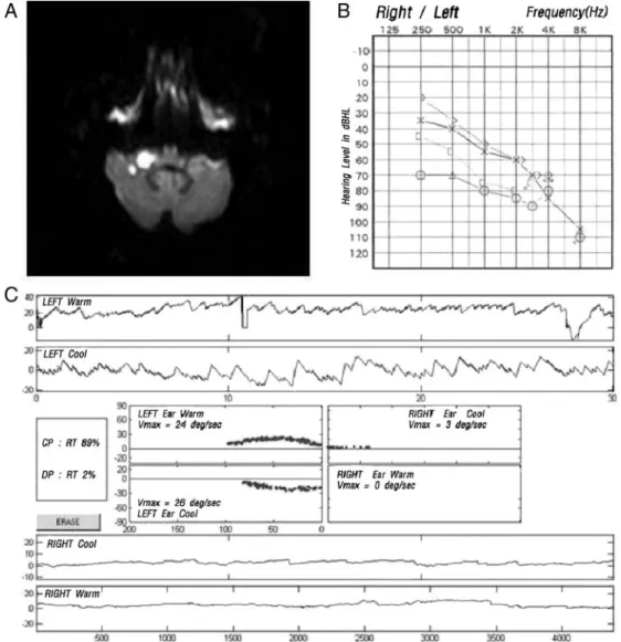
history of excessive exposure to noise, head trauma, menin- gitis, ototoxic drugs, or syphilis. Seven patients had a history of middle ear disease, but there was a clear aggravation of the hearing loss (SNHL on pure tone audiogram) at the time of infarction. MRI and audiovestibular findings of the represen- tative patients in Groups 1 and 3 are shown in Figures 1 and 2, respectively.
Topography of Infarcts Associated With Audiovestibular Loss and MRA Findings
The middle cerebellar peduncle was affected in 68 (83%), anterior inferior cerebellum in 38 (46%), and lateral inferior pons in 36 (44%) patients. AICA infarction was unilateral in 79 (96%) and bilateral in only 3 (4%) patients who showed lesions in both middle cerebellar peduncles. Complete AICA infarction involving the middle cerebellar peduncle, lateral pons, and anterior inferior cerebellum was found in only 13 (16%) patients. According to the findings on diffusion- weighted imaging, 3 categories of infarction were identified.
First, isolated AICA infarcts (n ⫽55) were restricted to the middle cerebellar peduncle (n ⫽44), dorsolateral pontine teg- mentum (n ⫽26), or anterior inferior cerebellum (n⫽27).
Second, AICA and other cerebellar infarcts (n ⫽14) involved AICA plus the posterior inferior cerebellar artery or superior cerebellar artery territory. AICA infarction was unilateral in the both groups. Third, AICA plus multiple infarcts (n ⫽13) showed multiple infarctions in the posterior circulation terri- tory in addition to AICA infarction. In 16 patients (20%), MRA disclosed focal (ie, middle or lower portion close to the AICA origin) or diffuse stenosis of the basilar artery. Thirty patients (37%) also showed diffuse or segmental narrowing of the vertebral artery ipsi- or contralateral to the AICA infarction. Another 41 (50%) patients showed normal verte- brobasilar artery on MRA. Patients with prodromal audioves- tibular disturbances showed 5 times higher prevalence of severe basilar artery occlusive disease than in the patients without prodromal audiovestibular disturbance (62% versus 13%, P ⬍0.001; Table 3). Patients with basilar stenosis also tended to have multiple posterior circulation infarcts than the patients without (5 of 16 [31%] versus 8 of 66 [12%],
P⬍0.05). Furthermore, severe basilar artery occlusive disease was more commonly observed in patients with AICA and multiple posterior circulation infarcts (6 of 13 [46%]) than in patients with isolated AICA or AICA plus other cerebellar infarcts (10 of 69 [14%], P ⬍0.017).
Discussion
To the best of our knowledge, this is the by far largest series of AICA infarction focused on the audiovestibular dysfunc- tions. In our series, almost all (98%) patients with AICA territory infarction presented with acute onset of prolonged ( ⬎24 hours) vertigo and had a vestibular dysfunction of peripheral, central, or combined origin. The most common pattern of vestibular dysfunction in our series was a combi- nation of peripheral (ie, unilateral CP) and central ocular motor or vestibular signs (ie, asymmetrically impaired smooth pursuit, bidirectional gaze-evoked nystagmus, or impaired modulation of the vestibular responses using visual input) that is observed in approximately 65% (53 of 82) of
Table 2. Frequencies of Audiovestibular Dysfunctions in 82Patients With AICA Territory Infarction
Frequency (n⫽82) Vertigo as a presenting or main symptom at the
time of AICA infarction
98% (80/82)
Central ocular motor or vestibular signs* 96% (79/82) Vestibular labyrinth infarction 65% (53/82)
Cochlear infarction 63% (52/82)
Combined vestibulocochlear infarction 60% (49/82) No auditory or vestibular infarction 32% (26/82) Isolated vestibular infarction without cochlear
involvement
5% (4/82)
Isolated cochlear infarction without vestibular involvement
3% (3/82)
Nonvertigo symptom as a presenting or main symptom at the time of AICA infarction
2% (2/82)
Isolated audiovestibular loss without central symptoms or signs
1% (1/82)
*Asymmetrical abnormalities of pursuit or optokinetic nystagmus, gaze- evoked bidirectional nystagmus, or impaired modulation of the vestibular response using visual input.
by guest on February 2, 2017http://stroke.ahajournals.org/Downloaded from
patients. These findings can be explained by the fact that AICA consistently supplies the peripheral vestibular struc- tures such as the inner ear and vestibulocochlear nerve in addition to the central vestibular structures.
2,5As a result, in contrast to other cerebellar artery territory infarction, com- plete AICA infarction usually results in combined peripheral and central vestibular damages in addition to hearing loss, facial weakness, limb and facial sensory loss, gait ataxia, and cerebellar dysmetria. Because ischemia of any structures supplied by AICA can lead to vertigo, a definite conclusion on the site(s) responsible for the prolonged vertigo seems difficult in the individual patient with AICA infarction.
However, 53 (65%) patients with AICA infarction had a unilateral weakness to caloric stimulation, suggesting that the vertigo was from the dysfunction of the peripheral vestibular structure, at least in part. On the other hand, 27 patients (33%) showed a normal caloric response, indicating that the vertigo may have resulted from ischemia to the central vestibular structures in these patients. Overall, our results showed that prolonged vertigo in AICA infarction mostly results from
ischemia to both the peripheral and central vestibular structures.
In our series, 60% (49 of 82) of patients with AICA infarction showed acute onset of audiovestibular loss charac- terized by CP and SNHL, which is in agreement with the previous reports that audiovestibular loss is an important sign for the diagnosis of AICA infarction.
3,12–16,17This finding can be explained by (1) the internal auditory artery, the principal artery for vascular supply to the inner ear, usually originates from AICA; and (2) the inner ear is particularly sensitive to transient ischemia because of its high energy requirements and lack of adequate collateral blood supply.
3,8 –10,16 –18Unlike a previous report
2that approximately 80% of patients with AICA infarction showed symptoms or signs indicative of lateral pontine dysfunction, facial weakness or crossed sen- sory loss suggesting pontine dysfunction was found in only 28% (23 of 82) of patients in our series, suggesting that although pontine signs are key features differentiating AICA infarction from a more common benign disorders involving the inner ear, it is less common than previously thought.
Figure 1. MRI and audiovestibular findings in a patient with AICA territory infarction and audiovestibular loss (Group 1). A, Axial diffusion-weighted MRI demonstrates acute infarct involving the right middle cerebellar peduncle and right anterior cerebellar hemi- sphere. B, Pure tone audiogram reveals severe degree of hearing loss on the right side. C, Video-oculographic recordings of bithermal caloric tests disclose right canal paresis (89%). Vmax, maximal velocity of slow phase of nystagmus; PTA, pure tone audiogram.
by guest on February 2, 2017http://stroke.ahajournals.org/Downloaded from
It is well known that anatomic variations are common in cerebellar vasculature. Even in normal persons, the AICA may dominate in one side, whereas the posterior inferior cerebellar artery mainly supplies the inferior cerebellum in the other side. At times, either the AICA or posterior inferior cerebellar artery is absent and one artery supplies the usual territories of both arteries. Rarely, one posterior inferior cerebellar artery may irrigate both sides of the
inferior cerebellum. Unfortunately, only a few of our patients underwent conventional angiography and the ex- act pathology of posterior inferior cerebellar artery is beyond the resolution of MRA.
It is interesting to compare MRA findings in the patients with prodromal audiovestibular disturbances (defined as ep- isode[s] of transient vertigo, hearing loss, and/or tinnitus within 1 month before the infarction) with those in the patients without prodromal audiovestibular disturbance; focal or diffuse stenosis of the basilar artery close to the origin of AICA was more common in the patients with prodromal audiovestibular disturbance (62% versus 13%, P ⫽0.000).
This finding may explain the high incidence of prodromal symptoms in the group of patients with basilar artery com- promise on MRA. Territorial strokes of the AICA have been associated with basilar artery branch occlusive disease.
2,5,6Because most of our patients with prodromal audiovestibular disturbances had evidence of a focal or diffuse segment of reduced blood flow in the basilar artery close to the AICA origin, an atheromatous plaque within the basilar artery may have extended into the AICA ostia. By this mechanism, decreased blood flow in the affected AICA might cause either transient episode of selective ischemia to the inner ear,
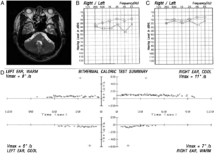
Figure 2. MRI and audiovestibular findings in a patient with AICA territory infarction and isolated auditory loss without vestibular loss (Group 3). A, Axial T2-weighted MRI demonstrates acute infarct involving the left dorsolateral pons. B, An initial PTA reveals mild hear- ing loss on the left side. C, Follow-up PTA 2 weeks later reveals normal hearing levels on both sides. D, Electro-oculographic record- ings of bithermal caloric tests disclose normal responses on both sides. Vmax, maximal velocity of slow phase of nystagmus; PTA, pure tone audiogram.Table 3. MRA Findings in Patients With (Group 2) and Without (Others) Prodromal Audiovestibular Loss
Group 2 (n⫽13)
Others (n⫽69)
P Value*
Basilar artery stenosis or occlusion†
62% (8/13) 13% (9/69) 0.000
Vertebral artery stenosis or occlusion‡
46% (6/13) 35% (24/69) 0.534
Normal vertebrobasilar system
23% (3/13) 55% (38/69) 0.067
*Based on2test. Significance was assumed at a value of P⬍0.05.
†Focal (lower or middle portion close to the origin of the anterior inferior cerebellar artery) or diffuse narrowing of the basilar artery.
‡Focal or diffuse narrowing of the vertebral artery.
by guest on February 2, 2017http://stroke.ahajournals.org/Downloaded from
resulting in isolated prodromal audiovestibular disturbance, or permanent damage to the widespread areas involving the middle cerebellar peduncle, lateral pons, and anterior cere- bellum giving rise to prolonged vertigo and hearing loss in addition to other central symptoms and signs. In our series, most patients (45 of 55 [82%]) with isolated AICA infarction showed normal MRA and severe basilar artery occlusive disease was more common in patients with posterior circula- tion infarcts in addition to AICA infarct, consistent with the previous report
2that isolated AICA infarcts are usually caused by basilar branch occlusive disease, whereas infarcts extending beyond AICA territory are mostly due to basilar artery occlusive disease.
Our results have practical implications. When patients with risk factors for stroke developed acute onset of isolated prolonged vertigo without accompanying hearing loss or other neurological symptoms, ischemic damage to the supe- rior vestibular labyrinth due to anterior vestibular artery infarction is reasonably suspected because the lumen of anterior vestibular artery is small and has little collateral circulation.
9,19,20A previous report
20also supported this assumption because approximately 50% of patients with isolated episodic vertigo of a vascular cause (ie, vertebrobasi- lar insufficiency) had unilateral CP, which is commonly localized to the inner ear (ie, superior vestibular labyrinth).
However, our finding did not support this assumption because only 4 (5%) patients showed isolated vestibular labyrinthine involvement at the time of AICA infarction. Thus, although isolated anterior vestibular artery infarction may be served as a mechanism of isolated vascular vertigo, the incidence would be low. Isolated involvement of the cochlea was also uncommon manifestation of AICA infarction in our series, which was observed in only 3% of patients. Based on our finding, we speculated that internal auditory artery ischemia seldom results in selective involvement of anterior vestibular artery or main cochlear artery. Unlike inner ear dysfunction of a viral cause, which can commonly present as an isolated vestibular (ie, vestibular neuritis) or cochlear loss (ie, sudden deafness), labyrinthine dysfunction of a vascular cause rarely results in isolated loss of vestibular or auditory function.
Thus, when sudden onset of isolated prolonged vertigo or hearing loss occurred in patients with vascular risk factors, vascular compromise to the inner ear was less likely consid- ered. However, when the combined audiovestibular loss occurred in patients with prolonged vertigo, the vascular cause was highly suspected. Our finding supported the assumption that sudden onset of isolated audiovestibular syndrome with vertigo and hearing loss can result from a vascular event (ie, labyrinthine infarction). Indeed, AICA territory infarction can produce a unique pattern of vestibu- loauditory loss in that the combined loss of both vestibular and cochlea function, rather than isolated vestibular or cochlear loss, would be more commonly expected if internal auditory artery is involved.
Although unilateral CP usually indicates a lesion in the peripheral vestibular structures from the ipsilateral labyrinth to vestibular nerve, including the root entry zone at the pontomedullary junction, it is well known that the root entry zone of the eighth cranial nerve has a rich network of
anastomotic vessels arising from the lateral medullary artery, AICA, and inferior lateral pontine artery.
21,22Thus, isolated focal infarction in that area is highly unlikely. Furthermore, to the best of our knowledge, there was no report of a vascular vertigo syndrome due to focal infarction in the root entry zone of the vestibular nerve. Indeed, we believe that the possibility of CP associated with a lesion in the root entry zone of the eighth cranial nerve was extremely low in our patients with AICA infarction.
Our study has several limitations. We only evaluated function of the superior vestibular labyrinth with horizontal semicircular canal by using caloric test and did not attempt to assess the function of the inferior vestibular labyrinth that includes the posterior semicircular canal and saccule with their afferent fibers. Second, because our study only included the patients with documented AICA territory infarct on MRI, the spectrum of audiovestibular dysfunction remains to be eluci- dated in isolated labyrinthine infarction.
Third, because some of our patients had also a lesion in the areas other than AICA territory, some of the neurological signs, including ocular motor abnormalities, limb ataxia, and severe gait disturbance with falling, may have resulted from dysfunction of the areas supplied by the arteries other than AICA. Finally, because MRA in most patients cannot ade- quately visualize the posterior inferior cerebellar artery/
AICA, and their smaller branches, further studies using conventional angiography are required to assess the vascular status of the AICA and posterior inferior cerebellar artery in AICA infarction.
In conclusion, infarction in the AICA territory can present with a broad spectrum of audiovestibular dysfunctions. Con- sidering the low incidence of selective cochlear or vestibular involvement in AICA infarction, vascular compromise ap- pears to give rise to combined loss of auditory and vestibular functions, whereas viral illness commonly presents as an isolated vestibular (ie, vestibular neuritis) or cochlear loss (ie, sudden deafness).
Disclosures
None.
References
1. Adams RD. Occlusion of the anterior inferior cerebellar artery. Arch Neurol Psychiatry. 1943;49:765–770.
2. Amarenco P, Rosengart A, Dewitt LD, Pessin MS, Caplan LR. Anterior inferior cerebellar artery territory infarcts. Mechanism and clinical features. Arch Neurol. 1993;50:154 –161.
3. Lee H, Sohn SI, Jung DK, Cho YW, Lim JG, Yi SD, Lee SR, Sohn CH, Baloh RW. Sudden deafness and anterior inferior cerebellar artery infarction. Stroke. 2002;33:2807–2812.
4. Fisher CM. Lacunar infarct of the tegmentum of the lower lateral pons.
Arch Neurol. 1989;46:566 –567.
5. Amarenco P, Hauw J-J. Cerebellar infarction in the territory of the anterior and inferior cerebellar artery. Brain. 1990;113:139 –155.
6. Amarenco P. The spectrum of cerebellar infarctions. Neurology. 1991;
41:973–979.
7. Roquer J, Lorenzo RJ, Pou A. The anterior inferior cerebellar artery infarcts: a clinical–magnetic resonance imaging study. Acta Neurol Scand. 1998;97:225–230.
8. Hinojosa R, Kohut RI. Clinical diagnosis of anterior inferior cerebellar artery thrombosis: autopsy and temporal bone histopathology study. Ann Otol Rhinol Laryngol. 1990;99:261–271.
9. Oas JG, Baloh RW. Vertigo and the anterior inferior cerebellar artery syndrome. Neurology. 1992;42:2274 –2279.
by guest on February 2, 2017http://stroke.ahajournals.org/Downloaded from
10. Lee H, Whitman GT, Lim JG, Lee SD, Park YC. Bilateral sudden deafness as a prodrome of anterior inferior cerebellar artery infarction.
Arch Neurol. 2001;58:1287–1289.
11. Lee H, Cho YW. Auditory disturbance as a prodrome of anterior inferior cerebellar artery infarction. J Neurol Neurosurg Psychiatry. 2003;74:
1644 –1648.
12. Lee H, Ahn BH, Baloh RW. Isolated sudden deafness with vertigo as a sole manifestation of anterior inferior cerebellar infarction. J Neurol Sci.
2004;222:105–107.
13. Murakami T, Nakayasu H, Doi M, Fukada Y, Hayashi M, Suzuki T, Takeuchi Y, Nakashima K. Anterior and posterior inferior cerebellar artery infarction with sudden deafness and vertigo. J Clin Neurosci.
2006;13:1051–1054.
14. Son EJ, Bang JH, Kang JG. Anterior inferior cerebellar artery infarction presenting with sudden hearing loss and vertigo. Laryngoscope. 2007;
117:556 –558.
15. Ito H, Hibino M, Iino M, Matsuura K, Kamei T. Unilateral hearing disturbance could be an isolated manifestation prior to ipsilateral anterior inferior cerebellar artery infarction. Intern Med. 2008;47:795–796.
16. Lee H, Kim HJ, Koo JW, Kim JS. Progression of acute cochleovestibu- lopathy into anterior inferior cerebellar artery infarction. J Neurol Sci.
2009;278:119 –122.
17. Kim JS, Cho KH, Lee H. Isolated labyrinthine infarction as a harbinger of anterior inferior cerebellar artery territory infarction with normal diffusion-weighted brain MRI. J Neurol Sci. 2009;278:82– 84.
18. Matsushita K, Naritomi H, Kazui S, Watanable Y, Okazaki H, Kuriyama Y, Sawada T. Infarction in the anterior inferior cerebellar artery territory: magnetic resonance imaging and auditory brain stem responses. Cerebrovasc Dis. 1993;3:206 –212.
19. Kim JS, Lopez I, DiPatre PL, Liu F, Ishiyama A, Baloh RW. Internal auditory artery infarction: clinical–pathologic correlation. Neurology.
1999;52:40 – 44.
20. Grad A, Baloh RW. Vertigo of vascular origin: clinical and electronys- tagmographic features in 84 cases. Arch Neurol. 1989;46:281–284.
21. Mazzoni A. Internal auditory canal arterial relations at the porus acusticus. Ann Otol Rhinol Laryngol. 1969;78:797– 814.
22. Mazzoni A. Internal auditory artery supply to the petrous bone. Ann Otol Rhinol Laryngol. 1972;81:13–21.
by guest on February 2, 2017http://stroke.ahajournals.org/Downloaded from
Je-Young Shin
Hyung Lee, Ji Soo Kim, Eun-Ji Chung, Hyon-Ah Yi, In-Sung Chung, Seong-Ryong Lee and
Print ISSN: 0039-2499. Online ISSN: 1524-4628
Copyright © 2009 American Heart Association, Inc. All rights reserved.
is published by the American Heart Association, 7272 Greenville Avenue, Dallas, TX 75231 Stroke
doi: 10.1161/STROKEAHA.109.564682
2009;40:3745-3751; originally published online September 24, 2009;
Stroke.
http://stroke.ahajournals.org/content/40/12/3745
World Wide Web at:
The online version of this article, along with updated information and services, is located on the
http://stroke.ahajournals.org//subscriptions/
is online at:
Stroke Information about subscribing to Subscriptions:
http://www.lww.com/reprints
Information about reprints can be found online at:
Reprints:
document.
Permissions and Rights Question and Answer process is available in the
Request Permissions in the middle column of the Web page under Services. Further information about this Once the online version of the published article for which permission is being requested is located, click
can be obtained via RightsLink, a service of the Copyright Clearance Center, not the Editorial Office.
Stroke in
Requests for permissions to reproduce figures, tables, or portions of articles originally published Permissions:
by guest on February 2, 2017http://stroke.ahajournals.org/Downloaded from