간섬유화 ( 경화)를 유도한 실험동물에서 생약 추출물 (LH )의
항산화와 항섬유화 효과 검색
임진아 * ■ 김기영 #
*원광대학교 한의학전문대학원
,의과대학 병리학교실
(Received February 7,
2002; Revised March 14,
2002)The Screening of Antioxidant and Antifibrotic Effect from Water Extracts of Herbs(LH) in Biliary Liver Fibrosis (Cirrhosis) Induced Rsts
Jin-A Lim * and Ki-young Kim#
^Professional Graduate School of Oriental Medicine Wonkwang University, Iksan, Korea Dep. of Pathology, Medical School Wonkwang University, Iksan, Korea
Abstracts —— The pathogenesis of cholestatic liver injury as well as the modulation of hepatic fibrogenesis is causally asso
ciated with involvement of reactive oxygen species (ROS) and free radical reactions. In this study, we investigated whether dried extracts of oriental medicine (LH) have antioxidant and antifibrotic effect under the biliary liver fibrosis (cirrhosis) c on- dition. The female Sprague-Dawley rats were divided in six groups (Normal, N-LH, op-2, op-4, opLH-2, opLH-4) and were observed in 2 weeks or 4 weeks. For this purpose the rats were operated by bile duct ligation/scission (BDL/S), which induced to liver fibrosis and cirrhosis. After surgery, the prepared LH was administered p.o. 2 m //day/rat in 2 weeks or 4 weeks for opLH groups. During the observation period, jaundices appeared in eyes, ears and tail of all BDL/S operated rats.
And at the time of sacrifice, cholestasis was observed in proximal bile duct, especially the color of bile juice and urine in opLH-4 group showed more clear than op-2, op-4 and opLH-2 group. The value of clinical parameters and product of lipid peroxidation (MDA) in sera and the hydroxyproline (hyp) content in liver tissue were significantly increased in all liver fibro
sis (cirrhosis) developed rats (p<0.001
〜
0.05). Among the clinical parameters of sera, value of BUN, ALP in opLH-4 group showed significantly lower than in op-4 group (p<0.05, p<0.001). The content of hyp in opLH-2, opLH-4 group (478.0 ± 134.3 (ag/g, 897.5士
118.2 (ig/g) showed lower than in op-2, op-4 group (528.9土
220.7 |ig/g, 1023.8士
277.1 jag/g) and then the value of MDA in opLH-4 was also significantly reduced to 59.4% of that in op-4 group (p< 0.001). The histological change (bile duct proliferation, fibrosis, collagen bundle) was similarly observed in op-2 group and in opLH-2 group, but the weak fibrosis and bile duct proliferation were observed in opLH-4 group compared with in op-4 group. Our data indicate that the 4 weeks treatment with LH extract suppressed lipid peroxidation and inhibited fibrotic (cirrhosis) process, and experimental cholestatic liver disease is associated with increased lipid peroxidation in BDL/S operated rats. Hence we concluded that the measurement of MDA and hyp can be useful monitor for the screening of antioxidant and antifibrotic effect in exper
imental liver fibrosis (cirrhosis), and LH has been shown to have hepatoprotective effect, antifibrotic effect and antioxidant effect.
Keywords □ Liver fibrosis, lipid peroxidation, MDA, antioxidant & antifibrotic effect
세포질에서
free radical-mediated과정은 산화■환원균형의 변 화로서
oxidative stress로 알려진 산화반응과 연관성이 있으며
, free radical은 세포막지질의 산화적 분해
(oxidative breakdown)를 유도하는 과정에서
reactive oxygen species(ROS)와
organic radical intermediate의 생성을 증가시킨다
.2’3)간과 신장의 조직
#본 논문에 관한 문의는 저자에게로 (전화) 0.63-850-6775 (팩스) 063-850-5179 (E-mail) [email protected]
에서
oxidative stress에 의한
ROS복합체의 생성은
growth factor(TGF-(3),4’5) lipid peroxides(LP)에 의해서 매개된다
.6’7)또
한 실험 동물의 혈장에서
superoxide anion과
hydroxyl radical과 같은
ROS의 생성이 촉진되고
,8’9)간질환 환자의 혈장에서 지
질과산회물의 증가가 나타난다
.10)이렇게 혈장게서 증가되는
ROS와 지질과산화물
(malondialdehyde)은
hepatic tissue뿐만 아니라
신장,심장,기타 다른 조직에서도 매우 유해하게 작용할 수 있
다는 보고가 많다
.11_15)최근 몇몇 연구자들은 담즙울체성 간질
환에서
oxidative stress노줄에 의한 지질괴산화물의 증가가 나
130 임진아 • 김기영
타났고,16'17) 실험적 담즙울체 rat에서 oxidative stress 방어효과가 감소되었을 뿐만 아니라18) 간조직에서 분리한 mitochondria에서 지질과산화물이 검출된다고 보고하고 있다.19’2(|) In vitro 실험에서 지 질과산화는 fibroblast와 hepatic stellate cell(HSC) 을 자극하여 collagen
생성을 촉진한다고 알려져 있으녀
,21} oxidative stress와
지질과산화의 산물인 MDA는 cytopathic change와 triggercollagen g e n e transcrip tio n
의
원인이 된다고 보고하고있 다
.22) 지질과산화에 의해 활성화되는 collagen 생성은 간섬유화와 연 관성이 있다. 간섬유화(liver fibrosis)는 tissue formation과 tissue remodeling 과정으로 간구성 세포의 정량적인 변화와 형 태학적인 변화를 수반하고, extracellular matrix(ECM) 구성요소, 특히 collagen의 지속적인 축적에 의한 만성 간질환으로 설명된 다.功 Collagen의 과도한 침착은 fibrogenesis(synthesis of collagen) 와 fibrolysis(degradation of new formed collagen) 의 불균형에 의한 fibrosis 형성으로 나타나고,24> HSC이 활성화되 면 collagen을 합성하는 myofibroblast와 같은 세포로 전환된다 고 알려져 있다.25’26) 최근 지질과산화를 억제 또는 방지할 수 있 는 기능, 즉 항산화 효과는 간보호 효과 및 항염증 작용이 있다 고 보고되고 있고,27’28) 항산화 물질(antioxidant compound)은 반 응성 산소중간생성물(reactive oxygen intermediate)에 의한 공 격에 대항해서 간과 간세포 보호의 목적으로 시용되고 있으며, 간질환에서 응용가능성에 대해서 연구되고 있다.29) 지금까지 천 연물에서 주줄된 flavonoid, silymarin 또는 vitamin E와 같은 항 산화제 (antioxidant agent)는 지질과산화와 간섬유화에 효과가 있는 물질로 보고되고 있고,30'31) N-acetylcysteine(NAC)은 항산 화활성을 통해 간섬유화의 초기단계에서 간섬유화와 oxidative stress를 저해한다고 알려 져 있다. 32> 또한 Picrorhiza kurroa (kutkin)는 지질괴신:화와 free radical에 의한 손상을 감소시켜 간 보호 효과를 도와주는 항산화 효과가 있는 천연물로 보고되고 있 다제 그러나 collagen의 불균형과 oxidative stress 에 의한 지질 과산화를 조절할 수 있는 항산화의 역할에 대해서는 아직 연구 가 진행중에 있다.이에 본 연구에서는 항산화효과가 있는 약물(천연물)을 대상 으로 항산화와 항섬유화 효과를 알아보기 위하여 간섬유화를 유 도한 랫드에 복합생약 추출물을 2주, 4주간 투여하여 간기능의 지표인 생화학적 수치 및 결합조직 단백의 변화를 나타내는 collagen 양(hyp)과 지질 과산화정도를 나타내는 MDA를 측정 및 관찰하였다.
실 험 방 법
실험동물
10주령 Sprague-Dawley 흰쥐-3* 6개 군(정상군, 정상 LH군,
이>2군,op-4군, opLH-2군,opLH-4군)으로 구분하여 사육하였다.
사육환경은 밤과 낮의 리듬을 구분하였고 사료와 물은 지유롭게 공급하였다.
간섬유화 유도 및 약물투여
정상군을 제외한 군의 흰쥐에 Kountras 등:i4)의 방법에 따라 담도결찰(proximal, distal bile duct ligation/scission) ■§:하여 간 섬유화를 유도하였다. 한국 생약협회에서 구입한 LH [결명자(3 g), 차전자(3 g), 지구자(10 g)]는 물(500 m/)과 흔합하며 전제煎 劑)한 후 여과하였으며 2주와 4주간 경-Y■투여 (2 m//day/rat)하였다.
재료
동물을 2주, 4주간의 관찰 후, 부검시 심장에서 채혈하여 실온 에서 3시간 이상 방치한 다음 3000 rpm에서 10분간 원심분리하 여 얻은 혈청은 생화학적 검사에 사용하였고, 간의 일부는 hydroxyproline 측정을 위하여 -70oC에서 보관하였으며 나머지 간조직은 1 0% 숭성 포르말린에 고정하여 hematoxylin & eosin, Masson's trichrome 염색에 시용하였다.
혈청 생화학적 검사
Alanine transaminase(ALT), Aspartate transaminase(AST), Alkaline phosphatase(ALP), total bilirubin, BUN, creatinine 읍 MBL-kit를 사용하여 분석하였다.
총 coUagen(hydroxyproline>양 측정
간조직내 hy加 xyproline(hyp)양의 즉정은 Jamall 능'®의 방법 에 따라 간조직을 염산0■■로 가수분해시켜 isopropylalcohol을 넣 고 chloramine-T로 산화시킨 후 , Ehrlich's reagent solution(p- dimethylaminobenzaldehyde)으로 발색시켜 558 nm에서 흡광도 를 측정하여 계산하였다.
M DA(m alondialdehyde )측정
Okawa 등36J의 방법에 따라 혈청시료와 표준물질(tetrame- tooxypropane) 에 0.2% SDS, 20% acetic acid, 0.8% thiobabi- turate를 가하여 95"C에서 반응시킨 후 냉각시켰다. 그런 다음 butanol을 가하고 원심분리하며 532 nm에서 흡광도를 측정하여 농도를 계산하였다.
조직염색 및 소견
간조직을 H&E,Masson's trichrome 방법으로 염 색한 후, 광 학현미경으로 관찰하였다. 조직소견은 담관증식,섞유회-, 염증, scar formation의 정도를 +, + + , + + +_V:■표시하였다.
통계처리
Student's t-testU 사용하여 m ean±SD으로 표시하였고 p-
J. Pharm. Soc. Korea
Fig. 1 - The value of clinical biochem istry in sera of 6 different group anim als. *’**T he significantly different from no rm al (p < 0 .0 5 , p < 0.001
),
#,##T he significantly different from op-2 or op-4 (p < 0 .0 5 , p < 0.001)
,
(a) AST: A spartate transam inase, (b) ALT: A lanine transam inase, (c) A LP: A lkaline phosphatase, (d) BU N : Blood urea nitrogen, (e) t-bilirubin: total bilirubinNormal N -LH o p -2 o p -4 o p L H -2 opLH4
(e)
Normal N -LH o p -2 o p -4 o pL H -2 o p나-14 Normal N -LH o p -2 o p -4 o p L H -2 ᄋpLH4111ᅳ ᅳC lᅳ
Normal N -LH o p -2 Dp-4 opL H -2 opLH4
■ * I * 7* *
value를 구하여 유의성을 검증하였으며 각 parameter들 사이의 상관관계 (regression)를 조사하였다.
실 험 결 과
일반적인 관찰 - 간섬유화(경화)를 유도한 op-2, op-4군 그리고
L H경구-Y-여군인 opLH-2, opLH-4군 모두에서 정상군과 비교했 을 때 귀와 눈주위에7、1 황달이 줄현하였고, proximal bile duct 에서는 밖쥬울체가 관찰되었0ᅵ녀, opLH-2, opLH-4군의 담즙색 은 op- 2,op-4군보다 맑은 짓으로 나타났다.
체중, 장기 무게 및 장기/체중 변화 - 정성'군에 비해서 간섬유
화를 i t 도한 op군과 opLH군에서 간方대와 비方대가 나타났다 (Table I). 간무게는 정상군보다 op-2, op-4, opLH-2, opLH-4군 에서 모두 유의성 있게 높게 나타났고(p<0.005~0.05), 또한 간 /체중비는 op-4군, opLH-4군 뿐''''I- 이-니라 op-2, opLH-2군에서도 정상군보다 유의성 있게 높게 관찰되었다(p<0.005, Table I). Op 군과 opLH군을 비교했을 때 신장과 비장의 무게는 op-2군보다 opLH-2군에서 낮게 나타났으나, 간무게와 간/체중비는 opLH-2 군에서 op-2군보다 높게 나타났고, opLH-4군에서는 간, 신장, 비 장의 무게와 장기/체중비 모두 op-4군보다 낮거나 비슷하였다 (Table I).
혈청 생화학적 변화 - 혈청 생화학적 결과에서 op-2,op-4,
Table I - T he w eight of organ and the ratio organ/body w eight in 6 different group anim al
Group norm al normal-LH op-2 op-4 opLH-2 opLH-4
Liver W eight(g) K idney W eight(g) Spleen W eight(g) R atio of LW /B W (% ) R atio of K W /B W (% ) R atio of S W /B W (% ) N um b e r o f anim al
9.66 ± 1.30 1.92 ± 0.21 0.60 ± 1.10 3.75 ± 0.24 0.75 ± 0.05 0.24 ± 0.05
6
8.57 ± 0.55 1.69 ± 0.12 0.49 ± 0.05 3.59 ± 0.23 0.71 ± 0.02 0.20 ± 0.03
6
11.66 ± 0.98*
2.23 ± 0.46 1.13 ± 0.31*
5.89 ± 0.45**
1.13 ± 0.29*
0.58 ± 0.19*
6
21.10 ± 1.90**
2.34 ± 0.18**
1.42 ± 0.21**
7.66 ± 0.69**
0.85 ± 0.06**
0.52 ± 0.08**
11
13.86 ± 1.96**
2.00 ± 0.23 1.08 ± 0.34*
6.54 ± 0.82**
0.95 ± 0.09**
0.51 ± 0.15*
7
20.85 ± 4.14*
2.35 ± 0.25*
1.41 ± 0.31**
7.34 ± 1.44**
0.83 ± 0.07 0.50 ± 0.10**
10
* ,**T h e significantly different from n o rm a l(p < 0 .0 5 , p < 0.005) op-2: T he 2 w eeks observed rats group after B D L /S operation op-4: T he 4 w eeks observed rats group after B D L /S operation
opLH-2: T he 2 w eeks observed rats group after B D L /S operation and tre a tm e n t w ith L H opLH-4: T he 4 w eeks observed rats group after B D L /S operation and tre a tm e n t w ith L H
(lp/eE)u!qruH!ql
5
ᄋ
5 Q 5 P 5 0
5
0
4 4 3 3 2 3 1 1
2"
l / n oi
_lv
50 00 50 8 50 00 50 00 50 G
4
4
3
3
2121
1
(IE/nlusv
5 0 5 0 5 0 5
c
3 3
2 2
1 1
d )( l p/ o)E ) N n CD ( c ) 350
300 ᅳ 250
<i 200 a: 150
< 100 50
0
Normal N-LH op-2 op-4 ᄋp나H-2 opLH-4
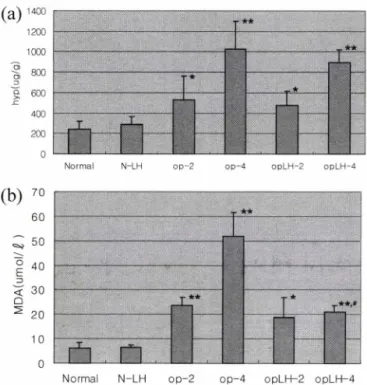
Fig. 2 - The diagrams of (a) hyp in liver tissue and (b) MDA in sera of rats. *’**The significantly different from normal (p<0.05, p< 0.005),#The significantly different from op-4 group (pcO.OOl).
J. Pharm. Soc. Korea opLH-2, opLH-4군에서 AST, ALT, ALR t-bilirubin의 수치가 정
상군과 비교했을 때 유의성 있게 높게 관찰되었다(p<0.001~0.05,
Fig. 1). OpLH-4군에서 AST, ALT 수치는 op-4군보다 각각 16.6%, 21.7% 낮게 나타났으나 유의성은 없었다. 그러나 opLH- 4군의 BUN과 ALP수치는 op-4군과 비교했을 때 유의성 있게 낮 게 나타났다(p<0.05,p<0.001, Fig. 1).
H ydroxyproline(hyp)양의 변화 - 정상군과 비교했을 때 hyp 양은 op-2, op-4군과 opLH-2, opLH-4군에서 모두 유의성 있게 높게 관찰되었다(p<0.001~0.05, Fig. 2a). Op-2, op-4군과 비교 했을 때 opLH-2, opLH-4군에서 유의성은 관찰되지 않았으나 각 각 9.6%, 12.3%로 낮게 나타났다(Fig. 2a).
MDA
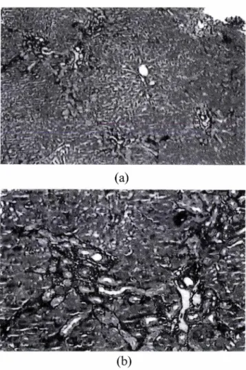
농도변화 - 혈청중 MDA 농도는 op-2, op-4군과 opLH- 2, opLH-4군 모두에서 정상군보다 유의성 있게 높게 나타났다 (p<0.05~0.001, Fig. 2b). 그러나 opLH-군의 MDA 농도는 op- 4군보다 59.4% 유의성 있게 낮게 관찰되었다(pcO.OOl, Fig. 2b).조직학적 변화 - 정상군보다 op-2, op-4군과 opLH-2, opLH-4 군에서 담관증식(+ + ~ + + +), 섬유화(+ + ~ + + + ), portal triads에서 과도한 collagen fiber축적 그리고 간세포 팽대와 핵붕 괴 등이 관찰되었다. 그러나 opLH-4군에서의 담관증식은 주로 portal triads에 편재되어 있고 stroma에서는 적게 나타났으나, op-4군에서는 portal triads 뿐만 아니라 stroma에서도 담관증식 이 관찰되었다(Fig. 3,Fig. 4). 특히 op-4군에서는 두꺼운 collagen bundle, portal- portal septum
과
scar fo rm a tio n이 관
(b)
Fig. 3 - Masson's trichrome stained liver tissue from fibrotic (cirrhotic) rat. (a) The liver tissue of BDL/S operated rat (x40), (b) The liver tissue of BDL/S operated rat (xlOO)
<= : collagen fiber, : bile duct proliferation
찰되었다. 그리고 opLH-4군에서 간세포의 종창은 op-4군보다 약 하게 나타났고,세포질의 파괴와 핵붕괴도 op-4군보다는 상태가 양호한 것으로 관찰되었다(Fig. 3, Fig. 4).
고 찰 및 결 론
Oxidative stress는 간손상이 유도된 동물의 혈청에서 지질과 산화물과 ROS생성 증가에 의해서 예측되며,37,38) HSC의 활성과 collagen gene expression의 조절과 연관성이 있다.32) 또한 지질 과산화물(aldehydic end product) 은 fibroblast 와 HSC 에서 collagen type I의 합성을 가속화시킬 뿐만 아니라,39) 조직 중 collagen 죽적을 자극하여 collagen 생성 증가와 지질과산화물인 malondialdehyde(MDA)의 증가에 따른 상호연관성이 보고되고 있다.40,41) 최근 지질과산화와 간섬유화사이의 연관성은 실험적 담즙울체 (bile duct ligation operation),42) 알코올 노출,43_47) CC14,31’48_50) iron 과부하5사 그리고 copper 과부하52)등의 임상과 실험에서 많이 연구되어지고 있고,이러한 지질과산화물의 정량
132 임진아 • 김기영
^ 避
(a)
ᄋᄋooᄋᄋo
7
6
5
4
3
2
1
b )(27loe3va^ 1400
1200
1000
800
600
400
200 0
( 6/ CDn QAL j
(b)
Fig. 4 - Masson's trichrome stained liver tissue of LH treated rat. (a) The liver tissue of in 4 weeks LH treated rat after BDL/S operation (x40), (b) The liver tissue of in 4 weeks LH treated rat after BDL/S operation (xlOO)
<= : collagen fiber, 4— : bile duct proliferation
적인 측정과 간섬유화의 지표 (collagen 축적량)의 측정은 항산 화와 항섬유화 효과를 동시에 검색할 수 있다는 점에서 큰 의미 로 대두되고 있다. 531
이에 본 연구에서는 복합생약 추출물(LH)의 항섬유화 효과와 항산화 효과를 검 색하기 위해 혈청 생화학적 검사,간조직중 hyp (총 collagen)양과 지질과산화물인 MDA 농도를 측정하여 효과 를 검색하였다. 그 결과, 장상군을 제외한 나머지군, 즉 간섬유 화(경화)를 유도한 수술군(op-2, op-4)과 LH를 투여한 수술군 (opLH-2, opLH-4) 모두에서 눈,귀, 꼬리에서 황달이 출현하였 고 담도결찰부위 (proximal bile duct)에서 담즙울체가 관찰되었 다. 특히 opLH-4군의 담즙과 소변의 색이 op-2, op-4군과 opLH-
2군보다 맑고 투명하였다. 간경화에 hepatorenal syndrome과 glomerular nephritis가 수반되는데 이때에 나타나는 변화는 단 백뇨(proteinurin)와 혈뇨(hematurin)이다.54’55) 단백뇨의 지표가 되는 혈청 중 BUN, ALP 등은 opLH-4군에서 op-4군보다 유의 성 있게 낮게 나타났다(p<0.05, pcO.OOl, Fig. 1). 이러한 결과
로 LH의 4주간 투여가 간경화에 의한 glomerular nephritis 의 발생을 예방하는 것으로 생각된다.
간섬유화 지표인 hyp양은 정상군보다 수 술군 (op-2,op-4, opLH-2, opLH-4)에서 3.7~8.2배 유의성 있게 높게 관찰되었다 (p<0.001~0.05). 그러나 op군과 opLH군을 비교하면 opLH-2, opLH-4군(478.0 ±134.3 ng/g, 897.5 ±118.2 收 /g) 의 hyp 양은 op-2, op-4군(528.9± 220.7 呢 /g, 1023.8±277.1 |Xg/g)보다 낮았 으나 유의성은 검증되지 않았다(Fig. 2a). Shimizn
등56)은
항산 화와 관련하여 dimethylnitrosamine(DMN)으로 유도한 간섬유 화 동물의 간조직에서 지질과산화물인 MDA수치의 유의성 없는 감소를 보고하였으나, 본 연구에서의 MDA농도는 op군과 opLH 군에서 정상군보다 3.8배~8.2배 모두 유의성 있게 높게 나타났 고(p<0.001~0.05),op-4군보다 opLH-4군에서 59.4% 유의성 있 게 낮았다(p<0.001, Fig. 2b). 이러한 결과는 BDL 실험동물에서 oxidative stress에 의해 혈청중 thiobarbituric acid-reactive substances(TBARs)와 지질과산화물인 MDA가 증가되었다는 보 고와 일치하고,42’57-59) common bile duct ligation으로 담즙울체 성 간손상을 유도한 랫드에서 UDCA를 24일간 투여하였을 때 혈청간기능 지표의 개선효과는 없었으나 지질과산화는 억제되었 다고 보고한 Ljubumcic 등60〉의 보고와 유사하다. 또한 간섬유화 실험동물에서 MDA의 생성은 hepatic fibrosis의 진행과정에서 일어나며,61) 지질과산화물인 MDA가 collagen합성과 HSC의 활 성개시를 자극한다는 연구결과와도 일치한다.40’41)그리고 opLH-4군을 opLH-2군과 비교했을 때,BUN과 ALR MDA의 수치는 LH투여 2주에는 증가하는 양상을 보였으나 4주 후에는 각각 19.1%, 37.6%, 59.4%로 유의성있게 감소했으며 (p<0.05~0.001), AST(16.6%<21.7% )와 hyp(9.6%<12.3% )는 LH투여 2주보다는 4주후에 많은 감소율을 나타냈다(Fig. 1,2).
또한 비투여군(op-4)과 비교했을 때 복합생약 추출액 LH 의 4주 투여군(opLH-4)에서 AST, ALT, hyp의 낮은 수치와 유의성 있게 낮은 ALR BUN 그리고 M DA 수치가 나타남으로써 적어도 4주 간 투여시 뚜렷하게 항산화,항섬유화 효능이 있다고 생각된다.
Parola 둥42]은 BDL 실험동물의 fibrotic liver에서 M DA의 함 량은 염증세포에 의한 tissue infiltration의 kinetic과 collagen의 축적에 좋은 상관성을 나타낸다고 보고하였는데, 본 연구에서 지 질과산화물인 M DA와 간섬유화의 지표인 hyp 함 량, 두 parameter간의 상관성은 나타나지 않았으나(r=0.337), opLH-4 군의 MDA와 hyp 함량은 op-4군에 비해서 59.4%, 12.3%로 현 저하게 낮게 관찰되었다. 이는 천연물 또는 약물이 항섬유화 및 항산화 효능이 있는 경우에는 각 지표들 사이에 상관성이 없을 수 있으며,간섬유화가 유도되었을 경우에는 간조직 중 hyp 함 량과 지질과산화 분해물인 MDA농도가 상관성을 나타낸다는 것 을 시사하는 결과이다.53)
본 연구결과로써 혈청 중 MDA의 측정과 간조직중 hyp 측정
134 임진아 • 김기영
은 항산화와 항섬유화 효과의 동시검색에 시병-할 수 있는 좋은 지표이며, 복합생약 추출액(LH)을 2주 보다는 4주간 투여하는 것이 바람직하고 L if e 간기능 개선과 항산화 및 항섬유화 효과 가 있을 7 1능성이 높다고 사료된다.
감 사 의 글
본 연구는 원광대학교 한의학전문대학원 BK21 사업비 및 미래 의료재단의 지원에 의해 수행된 것으로 이에 갑사 드립니다.
문 헌
1) Sies, H. ed.: Oxidative stress. Orlando. Academic Press (1985).
2) Slater, T. E ed. : Free radical mechanisms in tissue injury.
London. Pion Ltd (1972).
3) Slater, T. E : Free radical mechanisms in tissue injury. Biochem.
J. 222,1 (1994).
4) Herrera, B., Fernandez, M., Alvarez, A. M.,Roncero, C.,
Benito, M.,Gil, J., Fabregat, I. : Activation of caspases occurs downstream from radical oxygen species production, Bcl-xL down-regulation, and early cytochrome C release in apoptosis induced by transforming growth factor beta in rat fetal hepatocytes. Hepatology 34(3), 548 (2001).
5) Sanchez, A., Alvarez, A. M., Benito, M.,Fanregat, I. : Apoptosis induced by transforming growth factor- P in fetal hepatocyte primary cultures. J. Biol Chem. 271, 7416 (1996).
6) Bomzon, A., Holt, S., Moore, K. : Bile acid, oxidative stress and renal function in biliary obstruction. Semin. Nephrol. 17,
549 (1997).
7) Halliwell, B. : Reactive oxygen species in living system;
source, biochemistry and role in human disease. Am. Med.
suppl 3C, 14 (1991).
8) Chen, M. E, Mo, L. R.,Lin, R. C., Kuo, J. Y, Chang, K. K.,
Liao, C.,Lu, E J. : Increase of resting levels of superoxide anion in the whole blood of patients with decompensated liver cirrhosis. Free Radic. Biol. Med. 23(4),672 (1997).
9) Tsai, L. Y, Lee, K. T, Liu, T. Z. : Evidence for accelerated generation of hydroxyl radicals in experimental obstructive jaundices of rats. Free Radic. Biol. Med. 24,732 (1998).
10) Fabris, C., Pirisi, M.,Panozzo, M. R, Soardo, G.,Toniutto, R, Hocza, V,Bartoli, E. : Intensity of inflammatory damage and serum lipid peroxide concentrations in liver disease. J. Clin.
Pathol 46(4),364 (1993).
11) Ljubuncic, R, Tanne, Z.,Bomzon, A. : Evidence of a systemic phenomenon for oxidative stress in cholestatic liver disease.
Gut 47(5), 710 (2000).
12) Vaziri, N, D.,Ni, Z., Oveisi, E, Liang, K., Pandian, R. : Enhanced nitric oxide inactivation and protein nitration by
reactive oxygen species in renal insufficiency. Hypertension 39(1),135 (2002).
13) Pancewicz, S. A., Skrzydlewska, E.,Hermanowska-Szpakowicz, T., Zajkowska, J. M., Kondrusik, M. : Role of reactive oxygen species (ROS) in patients with erythema migrans, an early manifestation of Lyme borreliosis. Med. Sci. Monit 7(6),1230
( 2001 ).
14) Pawlak, W., Kedziora, J., Zolynski, K.,Kedziora-Kornatowska, K., Blaszczyk, J., Witkowski, R, Zieleniewski, J . : Effect of long term bed rest in men on enzymatic antioxidative defence and lipid peroxidation in erythrocytes. J. Gravit. Physiol. 5(1),163 (1998).
15) Kose, K., Yazici, C., Assioglu, O. : The evaluation of lipid peroxidation and adenosine deaminase activity in patients with Behcet's disease. Clin. Biochem. 34(2),125 (2001).
16) Tsai, L. Y., Lee, K. T., Tsai, S. M.,Lee, S. C.,Yu, H. S .: Change of lipid peroxide levels in blood and liver tissue of patients with obstructive jaundice. Clin. Chim. Acta. 215,41 (1993).
17) Ono, M.,Sekiya, C.,Ohhira, M.,Ohhira, M., Namiki, M., Endo, Y., Suzuki, K.,Matsuda, Y., Taniguchi, N. : Elevated level of serum Mn-superoxide dismutase in patients with primary biliary cirrhosis: Possible involvement of free radicals in the pathogenesis in primary biliary cirrhosis. J. Lab. Clin. Med.
118(5), 476 (1991).
18) Shinh, S., Shackleton, G.,Ah-Sing, E.,Chakraborty, J., Bailey, M. E. : Antioxidant defenses in the bile duct ligated rat.
Gastroenterology 103,1625 (1992).
19) Sokol, R. J., Deveraux, M., Khandwala, R. A .: Effect of dietary lipid and vitamin E on mitochondrial lipid peroxidation and hepatic injury in the bile duct-ligated rat. J. Lipid Res. 32,1349 (1991).
20) Deems, R. O., Skypala, R L.,Martinez-Hemandez, A., Friedman, L. S., Friedman, M. I . : Dietary fat exacerbates liver disease in bile duct-ligated rats. J. Nutr. 123, 1414 (1993).
21) Parola, M., Pinzani, M.,Casini, A., Albano, E., Poli, G., Gentilini, A., Gentilini, R, Dianzani, M. U .: Stimulation of lipid peroxidation or 4-hydroxynonenal treatment increases procollagen alpha 1(1) gene expression in human liver fat- storing cells. Biochem. Biophys. Res. Commun. 194,1044 (1993).
22) Bedossa, R, Houblum, K., Trautwein, C., Holstege, A., Chojkier, M. : Stimulation of collagen alpha 1(1) gene expression is associated with lipid peroxidation in hepatocellular injury: A link to tissue fibrosis? Hepatology 19(5), 1262 (1994).
23) Friedman, S. L., Roll, J. E, Boyles, J., Bissel, D. M. : Hepatic lipocytes: The principal collagen production cells of normal rat liver. Proc. Natl Acad. Sci. 82, 8681 (1985).
24) Zavoico, G. B. : New targets for the treatment of pathologic
J. Pharm. Soc. Korea
fibrosis, an unmet clinical need. Durg & Market Development 10(1),2 (1999).
25) Friedman, S. L. : Cellular sources of collagen and regulation of collagen production in liver. Semin. Liver Dis. 10,20 (1990).
26) Gressner’ A. M. and Bachem, M. G. : Cellular sources of noncollagenous matrix proteins: Role of fat-storing cells in fibrogenesis. Semin. Liver Dis. 10,30 (1990).
27) Das, D.,Pemberton, R W, Burrows, R C., Gordon, C.,Smith, A., McMahon, R. E, Wames, T. W : Antioxidant properties of colchicine in acute carbon tetrachloride induced rat liver injury and its role in the resolution of established cirrhosis. Biochem.
Biophys, Acta. 1502(3),351 (2000).
28) Lin, C. C., Huang, R C.,Lin, J. M. : Antioxidant and hepatoprotective effects of Anoectochilus formosanus and Gynostemma pentaphyllum. Am. J. Clin, Med. 28(1),87
( 2000 ).
29) Peres, W, Tunon, M. J., Collado, R S., Herrmann, S., Marroni, N.,Gonzalez-Gallego, J. : The flavonoid quercetin ameliorates liver damage in rats with biliary obstruction. J. Hepatol. 33(5),
742 (2000).
30) Mourelle, M., Muriel, R, Favari, L.,Franco, T.: Prevention of CC14- induced liver cirrhosis by silymarin. Fundam. Clin. Pharmacol 3,183 (1989).
31) Parola, M., Leonarduzzi, G.,Biasi, E,Albano, E.,Biocca, M. E., Poli, G.,Dianzani, M. U. : Vitamin E dietary supplementation protects against carbon tetrachloride-induced chronic liver damage and cirrhosis. Hepatology 16,1014 (1992).
32) Vendemiale, G.,Grattagliano, I., Caruso, M. L.,Serviddio, G., Valentini, A. M., Pirrelli, M.,Altomare, E. : Increased oxidative stress in dimethylnitrosamine-induced liver fibrosis in the rats:
Effect of N-acetylcysteine and interferon- a. Toxicol Appl Pharm. 175, 130 (2001).
33) Luper,S. : A review of plants used in the treatment of liver disease: Part 1. Altern. Med. Rev. 3(6),410 (1998).
34) Kountras, J., Billing, B. H., Scheuder,R J . : Prolonged bile duct obstuction: A new experimental model for cirrhosis in the rat.
Br. J. Exp. Path. 65, 305 (1984).
35) Jamall, I. S. and Finelli, V N. : A single method to determine nanogram levels of 4-hydroxyproline in biological tissue. Anal.
Biochem. 112,70 (1981).
36) Ohkawa, H.,Ohishi, N.,Yagi, K. : Assay for lipid peroxides in animal tissues by thiobarbituric acid reaction. Anal Biochem.
95,351 (1979).
37) Chen, M. E, Mo, L. R.,Lin, R. C.,Kuo, J. Y, Chang, K. K.,
Liao, C.,Lu, E J. : Increase of resting levels of superoxide anion in the whole blood of patients with decompensated liver cirrhosis. Free Radic. Biol. Med. 23, 672 (1997).
38) Fabris, C., Pirisi, M.,Panozzo, M. R, Soardo, G.,Toniutto, R, Hocza, V,Bartoli, E. : Intensity of inflammatory damage and
serum lipid peroxide concentrations in liver disease. J Clin Pathol. 46,364 (1993).
39) Poli, G. and Paroh, M. : Oxidative damage and fibrogenesis. Free Radic. Biol. Med. 22, 287 (1997).
40) Maher, J. J., Tzagarakis, C.,Gimenez, A. : Malondealdehyde stimulates collagen production by hepatic lipocytes only upon activation in primary culture. Alcohol & Alcoholism 29,605 (1994).
41) Lee, K. S., Buck, M.,Houglum, K., Chojkier, M .: Activation of hepatic stellate cells by TGF a and collagen type I is mediated by oxidative stress through c-myb expression. J. Clin. Invest 96,2461 (1995).
42) Parola, M., Leonarduzzi, G.,Robino, G.,Albano, E.,Poli, G., Dianzani, M. U. : On the role of lipid peroxidation in the pathogenesis of liver damage induced by long-standing cholestasis. Free Radic. Biol. Med. 20,351 (1996).
43) Dianzani, M. U. : Lipid peroxidation in ethanol poisoning: A critical reconsideration. Alcohol Acohol 20, 161 (1985).
44) French, S. W., Wong, K., Albano, E., Hagbjork, A. L.,Ingel- man-Sundberg, M. : Effect of ethanol on cytochrom P450 2E1(CYP2E1), lipid peroxidation and serum protein adduct formation in relation to liver pathology pathogenesis. Exp. Mol.
Pathol. 58’ 61 (1993).
45) Kamimura, S., Gall, K.,Britton, R. S., Triadafilopoulos, G.,
Tsukamoto, H. : Increased 4-hydroxynonenal levels in experimental alcoholic liver disease: Association of lipid peroxidation with liver fibrogenesis. Hepatology 16,448 (1992).
46) Nordmann, R.,Ribiere, C., Rouach, H. : Implication of free radical mechanisms in ethanol-induced cellular injury. Free Radic. Biol Med. 12,219 (1992).
47) Albano, E.’ Clot, R, Tabone, M., Arico, S., Ingelman- Sundberg, M. In., Poli, G.,Albano, E.,Dianzani, M. U.,eds. : Free radicals:
From basic science to medicine. Basel: Birkhauser Verlag. 310 (1993).
48) F^rola, M., Muraca, R.,Dianzani, I., Barrera, G., Leonarduzzi, G.,
Bendinelli, R, Piccoletti, R., Poli, G. : Vitamin E dietary supplementation inhibits transforming growth factor p i gene expression in the rat liver. FEBS Lett. 308,267 (1992).
49) Poli, G.,Parola, M., Leonarduzzi, G.,Pinzani, M. : Modulation of hepatic fibrogenesis by antioxidants. Mol. Aspects. Med. 14, 259 (1993).
50) Bedossa, R, Houghlum, K., Trautwein, C., Holstege, A., Chojkier, M. : Stimulation of collagen 1(1) gene expression is associated with lipid peroxidation in hepatocellular injury: A link to tissue fibrosis? Hepatology 19,1262 (1994).
51) Bacon, B. R.,Britton, R. S. : The pathology of hepatic iron overload: A free radical-mediated process? Hepatology 11, 127 (1990).
52) Sokol, R. J., Deveraux, M.,Mierau, G. W., Hambidge, K. M.,
136 임진아 . 김기영
Shikes, R. H .: Oxidant injury to hepatic mitochondrial lipids in rats with dietary copper overload: Modification by vitamin E deficiency. Gastroenterology 99, 1061 (1990).
53)
정재열
,임진아
,박선영
,서의석
,제갈승주
,김기영
. :실험동물에 서 복합천연물
(DW)의 지질과산화 억제와 간섬유화 저해 효과 검색
.약학회지
45, 513 (2001).54) Gerok, W ed.: Hepatologie. Urban & Schwarzenberg 547 (1987).
55) Cotran-Humar, R. ed. : Robbins pathologic basis of disease. 4th ed. 918 (1989).
56) Shimizu, I., Ma, Y. R., Mizobuchi, Y., Liu, E, Miura, T., Nakai, Y., Yasuda, M., Shiba, M., Hone, T., Amagaya,S., Kawada, N.’
Hori, H., Ito, S. : Effects of Sho-saiko-to, a Japanese herbal medicine on hepatic fibrosis in rats. Hepatology 29,149 (1999).
57) Singh S., Shackleton, G., Ah-Singh, E., Chakraborty, J., Bailey, M. E. : Antioxidant defenses in the bile duct-ligated rat.
Gastroenterology 103,1625 (1992).
58) Panozzo, M. P, Basso, D.,Balint, L., Biasin, M. R., Bonvicini, R, Metus, R, Plebani, M. : Altered lipid peroxidation glutathione ratio in experimental extrahepatic cholestasis. Clin. Exp.
Pharmacol Physiol. 22, 266 (1995).
59) Muriel, P, Suarez, O. R. : Role of lipid peroxidation in biliary obstruction in rat. J. Appl. Toxicol 14, 423 (1995).
60) Ljubuncic, R, Tanne, Z., Bomzon, A. : Ursodeoxycholic acid suppresses extent of lipid peroxidation in diseased liver in experimental cholestatic liver disease. Dig. Dis. Sci. 45(10), 1921 (2000).
61) Niemela, O., Parkkila, S., Yla-Herttuala, S., Villanueva, J., Ruebner, B., Halsted, C. H. : Squential acetaldehyde production, lipid peroxidation, and fibrogenesis in a micropig model of alcohol-induced liver disease. Hepatology 22,1208 (1995).
J. Pharm. Soc. Korea