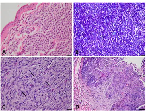
A retrospective study of feline cutaneous tumors in Korea from 2013 to 2018
7
0
0
전체 글
(2)
(3)
(4)
(5)
(6)
(7)
수치


관련 문서
3 Department of Internal Medicine, Korea University College of Medicine, Korea University Guro Hospital, Seoul; 4 Department of Internal Medicine, Seoul National University
Department of Internal Medicine, Jeju National University College of Medicine and Graduate School of Medicine, 15 Aran 13-gil, Jeju 63241, Korea.. Tel: +82-64-717-2833,
2 Brain Korea 21 Program for Veterinary Science, College of Veterinary Medicine, Seoul National University, Seoul 151-742, Korea.. 3 KRF Zoonotic Disease Priority
2 Cancer Research Institute, Seoul National University College of Medicine, Seoul, Korea. 3 Department of Pathology, Seoul National University College of Medicine,
College of Veterinary Medicine, Kyungpook National University, Daegu 41566, Korea Stem Cell Therapeutic Research Institute, Kyungpook National University, Daegu 41566,
Department of Biochemistry, School of Medicine, Institute of Medical Science, Jeju National University, Jeju 690-756, Republic of Korea. *Corresponding
3 Department of Surgery, Asan Medical Center, University of Ulsan College of Medicine, Seoul, Korea.. 4 Department of Surgery, Seoul National University College of Medicine,
1 Department of Radiology, Seoul National University College of Medicine, and Institute of Radiation Medicine, Seoul National University Medical Research Center, Seoul 03080, Korea;
