Giant chordoma of the upper thoracic spine with mediastinal involvement
Asian Spine Journal
Asian Spine Journal
353Copyright Ⓒ 2014 by Korean Society of Spine Surgery
This is an Open Access article distributed under the terms of the Creative Commons Attribution Non-Commercial License (http://creativecommons.org/licenses/by-nc/3.0/) which permits unrestricted non-commercial use, distribution, and reproduction in any medium, provided the original work is properly cited.
Asian Spine Journal • pISSN 1976-1902 eISSN 1976-7846 • www.asianspinejournal.org
Received Feb 13, 2013; Revised Jul 27, 2013; Accepted Jul 29, 2013 Corresponding author: Fabio Davoli
Thoracic Surgery Unit, Azienda Ospedaliero-Universitaria “Maggiore della Carità”-Novara, University of Eastern Piedmont, Corso Mazzini 18, 28100 Novara, Italy
Tel: +39-0321-3733076, Fax: +39-0321-3733578, E-mail: [email protected]
*This study was performed during the Research Fellowship “Dottorato di Ricerca, XXVI ciclo, Scienze Chirurgiche, Alma Mater Studiorum Università degli Studi di Bologna”.
Giant Chordoma of the Upper Thoracic Spine with Mediastinal Involvement: A Surgical Challenge
Ottavio Rena
1, Fabio Davoli
1, Giuliano Allegra
2, Caterina Casadio
1, Davide Turello
11Thoracic Surgery Unit, Azienda Ospedaliero-Universitaria “Maggiore della Carità”, University of Eastern Piedmont, Novara, Italy
2Neurosurgery Unit, Azienda Ospedaliero-Universitaria “Maggiore della Carità”, University of Eastern Piedmont, Novara, Italy
Thoracic chordomas are very rare malignant tumours originating from notochordal remnants. These tumours develop within a ver- tebral body and enlarge involving the mediastinal compartment. Because of their slow-growing attitude, they become symptomatic only when they invade or compress the spinal cord and/or mediastinal organs. We present a rare case of a thoracic spine chordoma presenting with increasing paraparesis with a huge mediastinal component which was surgically debulked to decompress the spinal cord and medistinal organs.
Keywords: Chordoma; Spine; Bone neoplasms; Mediastinum; Surgery
Technical Note Asian Spine J 2014;8(3):353-356 • http://dx.doi.org/10.4184/asj.2014.8.3.353
ASJ A SJ
Asian Spine Journal Asian Spine Journal
Introduction
Chordomas are slowly growing malignant tumours aris- ing from the remnants of the notochord. Normally, the notochord persists only in the nucleus pulposus of the intervertebral disc.
Chordomas are the only embryonic neoplasms that ap- pear in the later decades of life, typically in the fourth to fifth decades.
The most prevalent location of chordoma is the sa- crococcygeal region (50%–55%), craniooccipital region (25%–30%), cervical spine (8%) and lumbar spine (5%).
Thoracic spine chordomas account for only 1% to 2% of all chordomas [1].
A recent study described some cases of thoracic spine chordoma with mediastinal involvement from direct invasion to metastatic spread [2]; to our knowledge, the
present case represents a rarity because of its dimension [3-5].
Technical Note
A 69-year-old female was referred to our hospital because of acute back pain associated with increasing paraparesis.
Eight year before, the patient was found to be affected by a left paravertebral mass on routine chest X-ray (Fig. 1).
At that time, computed tomography of the chest re- vealed the presence of a vertebral tumour invading the posterior mediastinum. Histological confirmation through left minithoracotomy revealed a classic chordo- ma. At the time of diagnosis, the patient was 61 years old, and she refused surgical removal of the tumour.
Upon admission, thoracic computed tomographic scan was done. Destroyed not-collapsed second and third
Ottavio Rena et al.
354 Asian Spine J 2014;8(3):353-356
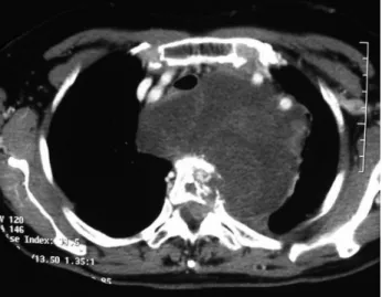
thoracic (T2 and T3) vertebral bodies contiguous with a giant posterior mediastinal soft tissue with anterior displacement of the visceral compartment of the medi- astinum and partial incorporation of mediastinal organs were demonstrated. Invasion of the spinal canal at the T3 level with compression of the spinal cord was confirmed (Fig. 2).
Staged decompressive laminectomy and thoracotomy
were planned. Spinal cord decompression through pos- terior access by T2, T3 and partial T4 laminectomy and resection of the endocanalar component of the tumour were done. The patient showed good recovery of her symptoms.
Ten days after the first operation, the patient underwent left postero-lateral thoracotomy in the third intercostal space with a transverse section of the neck of the second and third ribs, confirming the giant mediastinal mass covered by a fibrous capsule not dissociable from the me- diastinal vessels and other organs. Incomplete resection of the mediastinal mass was done with residual disease adherent to the great vessels and posterior tracheal wall.
T2 and T3 corporectomy, tumour resection and spinal cord decompression were then carried out followed by placement of a vertebral mesh and application of a long thoracic plate. The postoperative period was character- ised by left pleural effusion after removal of the chest tubes, which was resolved by thoracentesis (1,500 serum- haemorrhagic fluid). The patient had good recovery with- out neural deficits. A 30-day postoperative chest X-ray radiograph showed good surgical results (Fig. 3).
Macroscopically, the extrinsic part of the tumour out- side the bone appeared yellow and fatty, and the involved bone was yellow and cheese-like in consistency. Histo-
Fig. 1. Chest X-ray demonstrating a large mediastinal tumour taken at its clinical presentation, eight years before the surgical operation. At that time, the patient refused surgical treatment.
Fig. 2. Preoperatory chest computed tomographic scan. Vertebral de- struction, a huge posterior mediastinal tumour invading the structures of the visceral medistinum and spinal cord compression are demon- strated.
Fig. 3. A 30-day postoperative chest X-ray showing surgical results with placement of a vertebral mesh and application of a long thoracic plate.
Giant chordoma of the upper thoracic spine with mediastinal involvement
Asian Spine Journal
Asian Spine Journal
355logical examination confirmed the diagnosis of classical chordoma based on morphological aspects characterised by large epitheloid cells arranged in a cord-like fashion in a mucinous stroma with occasional cells containing mucinous material. The lobulated appearance along with cording of the cells and occasional physaliphorous cells suggested the diagnosis.
Discussion
Chordoma is a malignant bone tumour which exhibits notochord differentiation. The tumour grows slowly, recurs locally and metastasizes late. It is a relatively rare tumour, accounting for 3% to 55% of primary malignant bone tumours in the major series. Involvement of the thoracic spine is very rare [1].
Clinically the tumour may be asymptomatic but usu- ally presents with symptoms related to compression or involvement of adjacent structures, including the trachea, oesophagus and spinal cord [6]. In our case, even if gi- ant in its mediastinal component, the tumour manifested itself with symptoms of spinal cord acute compression many years after its first demonstration.
Radiographically, vertebral destruction is associated with the mediastinal mass because of the bony origin of the tumour. Lesions are usually lytic with calcifications within the tumour mass. Computed tomography scan and magnetic resonance imaging of the chest are manda- tory to evaluate the extent of both the tumour and in- volvement of adjacent organs.
The more accepted approach is aggressive surgery to resect the tumour as far as possible and prevent dissemi- nation into the surrounding tissues [3,4]. Thoracic chor- domas are more difficult to resect totally than analogous lesions affecting other sites since most patients like the present case have extradural and paraspinal tissue exten- sion at the time of diagnosis.
Surgical treatment considering posterior debulking ap- proaches has been reported in the past, but recently, they have been replaced by anterior or lateral approaches to ensure complete resection when possible and to achieve contemporary interbody fusion immobilization of the spine [7,8]. Completion of surgical removal of the me- diastinal component is directly related to its dimension:
in the past, few cases of complete resection of relatively small tumours have been described [3,5].
In our case, complete resection was not feasible because
of the large and strict adhesion of the tumour capsule to the neighbouring organs. Thus, tumour debulking and column stabilization were performed.
Recurrence after complete surgery is a common fea- ture (28%–68%) and is considered to be related to the violation of tumour margins at surgical time [9]. Even if not radiosensitive, radiotherapy has been used in some patients after total resection or palliative debulking. Ra- diotherapy does not prevent recurrence but seems to in- crease disease-free survival [10].
Five-year survival rate is reported to be 10% to 70%.
Thoracic chordomas with mediastinal involvement are rare slow-growing entities, which often present with neurological symptoms due to spinal cord compression.
Surgical complete removal is rare, and it depends on the localisation of the tumour and the dimension of the mediastinal component. Surgical procedures are often carried out to debulk the mediastinum and/or the spinal channel and to stabilize the affected spine to prevent ver- tebral collapse.
Conflict of Interest
No potential conflict of interest relevant to this article was reported.
References
1. Maesen F, Baur C, Lamers J, Versteege C, Willigha- gen R. Chordoma of the thorax. Eur J Respir Dis 1986;68:68-72.
2. Lee CS, Jung CH. Metastatic spinal tumor. Asian Spine J 2012;6:71-87.
3. Selvaraj A, Wood AJ. Superior mediastinal chordoma presenting as a bilobed paravertebral mass. Eur J Cardiothorac Surg 2003;23:248-50.
4. Topsakal C, Bulut S, Erol FS, Ozercan I, Yildirim H.
Chordoma of the thoracic spine: case report. Neurol Med Chir (Tokyo) 2002;42:175-80.
5. Roeyen G, Van Schil P, Somville J, Colpaert C, Van Oosterom A. Chordoma of the mediastinum. Eur J Surg Oncol 1999;25:224-5.
6. Cury JD, Peterson RJ, Lacy GD, Khaled AS, DeVane PT. Tracheal deviation from an atypical mediastinal mass. Chest 1997;111:503-5.
7. Hester TO, Valentino J, Strottmann JM, Blades DA, Robinson MC. Cervicothoracic chordoma presenting
Ottavio Rena et al.
356 Asian Spine J 2014;8(3):353-356
as progressive dyspnea and dysphagia. Otolaryngol Head Neck Surg 1999;120:97-100.
8. Murali R, Rovit RL, Benjamin MV. Chordoma of the cervical spine. Neurosurgery 1981;9:253-6.
9. Krol G, Sze G, Arbit E, Marcove R, Sundaresan N.
Intradural metastases of chordoma. AJNR Am J Neu- roradiol 1989;10:193-5.
10. Amendola BE, Amendola MA, Oliver E, McClatchey KD. Chordoma: role of radiation therapy. Radiology 1986;158:839-43.