CASE REPORT
Copyright © 2011, the Korean Surgical Society J Korean Surg Soc 2011;81:S85-88
http://dx.doi.org/10.4174/jkss.2011.81.Suppl1.S85
JKSS
Journal of the Korean Surgical Society pISSN 2233-7903ㆍeISSN 2093-0488
Received March 24, 2011, Revised April 21, 2011, Accepted May 6, 2011 Correspondence to: Yeon Jun Jeong
Division of Pediatric Surgery, Department of Surgery, Chonbuk National University Hospital, Chonbuk National University Medical School, 634-18 Geumam-dong, Deokjin-gu, Jeonju 561-712, Korea
Tel: +82-63-250-1977, Fax: +82-63-271-6197, E-mail: surgeon@chonbuk.ac.kr
cc Journal of the Korean Surgical Society is an Open Access Journal. All articles are distributed under the terms of the Creative Commons Attribution Non-Commercial License (http://creativecommons.org/licenses/by-nc/3.0/) which permits unrestricted non-commercial use, distribution, and reproduction in any medium, provided the original work is properly cited.
Choledochal cyst with ectopic distal location of the papilla of Vater
Sung Kang Kim
1, Yeon Jun Jeong
1,2, Jae-Chun Kim
11Division of Pediatric Surgery, Department of Surgery, Chonbuk National University Medical School, 2Research Institute of Clinical Medicine, Chonbuk National University Hospital, Jeonju, Korea
In cholangiographic techniques, the close relationship between choledochal cyst and anomalous union of pancreaticobiliary duct has attracted medical attention. There have been rare cases in which the papilla of Vater was found in a position other than its normal position, and such cases have been reported sporadically. However, such cases are interesting in the anatomi- cal context. In this review, we present our experience of choledochal cyst in a 30-month-old boy in whom the papilla of Vater was positioned in the third portion of the duodenum.
Key Words: Common bile duct cyst, Choledochal cyst, Cholangiography, Papilla of Vater
INTRODUCTION
The choledochal cyst can be defined as a congenital anomaly of the biliary system, characterized by cystic dila- tation of the intrahepatic duct and the extrahepatic biliary tree individually, or cystic dilatation of both these parts si- multaneously [1]. It has been reported that the incidence of choledochal cyst in Western countries ranges from 1:100,000 to 1:150,000 live births, and its incidence in Japan has been frequently reported to be 1:1,000 live births [2].
However, the precise incidence of choledochal cyst in Korea has not been reported so far. It has been known that choledochal cysts are caused by two main factors, namely, weakening of the bile duct walls and an obstruction distal to it. In addition to the causes of this disease, a close rela-
tionship between the choledochal cyst and pancreatico- biliary malunion (PBMU) has also been revealed [3]. Also, the probability of occurrence of PBMU related to the posi- tion of the opening of the papilla of Vater and the proba- bility of the occurrence of choledochal cyst related to such a situation has been considered to be one of the causes, and recently a series of reports have been made available [3,4].
In this connection, we report our experience of chol- edochal cyst with PBMU and the papilla of Vater posi- tioned in the third portion of the duodenum, in a 30-month-old boy.
Sung Kang Kim, et al.
S86 thesurgery.or.kr
Fig. 1. Abdominal computed tomography findings. There were some signs of choledochal cyst with dilatation of intrahepatic bile duct accompanied by dilatation of the common bile duct.
Fig. 2. Magnetic resonance cholangiopancreatographic findings.
Extrahepatic bile duct dilatation and intrahepatic bile duct dilatation at the center was observed, however the obvious part of the pancreaticobiliary union was not identified.
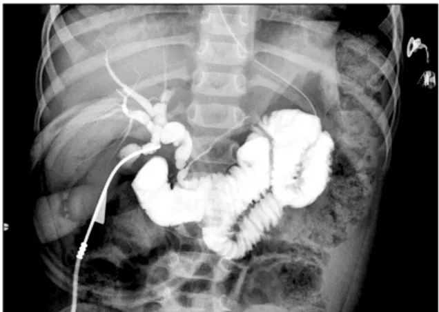
Fig. 3. Operative cholangiogram. P-B type pancreaticobiliary malunion, in which the pancreatic duct joins the common bile duct, and the papilla of Vater is positioned at the third portion of the duodenum was identified.
CASE REPORT
A 30-month-old boy, who was suffering from vomiting and abdominal pain since the past 20 days, underwent ab- dominal computed tomography (CT) scans at another hospital and was then transferred to our hospital under the impression of choledochal cyst. There was no partic- ular past medical history or family history, except for the history of hospitalization for treatment of pneumonia one month prior to being admitted to our hospital. Physical ex- amination revealed mild tenderness with rigidity in the right upper abdomen. Laboratory data including com-
plete blood count and urinalysis were within the normal limits, but blood biochemistry revealed the following: as- partate aminotransferase 121 IU/L, alanine aminotrans- ferase 155 IU/L, alkaline phosphatase 499 IU/L, gam- ma-glutamyl transpeptidase 168 IU/L, amylase 292 IU/L and lipase 129 U/L, showing elevated levels and T-bilir- ubin/D-bilirubin were 0.59 mg/dL and 0.16 mg/dL re- spectively, showing normal levels. Abdominal CT demon- strated dilatation of the extrahepatic bile duct and central dilatation of the intrahepatic bile duct (Fig. 1). Also mag- netic resonance cholangiopancreatography demonstrated findings similar to abdominal CT but it could not identify the PBMU (Fig. 2). On biliary scan, the hepatic uptake ap- peared to be within the normal range, however, the accu- mulation of isotope at the common bile duct (CBD) and delayed excretion into the duodenum were confirmed based on the findings.
Under the diagnosis of choledochal cyst the operation was carried out, and the P-B type of PBMU, in which the pancreatic duct joins the CBD, and the papilla of Vater is positioned in the third portion of the duodenum were identified through the operative cholangiography (Fig. 3).
Based on the operative findings, the diagnosis of a type I choledochal cyst was confirmed, and hence excision of the choledochal cyst and, Roux-en-Y hepaticojejunostomy were carried out. The patient was discharged on post- operative day 11 without any problems and in good gen- eral health condition.
Ectopic distal location of the papilla of Vater
thesurgery.or.kr S87
DISCUSSION
Choledochal cysts are congenital anomalies of the bili- ary system and consist of cystic dilatation of the extra- hepatic biliary tree, intrahapatic biliary ducts or both [2].
Based on the clinical and anatomic findings of choledochal cysts, Todani and others argued that choledochal cysts can be divided into 5 major types and these types can be fur- ther divided into 3 subcategories. Despite their classi- fication into many types, the surgical method of treatment for each type is not far different from the conventional method of treatment for choledochal cysts [2]. Many stud- ies on the pathogenesis of various types of choledochal cysts and their relationship with the adjacent structures are in full swing and are being reported. And in cholangio- graphic techniques, there has been a series of reports which suggest that there is a high possibility of a relation- ship between the choledochal cyst and PBMU [3]. A hep- atic diverticulum appears in the ventral wall of the primi- tive midgut early in the 4th week of intrauterine life in the development of the human embryo. The extrahepatic bile duct system and the ventral pancreas arise from the hep- atic diverticulum during the 4th to 6th week of intra- uterine life. The dorsal pancreatic bud appears opposite the hepatic diverticulum [4,5]. The main duct of the ven- tral pancreatic bud develops near the entry of the CBD into the duodenum. As the duodenum rotates to the right and become C-shaped, the ventral pancreatic bud is carried dorsally with the bile duct. The superior ventral branch of the ventral pancreas joins the distal portion of the dorsal pancreatic duct to form the main pancreatic duct, which merges with the CBD, inserting into the duodenum via the papilla of Vater [4]. The papilla of Vater is generally situ- ated halfway down the medial wall towards the posterior aspect of the descending or second part of the duodenum [3]. There have been rare cases in which the papilla of Vater was found in a position other than its normal position and such cases have been reported sporadically. However, such cases are interesting in the anatomical context [6,7].
Especially, very few cases have been reported in which the papilla of Vater opens in the third portion of the duode- num [8-10]. In a series of recent studies, it has been re- ported that the frequency of choledochal cyst in patients
was higher when the papilla of Vater was positioned distal to its normal position [3]. In this context from the anatomi- cal viewpoint, the ectopic distal location of the papilla of Vater represents the ectopic distal budding of the hepatic diverticulum during early embryonic life, it is straightfor- ward to postulate that the longer distance between the ventral and dorsal pancreatic buds may result in a delay in communication of the ventral duct with the dorsal duct or failure of communication [4]. And also the formation of PBMU at the early embryonic stage is caused by canal- ization of ventral pancreatic duct and the anomalous un- ion of pancreaticobiliary duct, which originate from the dislocation of the ventral pancreas, and it is also caused by the stretching of primitive common channel and CBD in accordance with the lengthening, which will result in a long CBD and a long common channel. Therefore, if growth of the epithelium in the CBD, the common channel and the pancreatic duct fails keep up with this anomalous elongation during fusion, the ducts may become attenu- ated, resulting in stenosis and weakness of the duct walls, and subsequent dilatation of the ducts, a phenomenon that has been observed clinically [4]. This suggests that the lengths of the common channel and the CBD in patients with choledochal cyst correlate strongly with the location of the papilla of Vater, i.e., the more distal the papilla of Vater is, the longer the common channel and the CBD [3].
In conclusion, we have reported a case of a 30- month-old child with choledochal cyst, in whom the form of PBMU occurred simultaneously and the papilla of Vater was positioned in the third portion of the duodenum.
There is no difference in the conventional surgical method of treatment in such cases despite the variation in the opening position of the papilla of Vater. But the likely rela- tionship between the opening position of the papilla of Vater and choledochal cyst should be considered when a variation in the opening position of the papilla of Vater is detected before surgery.
CONFLICTS OF INTEREST
No potential conflict of interest relevant to this article was reported.
Sung Kang Kim, et al.
S88 thesurgery.or.kr
ACKNOWLEDGEMENTS
This study was presented at the 62th annual meeting of Korean Surgical Society.
REFERENCES
1. de Vries JS, de Vries S, Aronson DC, Bosman DK, Rauws EA, Bosma A, et al. Choledochal cysts: age of presentation, symptoms, and late complications related to Todani's classification. J Pediatr Surg 2002;37:1568-73.
2. Sadiq J, Nandi B, Lakhoo K. An unusual variant of chol- edochal cyst: a case report. J Med Case Reports 2009;3:54.
3. Li L, Yamataka A, Yian-Xia W, Da-Yong W, Segawa O, Lane GJ, et al. Ectopic distal location of the papilla of vater in congenital biliary dilatation: Implications for pathoge- nesis. J Pediatr Surg 2001;36:1617-22.
4. Li L, Yamataka A, Wang YX, Wang DY, Wang K, Li ZX, et al. Anomalous pancreatic duct anatomy, ectopic distal lo- cation of the papilla of Vater and congenital biliary dilata- tion: a new developmental triad? Pediatr Surg Int 2003;
19:180-5.
5. Dowdy GS Jr, Waldron GW, Brown WG. Surgical anatomy of the pancreatobiliary ductal system. Observations. Arch Surg 1962;84:229-46.
6. Quintana EV, Labat R. Ectopic drainage of the common bile duct. Ann Surg 1974;180:119-23.
7. Disibeyaz S, Parlak E, Cicek B, Cengiz C, Kuran SO, Oguz D, et al. Anomalous opening of the common bile duct into the duodenal bulb: endoscopic treatment. BMC Gastroen- terol 2007;7:26.
8. Lindner HH, Peña VA, Ruggeri RA. A clinical and anatom- ical study of anomalous terminations of the common bile duct into the duodenum. Ann Surg 1976;184:626-32.
9. Keddie NC, Taylor AW, Sykes PA. The termination of the common bile duct. Br J Surg 1974;61:623-5.
10. Wilkinson LH, Simms AG, Floyd VT. Operative cholan- giography. Arch Surg 1966;92:677-88.