http://dx.doi.org/10.3988/jcn.2012.8.3.170 J Clin Neurol 2012;8:170-176
Introduction
Strokes are more severe, cause greater disability, and have worse outcomes in individuals with atrial fibrillation (AF) than in individuals without AF.1,2 Nonvalvular AF (NVAF) is asso- ciated with a fivefold increase in the risk of stroke, and st- roke associated with this condition accounts for approximate-
ly 15% of all strokes.3 Moreover, the likelihood of develop- ing NVAF increases with age.4 It is therefore important to minimize the risk of stroke in patients with NVAF.
Oral anticoagulant therapy can prevent thromboembolic events, but such therapy increases the risk of bleeding. Th- erefore, stroke risk-stratification schemes have been proposed to identify high-risk patients among individuals with NVAF.
Stroke Prevention in Atrial Fibrillation III (SPAF-III) inves- tigators have reported that the incidence of thromboembolism is correlated with the presence of transesophageal echocar- diography (TEE) risk markers in patients with NVAF.5 More- over, the National Registry of Atrial Fibrillation reported that
Transesophageal Echocardiographic Findings Are Independent and Relevant Predictors of Ischemic Stroke in Patients
with Nonvalvular Atrial Fibrillation
Shutaro Takashima,a Keiko Nakagawa,b Tadakazu Hirai,b Nobuhiro Dougu,a Yoshiharu Taguchi,a Etsuko Sasahara,b Kazumasa Ohara,b Nobuyuki Fukuda,b Hiroshi Inoue,b Kortaro Tanakaa
aDepartment of Neurology and bSecond Department of Internal Medicine, Toyama University Hospital, Toyama, Japan
Received October 3, 2011 Revised February 29, 2012 Accepted February 29, 2012 Correspondence Shutaro Takashima, MD, PhD Department of Neurology, Toyama University Hospital, 2630 Sugitani, Toyama 930-0194, Japan
Tel +81-76-434-7309 Fax +81-76-434-5033
E-mail [email protected]
Background and PurposezzNot only clinical factors, including the CHADS2 score, but also echocardiographic findings have been reported to be useful for predicting the risk of ischemic st- roke in patients with nonvalvular atrial fibrillation (NVAF). However, it remains to be determin- ed which of these factors might be more relevant for evaluation of the risk of stroke in each patient.
MethodszzIn 490 patients with NVAF who underwent transesophageal echocardiography (TEE), we examined the long-term incidence of ischemic stroke events (mean follow-up time, 5.7±3.3 years). For each patient, the predictive values of gender, the CHADS2 risk factors (congestive heart failure, hypertension, age ≥75 years, diabetes mellitus, history of cerebral ischemia), the CH- ADS2 score, and the findings on echocardiography, including TEE risk markers, were assessed.
ResultszzThe ischemic stroke rate was significantly correlated with the CHADS2 score (p<0.05).
According to the results of univariate analyses, age ≥75 years, history of cerebral ischemia, CH- ADS2 score ≥2, and presence of TEE risk were significantly correlated with the incidence of isch- emic stroke. Cox proportional hazards regression analyses identified age ≥75 years and presence of TEE risk as significant predictors of subsequent ischemic stroke events in patients with NVAF.
As compared with that in persons below 75 years of age without TEE risk, the ischemic stroke rate was significantly higher in persons who were ≥75 years of age with TEE risk (4.3 vs. 0.56%/year, adjusted hazard ratio=8.94, p<0.001).
ConclusionszzTEE findings might be more relevant predictors of ischemic stroke than the CH- ADS2 score in patients with NVAF. The stroke risk was more than 8-fold higher in patients aged
≥75 years with TEE risk. J Clin Neurol 2012;8:170-176
Key Wordszz stroke, risk, atrial fibrillation, transesophageal echocardiography.
Open Access
cc This is an Open Access article distributed under the terms of the Cre- ative Commons Attribution Non-Commercial License (http://creative- commons.org/licenses/by-nc/3.0) which permits unrestricted non-com- mercial use, distribution, and reproduction in any medium, provided the ori- ginal work is properly cited.
CHADS2, a stroke risk index, could be used to quantify the risk of stroke in patients with NVAF, and perhaps even aid in the selection of antithrombotic therapy.6 It has been confirm- ed that CHADS2 is statistically significantly associated with the risk of stroke. However, in the clinical setting, whether the CHADS2 index is the most relevant among the various exist- ing indices for evaluating the risk of stroke in individual pa- tients remains to be determined. The aim of the present study was therefore to clarify which factors among not only the ba- seline clinical characteristics (including the CHADS2 risk fac- tors), but also echocardiographic findings might most accu- rately predict the risk of stroke in patients with NVAF.
Methods
Study population
TEE was performed in 745 patients with AF who were admit- ted to our department at Toyama University Hospital (Toyama, Japan) between November 1994 and May 2007 for the man- agement of cardiovascular disease. TEE is routinely perform- ed in patients admitted to our department with AF in order to determine the risk of future embolic events. However, it is not performed in patients in the acute phase of cardiovascular events, including ischemic stroke or myocardial infarction, or in those suffering from infection or critical illness. Ultimately, 608 patients (82%) were enrolled for the present study after obtaining their written consent to participate. This retrospec- tive study was conducted with the approval of the Toyama University Ethics Committee.
Among the 608 patients with AF, 82 had valvular AF and 526 had NVAF. TEE was performed in all of the NVAF pa- tients, but technically adequate TEE data were not obtained from 36 patients. Therefore, only the data from the remain- ing 490 patients (men : women=357 : 133; age 67±11 years, mean±SD) were included in the analysis for the present study.
The following baseline clinical characteristics parameters were determined from the medical records: including age, gender, presence/absence of recent congestive heart failure, hypertension, diabetes mellitus, and history of cerebral isch- emia, the CHADS2 score, history of antiplatelet therapy or anticoagulant therapy with warfarin, and the prothrombin time-international normalized ratio (PT-INR). Routine labo- ratory data were also determined from the medical records.
Echocardiography
Transthoracic echocardiography was performed using an ul- trasound imaging system. The left atrial dimension (LAD), left ventricular diastolic dimension, and the left ventricular ejection fraction (LVEF) were determined from the M-mode images.
TEE was performed with a 5-MHz multiplane transducer connected to an ultrasound system. Each patient was studied in the fasting state without premedication. Multiple standard tomographic planes were imaged. The left atrial appendage peak flow velocity (LAAPV), presence of thrombi in the left atrium (LA), and severity of spontaneous echo contrast (SEC) in the LA (LASEC) were determined. LA appendage flow- velocity profiles were obtained by pulsed-wave Doppler echo- cardiographic imaging at the orifice of the appendage. The peak outflow velocity signals within each R-R interval were averaged over a minimum of six cardiac cycles. Atrial cavity or appendage thrombi were considered to be present when well-circumscribed, echodense, intracavitary masses that were acoustically distinct from the underlying endocardium and pectinate muscles were identified. LASEC was diagnosed in the presence of dynamic smoke-like echoes within the LA, and its severity was defined using the criteria of Fatkin et al.7 Aortic plaques were classified as either simple or complex (with the latter defined as any combination of mobile, pedun- culated, and ulcerated morphologies, or a plaque thickness of
≥4 mm).8
LA abnormality was defined as a slow LAAPV (less than 20 cm/sec) and/or the presence of LA thrombi or dense LASEC (≥grade 3+). Aortic atherosclerosis was defined as the pres- ence of complex aortic plaques. According to the SPAF-III study, TEE risk was defined as the existence of LA abnorma- lity and/or aortic atherosclerosis.5
The LAAPV, presence of LA thrombi, severity of LASEC, and presence of complex aortic plaques were determined by two independent observers (K.N. and T.H.). Any differences in the image interpretation were resolved by the opinion of a third observer (K.O.).
Outcomes
The composite endpoints of death from any cause, ischemic stroke or systemic embolism, and cardiac events (myocardial infarction or hospitalization for worsening heart failure) were determined in October 2008. Information on the endpoints was collected from the hospital databases and responses to questionnaires by the patients themselves or their family mem- bers. Stroke was defined as sudden onset of neurological defi- cit lasting for >24 hours. Whether a stroke was ischemic or hemorrhagic was confirmed by brain computed tomography and/or magnetic resonance imaging. Our evaluation of the in- cidence of ischemic stroke or systemic embolism indicated that none of the patients had a symptomatic systemic embolism.
Statistical analysis
Data are expressed as either mean±SD or percentage values.
All analyses were performed using SPSS software (SPSS
11.0J, Chicago, IL, USA). The mean values and proportions of variables were compared by the nonpaired t-test for con- tinuous variables and the χ2 test for categorical variables. The outcomes were assessed using Kaplan-Meier survival curves and compared using the log-rank test. Cox proportional-haz- ards regression analysis was used to identify independent pre- dictors of ischemic stroke. The level of statistical significance was set at p<0.05.
Results
Among the 490 patients with NVAF who were enrolled in this study, 143 (29%) died, 89 (19%) had cardiac events (11 with
myocardial infarction and 78 hospitalized for worsening he- art failure), and 42 (9%) developed ischemic stroke during the follow-up period (5.7±3.3 years). None of the patients had symptomatic mesenteric or peripheral emboli. In the present study, the ischemic stroke risk was estimated to be 1.5% per year. On the other hand, ten of the patients (2% of the entire cohort) had hemorrhagic stroke, of which nine were on war- farin therapy. Therefore, the risk of hemorrhagic stroke was estimated to be 0.46% per year in patients on warfarin thera- py. Table 1 compares the clinical characteristics and echocar- diographic markers in the patients showing subsequent de- velopment/no development of ischemic stroke. On average, the patients who suffered from ischemic stroke were older Table 1. Comparison of the baseline clinical characteristics and echocardiographic markers according to subsequent development/no de- velopment of ischemic stroke
Variable Overall (n=490) Subsequent ischemic stroke
p-value*
Absent (n=448) Present (n=42)
Age (years) 67.0±11.2 66.5±11.3 72.5±8.7 <0.001
Men 72.9% 73.4% 66.7% 0.34
Heart failure 21.8% 23.0% 11.9% 0.10
Hypertension 39.8% 39.7% 40.5% 0.92
Age ≥75 years 25.9% 24.3% 42.9% <0.01
Diabetes mellitus 15.3% 15.0% 19.0% 0.48
Prior stroke/TIA 25.5% 24.6% 35.7% 0.11
CHADS2 score 1.5±1.4 1.5±1.4 1.8±1.4 0.14
CHADS2 score ≥2 43.5% 42.6% 52.4% 0.22
Antiplatelet drug 23.6% 23.4% 25.7% 0.76
Warfarin 70.7% 70.8% 69.0% 0.80
PT-INR 1.7±1.2 1.7±1.2 1.7±0.7 0.96
LAD (mm) 41.4±8.3 41.3±8.3 42.5±8.3 0.43
LAD ≥40 mm 59.1% 58.3% 66.7% 0.35
LVDd (mm) 49.5±7.9 49.3±7.8 50.5±9.2 0.52
LVDd ≥50 mm 44.3% 43.8% 47.6% 0.74
LVEF (%) 59.0±14.2 58.6±14.2 62.4±14.5 0.13
LVEF <40% 9.3% 9.0% 11.8% 0.60
TEE risk 62.7% 60.9% 81.0% <0.05
LA abnormality 38.2% 37.1% 50.0% 0.09
LAAPV (cm/sec) 40.1±25.3 40.5±25.7 35.4±21.1 0.21
LAAPV <20 cm/sec 27.1% 26.5% 33.3% 0.34
LASEC 1.5±1.3 1.4±1.3 1.7±1.3 0.24
LASEC ≥3 26.2% 25.5% 34.1% 0.22
LA thrombi 12.5% 12.5% 11.9% 0.91
Aortic atherosclerosis 44.1% 43.8% 47.6% 0.62
Ao-IMT (mm) 3.4±2.0 3.4±2.1 3.8±1.9 0.24
Ao-IMT ≥4 mm 32.7% 31.9% 41.2% 0.27
Complex aortic plaque with Ao-IMT <4 mm 11.4% 11.9% 6.4% 0.51
TEE risk, LA abnormality, aortic atherosclerosis and complex aortic plaque are described in “Method”.
*p-value for ischemic stroke absent versus present.
Ao-IMT: aortic intima-media thickness, LA: left atrium, LAAPV: left atrial appendage peak flow velocity, LAD: left atrial dimension, LA- SEC: severity of spontaneous echo contrast in the left atrium, LVDd: left ventricular diastolic dimension, LVEF: left ventricular ejection fraction, PT-INR: prothrombin time-international normalized ratio (determined in patients receiving warfarin), TEE: transesophageal echocardiography, TIA: transient ischemic attack.
(p<0.001) and were more likely to be 75 years old or over (p<
0.01) relative to the patients that did not develop ischemic st- roke events. The frequency of warfarin use, PT-INR, and tr- ansthoracic echocardiographic findings did not differ signifi- cantly between the two groups. Although the prevalence rates of LA abnormality and aortic atherosclerosis also did not dif- fer between the two groups, the prevalence of TEE risk was significantly higher in the ischemic stroke group than in the
stroke-free group (p<0.01).
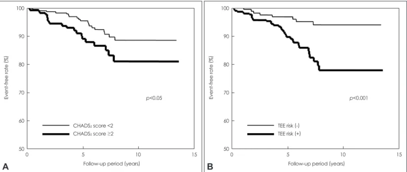
As shown in Fig. 1A, the incidence of ischemic stroke was significantly greater in patients with CHADS2 scores of ≥2 (p<0.05). Table 2 lists the results of the univariate analyses using Kaplan-Meier curves to identify the risk factors for isch- emic stroke events. According to the univariate analyses, age
≥75 years (p<0.001), a history of cerebral ischemia (p<0.05), a CHADS2 score of ≥2 (p<0.05), and the presence of TEE risk (p<0.001, Fig. 1B) were significantly positively correlated with the incidence of ischemic stroke.
Cox proportional-hazards regression analyses identified age ≥75 years [hazard ratio (HR), 3.17; 95% confidence inter- val (CI), 1.63-6.17; p<0.01] and the presence of TEE risk (HR, 2.96; 95% CI, 1.33-6.57; p<0.01) as significant predictors of ischemic stroke in patients with NVAF, after adjustments for a history of cerebral ischemia and CHADS2 score (Table 3).
The predicted annual ischemic stroke event rates based on a combination of age and presence/absence of TEE risk are given in Table 4. The rate of ischemic stroke events was sig- Fig. 1. A: Kaplan-Meier survival curves for ischemic stroke by the CHADS2 score in patients with NVAF. The incidence of ischemic stroke in patients with a CHADS2 score of less than 2 was 1.18% per year, while that in patients with a CHADS2 score of 2 or over was 2.19% per year (log-rank test, p<0.05). B: Kaplan-Meier survival curves for ischemic stroke according to the presence/absence of TEE risk in patients with NVAF. The incidence of ischemic stroke in patients without TEE risk was 0.67% per year, while that in patients with TEE risk was 2.26%
per year (log-rank test, p<0.001). NVAF: nonvalvular atrial fibrillation, TEE: transesophageal echocardiography.
100
90
80
70
60
50
100
90
80
70
60
50
Event-free rate (%) Event-free rate (%)
0 5 10 15 0 5 10 15
Follow-up period (years) Follow-up period (years)
p<0.05 p<0.001
CHADS2 score <2 CHADS2 score ≥2
TEE risk (-) TEE risk (+)
A B
Table 2. Results of univariate analyses using Kaplan-Meier curves performed to identify the risk factors for ischemic stroke events
Variable p-value
Male gender 0.48
Heart failure 0.10
Hypertension 0.62
Age ≥75 years <0.001
Diabetes mellitus 0.22
Prior stroke/TIA <0.05
CHADS2 score ≥2 <0.05
Antiplatelet drug 0.76
Warfarin 0.54
LAD ≥40 mm 0.16
LVDd ≥50 mm 0.88
LVEF <40% 0.75
TEE risk <0.001
LA abnormality <0.05
Aortic atherosclerosis 0.09
TEE risk, LA abnormality and aortic atherosclerosis are described in “Method”.
LA: left atrium, LAD: left atrial dimension, LVDd: left ventricular di- astolic dimension, LVEF: left ventricular ejection fraction, TEE: trans- esophageal echocardiography, TIA: transient ischemic attack.
Table 3. Results of the Cox proportional hazard regression analy- ses conducted for identifying predictors of ischemic stroke events in patients with non-valvular atrial fibrillation
Variable Hazard ratio 95% CI p-value
Age ≥75 years 3.17 1.63-6.17 0.001
Prior stroke/TIA 1.91 0.77-4.72 0.164 CHADS2 score ≥2 0.73 0.28-1.83 0.498
TEE risk 2.96 1.33-6.57 0.008
TEE risk is described in “Method”.
CI: confidence interval, TEE: transesophageal echocardiogra- phy, TIA: transient ischemic attack.
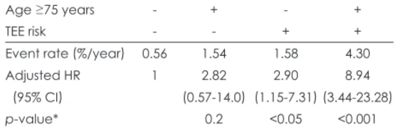
nificantly higher in persons who were 75 years of age or older with TEE risk than in persons under 75 years of age without TEE risk (4.3% vs. 0.56% per year; adjusted HR, 8.94; 95%
CI, 3.44-23.28; p<0.001). The adjusted HRs for ischemic stroke were comparable for those who were younger than 75 years but had TEE risk and those who were 75 years old or older without TEE risk (Table 4, Fig. 2).
Discussion
Whether the CHADS2 index might be the most useful of the various indices for evaluating the risk of stroke in individual patients in the clinical setting remains to be elucidated.9-11 In order to reevaluate the risk of stroke in patients with NVAF, we examined the long-term incidence of ischemic stroke in 490 patients with NVAF who had undergone TEE at our hos- pital. Our findings demonstrate that while the ischemic stroke
rate was significantly correlated with the CHADS2 score, the presence of TEE risk was also significantly correlated with the incidence of ischemic stroke. These results are consistent with those of previous studies.5,7 Cox proportional-hazards re- gression analyses identified age ≥75 years and presence of TEE risk as significant independent predictors of ischemic stroke events in patients with NVAF. Moreover, the risk esti- mation for ischemic stroke was more accurate for the combi- nation of age and TEE risk than for the CHADS2 score. Thus, our results indicate that TEE findings might be more relevant predictors of ischemic stroke than the CHADS2 score in pa- tients with NVAF, and that the stroke risk was more than eightfold higher in patients aged ≥75 years with TEE risk.
In fact, thromboembolism could occur even in patients with low CHADS2 scores. Kleemann et al.12 reported that 3% of their patients had LA thrombi and 8% had dense SEC despite having a low CHADS2 score of 0 or 1, and that the indepen- dent predictors of the presence of LA thrombi and dense SEC were an LVEF of <40% and an LAD of ≥50 mm. They con- cluded that echocardiography might be a useful tool for risk stratification in patients with low CHADS2 scores. On the other hand, in a subgroup analysis of the SPAF-III study, 382 patients with AF at high risk for thromboembolism under- went TEE at 18 echocardiography laboratories, and the cor- relation of the TEE findings with the subsequent develop- ment of ischemic stroke events was evaluated prospectively over a mean follow-up period of 1.1 years. The authors con- cluded that the subsequent occurrence of thromboembolism in the patients with AF was correlated with the presence of dense SEC, thrombus in the LA appendage, and/or aortic plaques.5 Thus, based on the results of the present study, it was concluded that the presence of TEE risk might serve as an in- dependent predictor of ischemic stroke in patients with NVAF.
While the severity of blood stasis in the LA and presence of aortic atherosclerosis have been reported to be correlated with an accumulation of clinical risk factors for thromboem- bolism in NVAF patients,13,14 the results of the present study suggest that the CHADS2 score alone is not sufficient to pre- dict the risk of ischemic stroke. The existence of complex aor- tic plaques is considered to be a marker of atherosclerosis, and is known to enhance hypercoagulability in patients with NV- AF.15 Therefore, the development of an accurate ischemic st- roke risk stratification scheme that would include certain types of laboratory data such as TEE risk markers that directly re- flect a prothrombotic condition of the LA is warranted.16,17
The invasiveness and requirement for operator proficiency of the TEE examination make it desirable to develop alterna- tive less-invasive tools that are at least as reliable as TEE.
Kleemann et al.12 reported that LVEF and LAD on transtho- racic echocardiography were independent predictors of the Table 4. Annual event rate and adjusted hazard ratio (95% CI) for
ischemic stroke according to the age and TEE risk
Age ≥75 years - + - +
TEE risk - - + +
Event rate (%/year) 0.56 1.54 1.58 4.30
Adjusted HR 1 2.82 2.90 8.94
(95% CI) (0.57-14.0) (1.15-7.31) (3.44-23.28)
p-value* 0.2 <0.05 <0.001
TEE risk is described in “Method”.
*p-value versus patients who were aged under 75 years old and without TEE risk.
Adjusted HR: as compared with patients who were aged un- der 75 years old and without TEE risk, CI: confidence interval, TEE: transesophageal echocardiography.
Fig. 2. Kaplan-Meier survival curves for ischemic stroke using the combination of age and TEE risk in patients with NVAF. The inci- dence of ischemic stroke was 0.56% per year in patients younger than 75 years of age without TEE risk, but 4.3% per year in pa- tients aged 75 years or older with TEE risk (log-rank test, p<0.001).
NVAF: nonvalvular atrial fibrillation, TEE: transesophageal echo- cardiography.
100
90
80
70
60
50
Event-free rate (%)
0 5 10 15 Follow-up period (years)
p<0.001
Age <75, TEE risk (-) Age ≥75, TEE risk (-) Age <75, TEE risk (+) Age ≥75, TEE risk (+)
presence of thrombus in the LA and dense SEC on TEE, wh- ereas the present study found that transthoracic echocardio- graphic markers were not significantly associated with these factors. Thus, further studies are warranted to evaluate the correlation between LVEF and LAD, and the presence of TEE risk. The addition of hemostatic markers, such as D-dimer levels, to the criteria for stroke risk stratification should also be considered.15,18,19
The ischemic stroke rate was 1.5% per year in the present study, which is slightly higher than that found in previous tri- als in which warfarin therapy was adjusted to maintain the PT-INR at 2-3. Japanese guidelines recommend low-intensi- ty adjusted-dose warfarin with a target PT-INR of 1.6-2.6 for NVAF patients aged 70 years or over.20 The mean PT-INR of 1.7 in the present study indicates that the treatment in most patients was consistent with those guidelines. Although war- farin evidently reduces the risk of stroke in patients with NV- AF, there are ethnic and racial differences in the risk of intra- cranial hemorrhage with warfarin treatment that should also be taken into consideration.21 In the present study, the rate of warfarin use or PT-INR did not differ between patients with and without subsequent development of ischemic stroke. Ac- cording to a Kaplan-Meier curve analysis, warfarin therapy was not associated with any significant reduction in the inci- dence of stroke. The present study had an observational rath- er than a randomized design, and more than 70% of the pa- tients were receiving warfarin therapy according to the ju- dgment of the attending physician. Therefore, the CHADS2
scores appeared to be higher in the patients on warfarin thera- py (1.60±1.37) than in those not on warfarin therapy (1.45±
1.38), although the difference was not statistically signifi- cant. Moreover, the percentages of patients with a history of congestive heart failure and with LA abnormality were signifi- cantly higher for those on warfarin therapy (26% and 42%, re- spectively) than for those not on warfarin therapy (12% and 28%), which made it difficult to evaluate the reduction in st- roke occurrence related to warfarin therapy from the data ob- tained in the present study.
Hart and Pearce11 described several problems related to stroke risk stratification schemes for NVAF patients. Even NVAF patients with low CHADS2 scores may occasionally develop ischemic stroke events. In addition, it is necessary to consider the prevention of stroke recurrence in patients on warfarin therapy who develop an ischemic stroke. The results of the present study actually indicate that TEE risk might be a powerful independent predictor of ischemic stroke in indi- vidual patients with NVAF, and that the stroke risk was great- er than eightfold higher in patients aged ≥75 years with TEE risk. Furthermore, a patient with TEE risk may develop an ischemic stroke even if he or she is on usual-dose warfarin
therapy. Therefore, high-intensity warfarin therapy or treat- ment with a direct thrombin inhibitor should be considered in patients with TEE risk.
The present study was subject to several limitations. First, the study had a retrospective rather than a prospective design.
Second, the inclusion of a relatively small sample from one institution might have affected the statistical power of the analyses. Third, 70% of the patients were on warfarin therapy according to the judgment of the attending physician, and the status of treatment with antithrombotic drugs was not deter- mined systematically throughout the follow-up period. Fi- nally, selection bias might have been present, since the sub- jects were selected from among patients with NVAF who had undergone TEE for evaluation of the potential risk of th- romboembolism. This meant that the present study involved NVAF patients who already had a comparatively high risk of developing thromboembolic events.
Despite the aforementioned limitations, the results of the present study indicate that the presence of TEE risk may be a powerful and independent predictor of ischemic stroke events in individual patients with NVAF. Although the CHA2DS2- VASc score has recently been proposed,16 the present results suggest that certain parameters directly indicating a pro- thrombotic state in the LA should be added to any ischemic stroke risk stratification scheme for individual patients with NVAF. We believe that a prospective study should be per- formed in the future to confirm the results of our present study.
Conflicts of Interest
The authors have no financial conflicts of interest.
REFERENCES
1. Yokota C, Minematsu K, Hasegawa Y, Yamaguchi T. Long-term prog- nosis, by stroke subtypes, after a first-ever stroke: a hospital-based study over a 20-year period. Cerebrovasc Dis 2004;18:111-116.
2. Marini C, De Santis F, Sacco S, Russo T, Olivieri L, Totaro R, et al.
Contribution of atrial fibrillation to incidence and outcome of isch- emic stroke: results from a population-based study. Stroke 2005;36:
1115-1119.
3. Wolf PA, Abbott RD, Kannel WB. Atrial fibrillation as an indepen- dent risk factor for stroke: the Framingham Study. Stroke 1991;22:
983-988.
4. Go AS, Hylek EM, Phillips KA, Chang Y, Henault LE, Selby JV, et al. Prevalence of diagnosed atrial fibrillation in adults: national im- plications for rhythm management and stroke prevention: the AnTi- coagulation and Risk Factors in Atrial Fibrillation (ATRIA) Study.
JAMA 2001;285:2370-2375.
5. Stroke Prevention in Atrial Fibrillation Investigators Committee on Echocardiography. Transesophageal echocardiographic correlates of thromboembolism in high-risk patients with nonvalvular atrial fibril- lation. Ann Intern Med 1998;128:639-647.
6. Gage BF, Waterman AD, Shannon W, Boechler M, Rich MW, Rad- ford MJ. Validation of clinical classification schemes for predicting stroke: results from the National Registry of Atrial Fibrillation. JAMA
2001;285:2864-2870.
7. Fatkin D, Kelly RP, Feneley MP. Relations between left atrial ap- pendage blood flow velocity, spontaneous echocardiographic contrast and thromboembolic risk in vivo. J Am Coll Cardiol 1994;23:961-969.
8. Stroke Prevention in Atrial Fibrillation Investigators Committee on Echocardiography. Transesophageal echocardiography in atrial fibril- lation: standards for acquisition and interpretation and assessment of interobserver variability. J Am Soc Echocardiogr 1996;9:556-566.
9. Weir NU. An update on cardioembolic stroke. Postgrad Med J 2008;
84:133-142.
10. Stroke Risk in Atrial Fibrillation Working Group. Comparison of 12 risk stratification schemes to predict stroke in patients with nonval- vular atrial fibrillation. Stroke 2008;39:1901-1910.
11. Hart RG, Pearce LA. Current status of stroke risk stratification in pa- tients with atrial fibrillation. Stroke 2009;40:2607-2610.
12. Kleemann T, Becker T, Strauss M, Schneider S, Seidl K. Prevalence and clinical impact of left atrial thrombus and dense spontaneous echo contrast in patients with atrial fibrillation and low CHADS2 score.
Eur J Echocardiogr 2009;10:383-388.
13. Ohara K, Hirai T, Fukuda N, Sakurai K, Nakagawa K, Nozawa T, et al.
Relation of left atrial blood stasis to clinical risk factors in atrial fi- brillation. Int J Cardiol 2009;132:210-215.
14. Wysokinski WE, Ammash N, Sobande F, Kalsi H, Hodge D, McBane RD. Predicting left atrial thrombi in atrial fibrillation. Am Heart J 2010;159:665-671.
15. Nakagawa K, Hirai T, Sakurai K, Ohara K, Nozawa T, Inoue H. Tho- racic aortic plaque enhances hypercoagulability in patients with non-
rheumatic atrial fibrillation. Circ J 2007;71:52-56.
16. European Heart Rhythm Association; European Association for Car- dio-Thoracic Surgery, Camm AJ, Kirchhof P, Lip GY, Schotten U, et al. Guidelines for the management of atrial fibrillation: the Task Force for the Management of Atrial Fibrillation of the European Society of Cardiology (ESC). Eur Heart J 2010;31:2369-2429.
17. Fuster V, Rydén LE, Cannom DS, Crijns HJ, Curtis AB, Ellenbogen KA, et al. 2011 ACCF/AHA/HRS focused updates incorporated into the ACC/AHA/ESC 2006 guidelines for the management of patients with atrial fibrillation: a report of the American College of Cardiolo- gy Foundation/American Heart Association Task Force on practice guidelines. Circulation 2011;123:e269-e367.
18. Nozawa T, Inoue H, Hirai T, Iwasa A, Okumura K, Lee JD, et al. D-di- mer level influences thromboembolic events in patients with atrial fi- brillation. Int J Cardiol 2006;109:59-65.
19. Dougu N, Takashima S, Sasahara E, Taguchi Y, Toyoda S, Hirai T, et al. Differential diagnosis of cerebral infarction using an algorithm combining atrial fibrillation and D-dimer level. Eur J Neurol 2008;
15:295-300.
20. Yamaguchi T, for Japanese Nonvalvular Atrial Fibrillation-Embolism Secondary Prevention Cooperative Study Group. Optimal intensity of warfarin therapy for secondary prevention of stroke in patients with nonvalvular atrial fibrillation : a multicenter, prospective, randomized trial. Stroke 2000;31:817-821.
21. Shen AY, Yao JF, Brar SS, Jorgensen MB, Chen W. Racial/ethnic dif- ferences in the risk of intracranial hemorrhage among patients with atrial fibrillation. J Am Coll Cardiol 2007;50:309-315.