101
<접수일:2011년 1월 3일, 수정일:2011년 1월 19일, 심사통과일:
2011년 1월 20일>
통신저자:지 종 대
서울시 성북구 안암동 5가 126-1
고려대학교 의과대학 류마티스내과학교실 E-mail:[email protected]
Network Analysis을 이용한 류마티스관절염 활액 대식세포에서 유전자 발현 연구
지종대1ㆍ김태환2ㆍ이빛나라2ㆍ최성재1ㆍ이영호1ㆍ송관규1
고려대학교 의과대학 류마티스내과학교실1, 한양대학교 의과대학 류마티스병원2
Study of the Gene Expressions in Rheumatoid Arthritis Synovial Macrophages Using Network Analysis
Jong Dae Ji1, Tae-Hwan Kim2, Bitnara Lee2, Sung Jae Choi1, Young Ho Lee1, Gwan Gyu Song1
Department of Rheumatology, College of Medicine, Korea University University1, The Hospital for Rheumatic Diseases, College of Medicine, Hanyang University2, Seoul, Korea
Objective. We wanted to investigate the mechanisms that could account for the pathogenesis of rheumatoid arthritis, so we examined the different expressions of the genes in rheumatoid arthritis (RA) synovial fluid macrophages as compared with that of normal peripheral blood (PB) mon- ocyte-derived macrophages using microarray and bio- informatic analysis.
Methods. We examined the expression of genes by using a gene expression oligonucleotide microarray. The differ- ences of the gene expressions between the RA synovial macrophages and the normal PB monocytes-derived mac- rophages were analyzed using bioinformatic tools, includ- ing cytoscape and its plugin.
Results. In this study, we found that 899 genes (464 genes up-regulated and 435 genes down-regulated) were differ- entially expressed between the two groups. Among the 899
genes, 552 genes were included for gene ontology analysis and network analysis. Based on biological process ontology, they were categorised mainly into immune response proc- esses, responses to stimulus and signaling and regulation of biological processes. In addition to the genes related with STAT1 and AP-1 signaling, we found that the genes in- volved in the antigen processing and the cell cycle are abundantly expressed in RA synovial macrophages, sug- gesting that these genes may play an important role in the pathogenesis of RA.
Conclusion. Our study suggest that this approach using in- tegration of the gene expression profile with the protein interaction data may help to find several important patho- genic mechanisms in RA.
Key Words. Rheumatoid arthritis, Synovial macrophages, Microarray, Bioinformatics
서 론
류마티스관절염은 손, 발의 작은 관절을 일차적으로 침 범하는 전신성 염증질환으로 만성 염증과 함께 동반된 연
골 및 골 파괴로 심각한 장애를 일으키는 질환이다. 현재 까지 류마티스관절염의 병인을 밝히기 위한 많은 연구가 진행되어 T 림프구, B 림프구, 단핵구/대식세포, 섬유아세 포, 파골세포 등 다양한 세포와 사이토카인, 케모카인 등 의 다양한 염증물질들이 관여함이 밝혀졌다 (1). 특히 최근 에는 microarray 기법을 이용한 다양한 유전자 발현 프로파 일링(gene expression profiling) 연구가 류마티스관절염 활 막 조직이나 말초혈액 세포에서 진행되어 류마티스관절염 병인에 관여하는 중요한 유전자들이 밝혀지고 있다 (2-5).
류마티스관절염에서 전사 프로파일링(transcription profil-
ing)의 연구는 류마티스관절염의 병인에 관여하는 특징적 유전자발현의 발견 뿐 아니라 발현에 차이를 보이는 유전 자 조합에 따라 류마티스관절염을 여러군으로 세분화하여 항 TNF제제나 anakinra와 같은 특정 항류마티스제제에 대 한 치료반응을 예측하는 지표로도 활용될 가능성이 최근 의 연구들에 의해 제시되었다 (2,5-7). 그러나 면역계는 다 양한 면역세포와 그 세포들에서 생성되는 사이토카인, 케 모카인 등의 염증 물질들에 의해 매우 복잡하게 조절되기 때문에 류마티스관절염에 작동하는 병태 생리학적 이상은 단지 독립적인 유전자들의 발현변화만으로는 설명하기가 힘들다. 최근 류마티스관절염 등과 같이 복잡한 요인들이 작동하는 질환의 병인 연구에서 다양한 omics기법을 이용 하여 단백질 및 유전자 발현의 차이를 분석하고 이 들 유전 자들이 유기적으로 작동하는 생물학적 네트워크(biological network)를 밝혀내고 질환의 병인에 중요한 역할을 하는 공 통된 생리학적 기능을 가지는 유전자들의 군(regulatory module)을 밝혀냄으로써 질환의 병인을 규명하려는 연구 들이 진행되고 있다 (8,9).
다양한 유전자 및 단백질의 발현 profile정보와 단백질- 단백질 상호작용(protein-protein interaction), 단백질-DNA 상호작용(protein-DNA interaction) 등의 세포 network 정보 (cell network information)를 통합 분석함으로써 각각의 질 환에서 관찰되는 특징적 병태생리학적 기전을 찾으려는 연구가 있다. 이 같은 network 정보와 expression profile의 통합 분석을 목적으로 cytoscape을 비롯한 다양한 software 들이 개발되었다 (10). Cytoscape는 분자상호작용 네트워 크(molecular interaction network)를 보기 쉽게 표현하고 이 같은 네트워크와 gene expression profile을 통합 분석할 수 있도록 하는 생물정보학 소프트웨어(open source bioinfor- matic software)이다 (11). Cytoscape는 jActiveModulessk Bi- NGO같은 plugin 소프트웨어를 인터넷에서 내려 받아 실 행시킴으로써 gene ontology (GO) enrichment analysis를 하 거나 조절모듈(regulatory module)을 찾는데 사용될 수 있 다 (8,10).
본 연구자들은 microarray를 이용하여 류마티스관절염 환 자의 활액 대식세포와 정상인의 말초혈액 단핵구에서 분화 된 대식세포에서 유전자 발현을 관찰하고 그 병태생리학적 의의를 찾기 위해 cytoscape와 그 plugin을 이용하여 생물정 보학적 분석(bioinformatic analysis)를 시행하였다.
대상 및 방법 세포의 분리
건강 자원자로부터 혈액을 채취한 후 Ficoll (Invitrogen, Carlsbad, CA, USA)을 이용하여 말초 혈액 단핵세포를 얻 고 이 세포로부터 anti-CD14 magnetic beads (Miltenyi Biotec, Auburn, CA, USA)을 이용하여 단핵구를 얻었다.
10% 우태아혈청(fetal bovine serum, FBS)이 포함된 RPMI 1,640 배양액에서 단핵구를 M-CSF 20 ng/mL로 처리한 후
2일간 배양하여 대식세포로 분화 시켰다. 류마티스관절염 환자로부터 활액을 얻은 후 anti-CD14 magnetic beads (Miltenyi Biotec, Auburn, CA, USA)을 이용하여 CD14 양성 세포를 얻었다. 활액을 얻었던 류마티스관절염 환자들은 1987년 미국 류마티스학회가 제정한 류마티스관절염의 진 단기준을 만족하였고 prednisolone, methotrexate, sulfasala- zine, hydroxychloroquine, leflunomide 등의 항류마티스 제 제를 사용하였으나 병의 활성이 조절되지 않은 환자의 활 액을 대상으로 하였다. 이 연구의 계획은 한양대학교 류마 티스병원 임상시험심사위원회(institutional review board)의 승인을 얻어 시행하였고 혈액 및 활액을 채취한 건강 자원 자 및 환자들로부터 연구에 대해 설명하고 동의서를 받았 다.
류마티스관절염 환자의 활액 대식세포 및 건강 자원자의 말초 혈액 단핵구-유래 대식세포의 Gene expression pro- filing
류마티스관절염 환자의 활액 내 대식세포에서의 유전자 발현을 비교하기 위해 대조군으로 정상인 말초혈액에서 분리된 단핵구를 M-CSF로 처리하여 분화시킨 대식세포를 사용하였는데 이는 류마티스관절염 활액에 존재하는 CD14+세포가 대식세포로 생각되고 기존의 연구들에서 류마티스 관절염 활액 대식세포의 대조군으로 정상인 말 초혈액의 단핵구-유래 대식세포를 사용하였기 때문에 본 연구에서도 말초혈액 단핵구를 분화시킨 대식세포를 대조 군으로 사용하였다 (12,13). 2명의 건강 자원자의 말초 혈 액으로부터 분리된 단핵구에서 분화된 대식세포와 6명의 류마티스관절염 환자의 활액으로부터 분리된 대식세포로 부터 Trizol reagent (Invitrogen, Carlsbad, CA, USA)를 이용 하여 제조사의 지침에 따라 total RNA를 추출하였다. 추출 된 total RNA를 이용하여 mRNA microarray를 시행하였다.
mRNA microarray는 Illumina HumanRef-8 v3 Expression BeadChip (Illumina, Inc., San Diego, CA)을 이용, 분석은 Illumina BeadStudio v3.1.3 (Gene Expression Module v3.3.8) 을 이용, ArrayAssistⓇ 5.5.1 (Stratagene, La Jolla, USA)와 R statistical language v. 2.4.1를 이용하여 통계처리 하였다. 각 세포들간의 유전자의 발현 차이는 one-way analysis of var- iance (ANOVA)을 이용하여 분석하였고 통계적 유의 수준 은 0.05를 기준으로 하였다. 이상의 microarray를 이용한 mRNA profiling실험은 Macrogen (Seoul, Korea)에 의뢰하 여 진행하였다.
Microarray 결과의 검증
MicroArray의 결과를 확인하기 위해 total RNA (3명의 건 강 자원자의 말초 혈액과 12명의 류마티스관절염 환자의 활액으로부터 분리된 CD14 양성 세포)로부터 a First Str- and cDNA Synthesis kit (Fermentas)를 이용하여 cDNA를 만 들고 iQTM SYBR-GreenSupermix와 iCycler iQTM thermal
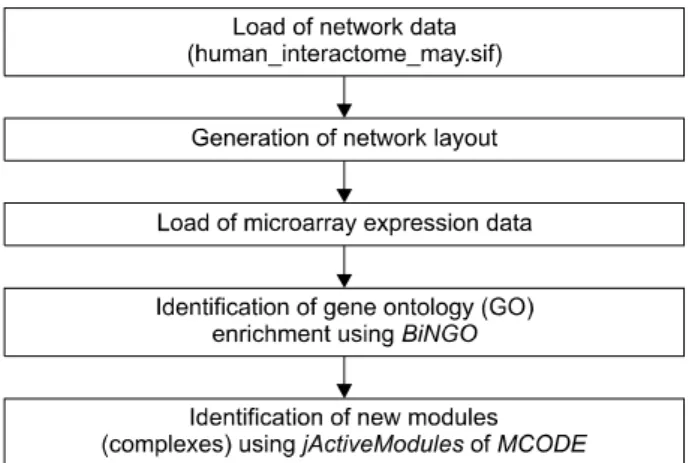
Figure 1. Overview of the bioinformatic analysis using cytoscape.
Figure 2. The array data for the gene expressions was validated by performing quantitative real-time PCR in the healthy volunteer PB monocyte-derive macrophages and the RA synovial macrophages *p<0.05 versus the healthy volun- teer PB monocyte-derive macrophages.
cycler (Bio-Rad Laboratories, Hercules, CA)를 이용한 re- al-time PCR법으로 유전자 발현을 재확인하였다. 검사한 유전자의 발현은 GAPDH의 발현과 비교하여 정량화하였 다. 유전자 발현 차이는 Student's t-test를 이용하여 분석하 였고 통계적 유의 수준은 0.05를 기준으로 하였다.
생물 정보학적 분석
Cytoscape를 이용한 생물 정보학적 분석은 2007년 Cline 등에 의해 보고되었던 논문의 방법을 이용하였다(그림 1).
간단히 살펴보면 Cytoscape에 불러온(import한) 네트워크 정보(network data)는 www.cytoscape.org에서 내려 받은 Human_Interactome_May.sif을 이용하였다. Human_Interac- tome_May.sif 파일은 yeast two-hybrid (Y2H) system과 co-affinity purification assay를 이용하여 찾아낸 protein-pro- tein interaction과 문헌 검색을 통해 찾아낸 literature-curated interaction (LCI)을 이용한 세편의 논문에 보고된 data set들 에 더하여 (14-16) IntAct molecular interaction database (IntAct), Database of Interacting Proteins (DIP), Biomolecular Interaction Network Database (BIND), Human Protein Refe- rence Database (HPRD)에 보고된 interaction들을 통합하여 만든 인간 interaction network data로 10,203개의 gene (node)와 61,262개의 interaction (edge)으로 구성되어 있다.
Cytoscape의 layout algorithms을 이용하여 network layout을 생성한 후 microarray를 이용하여 만든 gene expression data 를 불러왔다(import). Cytoscape plugin인 jActiveModules과 MCODE를 이용하여 류마티스관절염 활액 대식세포에서 특징적으로 작동하는 유전자들의 module (complex)들을 찾고 또 다른 plugin인 BiNGO를 이용하여 gene ontology
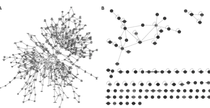
Figure 3. (A) Map of the biological processes associated with RA synovial macrophages. Darker nodes mean the more significant ontology terms. The size is proportional to the number of genes included in that ontology term. (B) Network of the genes included in the immune system processes (GO. 2376). A blue node means down-regulation of genes and a red node means up-regulation of genes in the RA synovial macrophages.
enrichment를 찾았다 (10). Protocol에 따라 jActiveModules의 경우는 score 3이상을 의미 있는 module로 간주하였고 MCODE에서는 score 2 이상, 적어도 node가 4개 이상을 의 미 있는 complex로 간주하였다.
결 과
류마티스관절염 환자의 활액 대식세포 및 건강 자원자의 말초 혈액 단핵구-유래 대식세포에서 유전자의 발현 비교 류마티스관절염 환자의 활액 대식세포의 병태생리학적 역할을 규명하기 위하여 말초 혈액 내 단핵구로부터 분화 된 대식세포와 류마티스관절염 환자의 활액 내 대식세포 에서 유전자들의 발현을 mRNA microarray 방법을 이용하 여 검사하여 비교하였다. microarray 분석결과를 보면 건강 한 자원자의 단핵구에서 분화된 대식세포와 비교하였을 때 류마티스관절염 활액 대식세포에서는 899개의 유전자 가 통계적으로 유의한 발현의 차이를 보였고 그 중 464개 유전자의 발현이 증가되었고 435개 유전자의 발현이 감소 되어 있었다.
Microarray 검사 결과에서 발현에 차이를 보인 파골세포 분화 관련 유전자의 검증
Microarray 검사에 사용되었던 검체를 포함하여 2명의 건 강 자원자의 말초 혈액과 12명의 류마티스관절염 환자의 활액으로부터 분리된 CD14 양성 세포로부터의 total RNA 를 이용하여 microarray에서 유의한 차이를 보였던 유전자 들을 real-time PCR 검사법으로 확인하였다. real-time PCR
검사에서도 microarray 결과와 같이 c-Fos와 IL-7R는 유의 하게 류마티스관절염에서 증가되었으나 CDKN1A의 경우 는 microarray 결과와는 반대로 류마티스관절염에서 현저 히 낮았다(그림 2).
류마티스관절염 활액 대식세포와 말초 혈액 내 단핵구-유 래 대식세포에서 microarray방법으로 확인된 유전자발현 차이에 대한 생물정보학적 분석
Microarray결과에서 보인 류마티스관절염 환자의 활액 대 식세포와 건강 자원자의 단핵구-유래 대식세포에서 유전 자 발현의 차이의 생물학적 의의를 규명하기 위해 cyto- scape와 그 plugin을 이용하여 gene ontology analysis등의 생 물정보학적 분석을 시행하였다. 류마티스관절염 환자의 활액 대식세포와 건강 자원자의 단핵구-유래 대식세포에 서 발현의 차이를 보였던 899개의 유전자 중 본 연구에 사 용된 네트워크 자료에서 상호작용이 보고된 522개의 유전 자(node)를 대상으로 분석을 진행하였다. 이들 552개의 유 전자(node)는 437개의 상호작용(edge)이 있었고 325개의 유전자가 증가되어 있었고 227개의 유전자가 감소되어 있 었다.
본 연구자는 류마티스관절염 환자의 활액 대식세포에서 유의하게 차이를 보이는 유전자들의 생물학적 기능 및 유 전자 발현의 변화가 류마티스관절염의 병태생리에 미치는 여향을 파악하기 위해 cytoscape의 plugin 프로그램인 BiNGO를 이용하여 gene ontology enrichment 분석을 시행 하였다. Biological process GO annotation을 가지는 인간 유
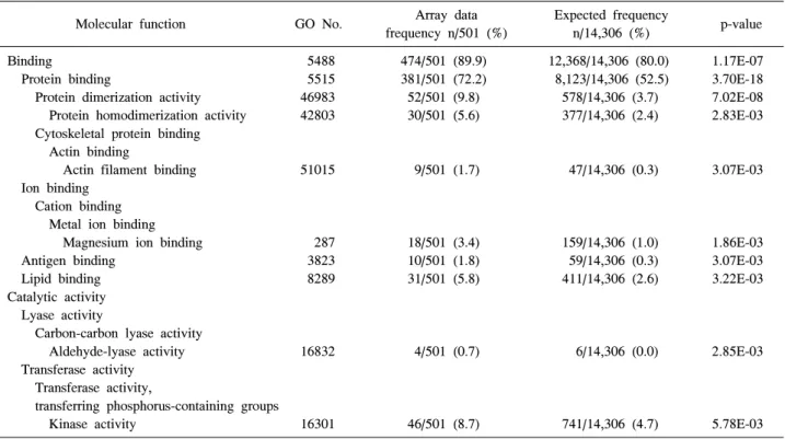
Table 2. The gene ontology analysis (molecular function) of the differentially expressed genes in RA synovial macrophages using the BiNGO plugin (The top ten GO terms were statistically significant)
Molecular function GO No. Array data
frequency n/501 (%)
Expected frequency
n/14,306 (%) p-value
Binding 5488 474/501 (89.9) 12,368/14,306 (80.0) 1.17E-07
Protein binding 5515 381/501 (72.2) 8,123/14,306 (52.5) 3.70E-18
Protein dimerization activity 46983 52/501 (9.8) 578/14,306 (3.7) 7.02E-08 Protein homodimerization activity 42803 30/501 (5.6) 377/14,306 (2.4) 2.83E-03 Cytoskeletal protein binding
Actin binding
Actin filament binding 51015 9/501 (1.7) 47/14,306 (0.3) 3.07E-03 Ion binding
Cation binding Metal ion binding
Magnesium ion binding 287 18/501 (3.4) 159/14,306 (1.0) 1.86E-03
Antigen binding 3823 10/501 (1.8) 59/14,306 (0.3) 3.07E-03
Lipid binding 8289 31/501 (5.8) 411/14,306 (2.6) 3.22E-03
Catalytic activity Lyase activity
Carbon-carbon lyase activity
Aldehyde-lyase activity 16832 4/501 (0.7) 6/14,306 (0.0) 2.85E-03 Transferase activity
Transferase activity,
transferring phosphorus-containing groups
Kinase activity 16301 46/501 (8.7) 741/14,306 (4.7) 5.78E-03
Table 1. The gene ontology analysis (biological process) of the differentially expressed genes in RA synovial macrophages using the BiNGO plugin (The top ten GO terms were statistically significant)
Biological process GO No. Array data
frequency n/501 (%)
Expected frequency
n/14,306 (%) p-value
Immune system processes 2376 96/501 (19.1) 948/14,306 (6.6) 2.41E-18
Regulation of immune system processes 2682 47/501 (9.3) 424/14,306 (2.9) 2.35E-09
Immune responses 6955 54/501 (10.7) 619/14,306 (4.3) 1.80E-07
Regulation of immune responses 50776 30/501 (5.9) 235/14,306 (1.6) 2.72E-07
Response to stimulus 50896 197/501 (39.3) 3,633/14,306 (25.3) 2.35E-09
Response to stress 6950 115/501 (22.9) 1,773/14,306 (12.3) 1.40E-08
Defense responses 6952 55/501 (10.9) 620/14,306 (4.3) 7.40E-08
Signaling 23052 172/501 (34.3) 3,131/14,306 (21.8) 2.78E-08
Regulation of biological processes
Positive regulation of biological processes 48518 133/501 (26.5) 2,208/14306 (15.4) 2.78E-08 Regulation of responses to stimulus 48583 50/501 (9.9) 524/14306 (3.6) 4.78E-08
전자의 수는 14,306개였고 류마티스관절염 환자의 활액 대식세포에서 유의하게 차이를 보이는 유전자들 중 GO annotation을 가지고 있어 분석에 포함된 유전자는 501개였 다. GO enrichment 분석의 결과를 보면 생물학적 과정(bio logical process) ontology의 경우는 274개 GO term에서 의미 있는 차이를 보였고 분자 기능 ontology는 38개, 세포 조성 ontology는 49개의 GO term에서 의미 있는 차이를 보였다 (그림 3A). 각각의 세부 ontology에서 유의한 차이를 보였 던 GO term을 보면 생물학적 과정의 범주에서는 면역 반 응 과정, 자극에 대한 반응, 생물학적 과정의 조절 등에서 유전자 발현의 차이를 보였고 분자 기능에서는 결합이나
촉매 활동에서 세포 조성은 세포 부분 범주 중 세포 내 부 분, cell leading edge, 세포 내, 세포 표면 등의 subcategory에 서 차이를 보였다(표 1, 2). 류마티스관절염의 병인과 관련 하여 분자 기능의 category중 면역계 과정에 속한 유전자 들을 보면 Toll-like receptor 2 (TLR2), Myeloid differentiation primary response gene 88 (MYD88), Interleukin-1 receptor- associated kinase 2 (IRAK2), IRAK3 등의 유전자 발현이 류 마티스관절염 환자의 활액 대식세포에서 현저히 증가되어 있어 TLR 신호전달과정이 류마티스관절염의 병인에 중요 한 역할을 할 것으로 생각할 수 있었고 LYN, FYN 등의 Src family kinases와 FCAR, FCGR1A, FCGR2B 등의 Fcγ 수용
Figure 4. Different expressions of the STAT1-related genes in the RA synovial macrophages. A blue node means down- regulation of genes and a red node means up-regulation of genes in the RA synovial macrophages.
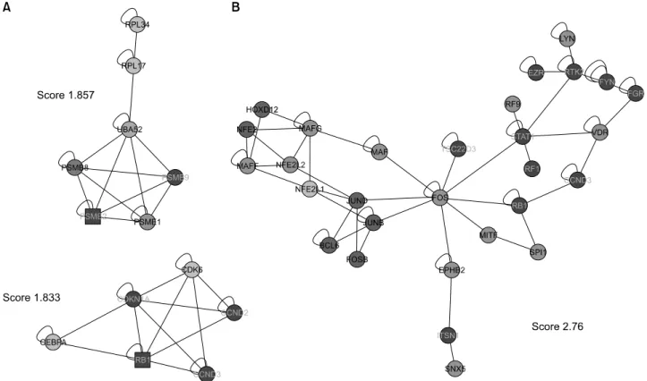
Figure 5. (A) Detection of densely connected regions in the network of genes differentially expressed in the RA synovial macrophages and the PB monocyte-derived macrophages from healthy volunteer using MCODE. A square node is a seed node. (B) Identification of the functional modules as highly connected regions with similar responses using jActiveModules. A blue node means down-regulation of genes and a red node means up-regulation of genes in the RA synovial macrophages.
체 유전자들이 유의하게 증가되어 있었다(그림 3B). micro- array를 이용한 기존의 류마티스 관절염의 병인연구들에서 (5,12,17) STAT1 관련 유전자들의 발현이 증가된 것과 마찬
가지로 본 연구에서도 STAT1이 증가되어 있었고 STAT1과 interaction을 가지는 IRF1, IRF9의 발현도 증가되어 있었으 며 FOS, JUNB, JUND등의 유전자가 STAT1과 상호작용 (interaction)을 보였다 (그림 4).
단백질-단백질 상호작용 네트워크에서 연결이 밀집된 부 분은 함께 기능하는 단백질 복합체를 의미한다. 연결이 밀 집된 부분을 찾기 위해 cytoscape plugin인 MCODE를 이용 하였다. MCODE에서는 score 2 이상, 적어도 node가 4개 이상을 의미 있는 complex로 간주하는데 이 기준에 합당 한 complex는 발견되지 않았다. 그러나 score 2에 근접한 두 개의 complex를 찾았는데 하나는 PSME1, PSMB8, RPL34, UBA52, RPL17, PSME2, PSMB9 등 7개의 node로 구 성된 complex고 다른 하나는 CCND2, RB1, CDKN1A, CCND3, CDK6, CEBPA 등 6개의 node로 구성된 complex였 다(그림 5A). 또한 jActiveModules을 이용하여 본 연구에서 사용한 네트워크에서 기능 모듈을 찾으려 했으나 score 3 이상의 module은 발견할 수 없었다. 그러나 3에 근접한 score를 보이는 module로 FOS와 STAT1이 중심이 되어 구 성된 module을 찾을 수 있었다(그림 5B).
고 찰
류마티스관절염은 임상양상이 환자에 따라 다양하고 치
료에 대한 반응의 정도도 매우 다르다. 그 동안의 많은 연 구에도 불구하고 류마티스관절염의 원인은 현재까지 명확 히 알려져 있지 않으며 작동하는 병태 생리학적 기전이 매 우 복잡하고 다양해서 독립적인 개개의 유전자나 단백질 발현 변화에 대한 연구만으로는 정확한 병인을 파악하고 질환의 원인을 찾아내기가 힘들 것으로 생각된다. 최근 microarray 등과 같은 분자 생물학적 방법의 진보의 결과로 류마티스 관절염에서 작동하는 유전자 발현이나 단백질 발현이상을 소수의 유전자에 대한 연구가 아닌 전체 유전 자 발현의 이상을 파악하고 생물 정보학적 기법으로 분석 하려는 시도가 이루어지고 있다. 본 연구에서는 microarray 기법을 이용하여 류마티스관절염 환자의 활액 대식세포와 정상인의 말초혈액 단핵구에서 분화된 대식세포에서 나타 나는 유전자 발현의 차이를 관찰하고 생물정보학적 기법 을 이용하여 류마티스관절염의 병인에 중요한 역할을 할 것으로 예상되는 단백질들의 네트워크를 밝혀내고자 하였 다. 본 연구에서는 899개의 유전자가 통계적으로 유의한 발현의 차이를 보였고 그 중 464개 유전자의 발현이 증가 되었고 435개 유전자의 발현이 감소되었으며 이들 유전자 중 본 연구에 사용된 network data에서 interaction이 보고된 522개의 유전자(node)를 대상으로 생물정보학적 분석을 시행하였다.
본 연구의 gene ontology enrichment 분석 결과를 보면 생 물학적 과정 ontology의 경우 면역 반응 과정, 자극에 대한 반응, 신호전달, 생물학적 과정의 조절 등에서 유전자 발 현의 차이를 보였다. 면역 반응 과정 GO subcategory에 속 한 단백질들 중 interleukin-1 receptor/TLR 신호전달에 관여 하는 TLR2, MYD88, IRAK2, IRAK3 등의 유전자 발현이 증 가되어 있었고 네트워크상에서 SP1과 TLR2사이의 단백질 -단백질 상호작용은 관찰되지 않았지만 인간 단핵구/대식 세포에서 TLR2의 주 전사인자로 작용하는 (18) SP1의 발 현이 증가됨이 관찰되어 interleukin-1 receptor/ TLR 신호전 달 과정에 관여하는 유전자들이 류마티스관절염의 병태생 리에 중요한 역할을 함을 시사하며 이는 TLR 신호전달 과 정이 류마티스관절염에 중요한 역할을 한다는 기존의 연 구들의 주장을 뒷받침하는 결과이다. 기존의 연구 결과를 보면 High Mobility Group Box chromosomal protein 1 (HMGB-1)이 류마티스관절염 환자의 활액에서 증가되어 있고 TLR2 ligand로 작용하며 본연구에서와 같이 정상인 의 말초혈액 단핵구-유래 대식세포에 비해 TLR2의 발현이 증가되 있음이 보고되었다 (13,19,20). 또한 기존의 연구 결 과와 유사하게 STAT1, IRF1, IRF9 등 STAT1 관련 유전자 들의 발현이 류마티스관절염 환자의 활액 대식세포에서 증가되어 있었다. 또한 FOS, JUNB, JUND 등 AP-1 관련 유전자들도 증가되어 있어 AP-1 신호전달계가 류마티스관 절염의 병인에 역할을 할 것임을 시사하였다. 본 연구에서 는 STAT1관련 유전자와 AP-1관련 유전자들이 류마티스관 절염 환자의 활액 대식세포에서 증가되어 있고 네트워크상
에서 단백질-단백질 상호작용이 관찰되므로 이는 STAT1과 AP-1이 상호작용에 의해 단백질 복합체(complex)를 이루 어 류마티스관절염 병인에 관여하는 중요한 유전자의 발 현을 유도할 가능성을 시사한다 하겠다. 실제로 기존의 연 구결과를 보면 인간 폐 상피세포에서 IFNγ에 의한 nitric oxide synthase-2 (NOS2)의 발현유도가 c-Fos/STAT1 복합체 가 NOS2의 γ-activated site (GAS)에 결합하여 일어남이 보 고되었다 (21).
저자들은 MCODE plugin을 이용하여 본 연구에서 사용된 네트워크에서 단백질들 사이의 연결이 밀집된 부분을 찾 으려 하였는데 이 부분은 서로 연관된 단백질들이 함께 기 능하는 복합체를 의미한다. 본 연구의 네트워크 모델에서 는 의미 있는 complex로 간주되는 score 2 이상이고 node가 4개 이상인 complex는 관찰하지 못하였으나 2에 근접한 score를 보이는 complex를 관찰할 수 있었다. 이들 중 score 1.857인 complex를 구성하는 유전자로 PSME1 (PA28α), PSME2 (PA28β), PSMB8 (immunoproteasome β5i), PSMB9 (immunoproteasome β1i) 등 proteasome subunit과 ubiquiti- nation에 관여하는 UBA52의 발현이 증가하였고 ribosomal protein의 유전자인 RPL34와 RPL17의 발현은 감소함을 보 였다. 이 complex에서 특히 흥미로운 것은 PSME1, PSME2, PSMB8, PSMB9은 IFNγ에 의해 유도되는 유전자로 class I MHC peptides의 생성에 관여하여 항원 제시에 중요한 역 할을 하는 것으로 알려져 있다 (22,23). 따라서 본 연구의 결과로 미루어 류마티스관절염 활액 대식세포에서 항원 제시 관련 유전자들 발현의 변화가 류마티스관절염 병인 에 관여하였을 가능성을 시사한다. 또한 score 1.833인 complex는 발현이 증가된 CDKN1A, CDK6, CCND2, CCND3 의 유전자와 발현이 억제된 RB1, CEBPA로 구성되어 있다.
이전의 섬유아세포를 이용한 여러 연구에서 CDKN1A (p21CIP1)는 세포주기의 진행을 억제하고 c-Jun의 활성을 감 소시켜 IL-6, IL-8, type I IL-1R, MCP-1 등 염증성 물질의 발현을 억제하여 동물 모델에서 관절염을 억제하는 효과 가 있음이 알려졌다 (24,25). 그러나 serum transfer-induced arthritis 모델에서는 p21CIP1이 단핵구의 발달 및 분화를 조 절하여 염증성 관절염의 발생에 중요한 역할을 함이 보고 되어 세포의 종류에 따라 p21CIP1이 세포주기의 조절에 서 로 다른 효과를 나타냄을 시사하였다 (26). 최근의 연구들에 서 RANKL나 TNFα에 의한 파골세포분화 유도에 있어 p21CIP1이 세포주기 억제를 통해 관여함이 보고되어 류마 티스관절염의 염증성 골파괴에 있어 p21CIP1의 발현 변화 가 영향을 줄 가능성을 생각할 수 있다 (27,28). 본 연구는 대식세포를 사용한 연구로 p21CIP1의 발현이 microarray 와 real-time PCR 실험에서 반대되는 결과를 보여 이 결과로 p21CIP1의 발현에 대한 결론은 낼 수 없고 보다 많은 대상 을 연구 후에 판단할 수 있을 것이다. 그러나 본 연구에서 MCODE를 이용한 분석은 유전자 발현의 증감은 고려하지 않으므로 p21CIP1의 의미 있는 발현 증가나 감소가 단핵구/
대식세포에 영향을 미쳐 관절염 병태 생리에 영향을 미쳤 을 가능성을 생각할 수 있으며 아울러 CDK6, CCND2, CCND3과 같은 세포 증식을 촉진하는 유전자들이 증가되 어 있고 RB1같은 세포 증식을 억제하는 유전자들이 감소 됨을 보여 이 유전자들의 변화에 의해 활막 대식세포의 증식이 촉진 되었을 가능성을 생각할 수 있으며 실제로 여러 연구에서 류마티스 관절염 환자의 활액 대식세포에 서 세포 사멸이 감소되어 있다는 보고가 있어 이들 유전 자들이 관여하였을 가능성을 시사한다 하겠다 (29,30). 또 한 류마티스관절염 활막 섬유아세포를 이용한 연구에서 cyclin-D-CDK4/6 complex가 MMP-3의 발현을 증가시키고 RB에 의해 MMP-3와 MCP-1의 생성이 감소됨을 보였다 (31). 이는 CDKN1A, CDK6, CCND2, CCND3, RB1 등의 유 전자발현의 변화가 세포증식이나 사멸에 대한 효과 외에 도 염증 물질의 발현을 조절하여 류마티스관절염의 병태 생리에 관여하였을 가능성을 시사한다. 본 연구자들은 jActiveModules을 이용하여 연구에 사용된 네트워크에서 functional module을 찾으려 했는데 했으나 score 3 이상의 의미 있는 module은 발견할 수 없었다. 그러나 3에 근접한 score를 보이고 FOS, FOSB, JUNB, JUND 등 AP-1 전사인 자를 구성하는 유전자와 STAT1, IRF1, IRF9과 같은 발현 이 증가된 유전자들로 이루어진 module을 찾을 수 있었 다. 이는 류마티스관절염의 병인에 있어 AP-1, STAT1이 중요한 역할을 할 것임을 시사하는 소견으로 기존의 연구 들의 결과와 유사한 소견이다.
결 론
본 연구자들은 류마티스관절염 활액 대식세포에서 정상 인 말초혈액 단핵구-유래 대식세포에 비해 immune re- sponse process에 관여하는 유전자들의 발현이 현저히 증가 되었음을 확인하였고 cytoscape와 그 plugin인 MCODE, jActiveModules를 이용한 생물정보학적 분석에서 proteaso- mal degradation과 항원 제시에 관여하는 유전자들의 com- plex와 세포 증식 및 분화에 관여하는 유전자들의 complex 를 찾을 수 있었고 AP-1 전사인자를 구성하는 FOS, FOSB, JUNB, JUND 등의 유전자들과 STAT1 신호전달 관련 유전 자들의 발현이 증가되어 있음을 확인하여 이들 유전자들 이 류마티스관절염의 병태생리에 중요한 역할을 할 가능 성을 발견하였다.
감사의 글
본 연구는 보건복지가족부 보건의료기술진흥사업의 지 원에 의하여 이루어진 것임(과제고유번호 A084224).
참고문헌
1. Firestein GS. Evolving concepts of rheumatoid arthritis.
Nature 2003;423:356-61.
2. Teixeira VH, Olaso R, Martin-Magniette ML, Lasbleiz
S, Jacq L, Oliveira CR, et al. Transcriptome analysis de- scribing new immunity and defense genes in peripheral blood mononuclear cells of rheumatoid arthritis patients.
PLoS One 2009;4:e6803.
3. Toonen EJ, Barrera P, Radstake TR, van Riel PL, Scheffer H, Franke B, et al. Gene expression profiling in rheumatoid arthritis: current concepts and future directions. Ann Rheum Dis 2008;67:1663-9.
4. van der Pouw Kraan TC, Wijbrandts CA, van Baarsen LG, Voskuyl AE, Rustenburg F, Baggen JM, et al.
Rheumatoid arthritis subtypes identified by genomic profiling of peripheral blood cells: assignment of a type I interferon signature in a subpopulation of patients. Ann Rheum Dis 2007;66:1008-14.
5. van Baarsen LG, Bos CL, van der Pouw Kraan TC, Verweij CL. Transcription profiling of rheumatic dise- ases. Arthritis Res Ther 2009;11:207.
6. Bansard C, Lequerré T, Derambure C, Vittecoq O, Hiron M, Daragon A, et al. Gene profiling predicts rheu- matoid arthritis responsiveness to IL-1Ra (anakinra).
Rheumatology (Oxford) 2011;50:283-92.
7. Lindberg J, Wijbrandts CA, van Baarsen LG, Nader G, Klareskog L, Catrina A, et al. The gene expression pro- file in the synovium as a predictor of the clinical re- sponse to infliximab treatment in rheumatoid arthritis.
PLoS One 2010;5:e11310.
8. Diez D, Wheelock AM, Goto S, Haeggström JZ, Paulsson-Berne G, Hansson GK, et al. The use of net- work analyses for elucidating mechanisms in car- diovascular disease. Mol Biosyst 2010;6:289-304.
9. Segal E, Shapira M, Regev A, Pe'er D, Botstein D, Koller D, et al. Module networks: identifying regulatory modules and their condition-specific regulators from gene expression data. Nat Genet 2003;34:166-76.
10. Cline MS, Smoot M, Cerami E, Kuchinsky A, Landys N, Workman C, et al. Integration of biological networks and gene expression data using Cytoscape. Nat Protoc 2007;2:2366-82.
11. Shannon P, Markiel A, Ozier O, Baliga NS, Wang JT, Ramage D, et al. Cytoscape: a software environment for integrated models of biomolecular interaction networks.
Genome Res 2003;13:2498-504.
12. Antoniv TT, Ivashkiv LB. Dysregulation of inter- leukin-10-dependent gene expression in rheumatoid ar- thritis synovial macrophages. Arthritis Rheum 2006;54:
2711-21.
13. Huang Q, Ma Y, Adebayo A, Pope RM. Increased mac- rophage activation mediated through toll-like receptors in rheumatoid arthritis. Arthritis Rheum 2007;56:2192- 201.
14. Ramani AK, Bunescu RC, Mooney RJ, Marcotte EM.
Consolidating the set of known human protein-protein interactions in preparation for large-scale mapping of the human interactome. Genome Biol 2005;6:R40.
15. Rual JF, Venkatesan K, Hao T, Hirozane-Kishikawa T, Dricot A, Li N, et al. Towards a proteome-scale map of the human protein-protein interaction network. Nature
2005;437:1173-8.
16. Stelzl U, Worm U, Lalowski M, Haenig C, Brembeck FH, Goehler H, et al. A human protein-protein inter- action network: a resource for annotating the proteome.
Cell 2005;122:957-68.
17. van der Pouw Kraan TC, van Gaalen FA, Kasperkovitz PV, Verbeet NL, Smeets TJ, Kraan MC, et al.
Rheumatoid arthritis is a heterogeneous disease: evi- dence for differences in the activation of the STAT-1 pathway between rheumatoid tissues. Arthritis Rheum 2003;48:2132-45.
18. Haehnel V, Schwarzfischer L, Fenton MJ, Rehli M.
Transcriptional regulation of the human toll-like receptor 2 gene in monocytes and macrophages. J Immunol 2002;168:5629-37.
19. Park JS, Svetkauskaite D, He Q, Kim JY, Strassheim D, Ishizaka A, et al. Involvement of toll-like receptors 2 and 4 in cellular activation by high mobility group box 1 protein. J Biol Chem 2004;279:7370-7.
20. Taniguchi N, Kawahara K, Yone K, Hashiguchi T, Yamakuchi M, Goto M, et al. High mobility group box chromosomal protein 1 plays a role in the pathogenesis of rheumatoid arthritis as a novel cytokine. Arthritis Rheum 2003;48:971-81.
21. Xu W, Comhair SA, Zheng S, Chu SC, Marks- Konczalik J, Moss J, et al. STAT-1 and c-Fos interaction in nitric oxide synthase-2 gene activation. Am J Physiol Lung Cell Mol Physiol 2003;285:L137-48.
22. Rivett AJ, Bose S, Brooks P, Broadfoot KI. Regulation of proteasome complexes by gamma-interferon and pho- sphorylation. Biochimie 2001;83:363-6.
23. Tanaka K. Role of proteasomes modified by interfer- on-gamma in antigen processing. J Leukoc Biol 1994;
56:571-5.
24. Nonomura Y, Kohsaka H, Nagasaka K, Miyasaka N.
Gene transfer of a cell cycle modulator exerts anti-in- flammatory effects in the treatment of arthritis. J Imm-
unol 2003;171:4913-9.
25. Perlman H, Bradley K, Liu H, Cole S, Shamiyeh E, Smith RC, et al. IL-6 and matrix metalloproteinase-1 are regulated by the cyclin-dependent kinase inhibitor p21 in synovial fibroblasts. J Immunol 2003;170:838-45.
26. Scatizzi JC, Hutcheson J, Bickel E, Woods JM, Klosowska K, Moore TL, et al. p21Cip1 is required for the development of monocytes and their response to se- rum transfer-induced arthritis. Am J Pathol 2006;
168:1531-41.
27. Kwak HB, Jin HM, Ha H, Kang MJ, Lee SB, Kim HH, et al. Tumor necrosis factor-alpha induces differentiation of human peripheral blood mononuclear cells into osteo- clasts through the induction of p21(WAF1/Cip1). Bio- chem Biophys Res Commun 2005;330:1080-6.
28. Sankar U, Patel K, Rosol TJ, Ostrowski MC. RANKL coordinates cell cycle withdrawal and differentiation in osteoclasts through the cyclin-dependent kinase in- hibitors p27KIP1 and p21CIP1. J Bone Miner Res 2004;
19:1339-48.
29. Perlman H, Pagliari LJ, Liu H, Koch AE, Haines GK 3rd, Pope RM. Rheumatoid arthritis synovial macro- phages express the Fas-associated death domain-like in- terleukin-1beta-converting enzyme-inhibitory protein and are refractory to Fas-mediated apoptosis. Arthritis Rh- eum 2001;44:21-30.
30. Liu H, Huang Q, Shi B, Eksarko P, Temkin V, Pope RM. Regulation of Mcl-1 expression in rheumatoid ar- thritis synovial macrophages. Arthritis Rheum 2006;54:
3174-81.
31. Nonomura Y, Nagasaka K, Hagiyama H, Sekine C, Nanki T, Tamamori-Adachi M, et al. Direct modulation of rheumatoid inflammatory mediator expression in reti- noblastoma protein-dependent and -independent path- ways by cyclin-dependent kinase 4/6. Arthritis Rheum 2006; 54:2074-83.