9
Radiation Induced Lung Damage: Mechanisms and Clinical Implications
Radiation therapy is one of most important therapeutic modalities for thoracic malignancies. However, radiation-induced lung damage, such as radiation pneumonitis or fibrosis, is a main dose-limiting factor when irradiating the thorax.
The radiation over threshold dose results in damage to pneumocytes and endothelial cells and the inflammatory changes following the damage lead to necrosis of damaged tissue, which are then replaced by fibrotic tissue. There is diffuse lung damage and edema on histopathologic inspection; however, the tissue damage and edema is not specific for radiation injury and we are far from a reliable pathogenic model. Many parameters have been evaluated for predicting radiation pneumonitis and the most consistent predictor is cumulative radiation dose to normal lung tissue. The combination of chemotherapy probably increases the incidence and severity of radiation pneumonitis; however, this is not clear. Efforts to reduce the radiation dose to normal lung tissue using new radiotherapy techniques can reduce the incidence and severity of radiation- induced lung damage. Many biological agents have been tried to prevent and treat radiation pneumonitis; however, more data is needed. (J Lung Cancer 2008;7(1):9 18)
Key Words: Lung cancer, Radiotherapy, Radiation pneumonitis, Radiation fibrosis
Young-Taek Oh, M.D.
Department of Radiation Oncology, Ajou University School of Medicine, Suwon, Korea
Received: May 22, 2008 Accepted: May 31, 2008
Address for correspondence Young-Taek Oh, M.D.
Department of Radiation Oncology, Ajou University School of Medicine, San 5, Woncheon-dong, Yeongtong- gu, Suwon 443-721, Korea
Tel: 82-31-219-5338 Fax: 82-31-219-5894 E-mail: [email protected]
서 론
폐의 방사선치료 후에 발생하는 폐 독성은 급성기의 방 사선 폐렴(radiation pneumonitis)과 만성기의 방사선 폐 섬유 화(radiation induced pulmonary fibrosis)로 구분할 수 있으며, 방사선치료가 중요한 역할을 담당하는 폐암의 방사선치료 에서 방사선량의 제한을 고려해야 하는 대표적인 요인이 다. 폐 조직에 필연적으로 방사선이 조사되는 폐암의 방사 선치료뿐만이 아니라 다른 종양의 방사선치료에서도 폐 조 직에 방사선이 조사된다면 방사선에 의한 폐 독성은 발생 할 수 있다. 실제로 유방암, 종격동 림프종, 흉선종 등에서 도 방사선치료가 시행되는 경우에는 방사선 폐 독성의 발 생이 가능하다(1∼4). 폐암으로 폐 절제술을 받아 폐 기능 이 저하되어 있는 경우에 수술 후 방사선치료가 방사선에 의한 폐 독성으로 인해 사망률을 높일 가능성이 있음이 알 려져 있으며(5), 전신방사선조사의 경우 방사선 폐렴은 가 장 심각한 부작용의 하나이다.
정상 폐 조직에 방사선이 조사되면 폐포 세포나 폐포 내 의 혈관 내막 세포의 손상이 초래되며, 이 후에 진행되는
염증 반응은 방사선 폐렴을 초래하게 된다. 방사선 폐렴의 발생 유무에 관계없이 방사선량이 집중되어 폐포 세포들이 파괴된 곳에서는 치유과정의 일환으로 섬유화가 진행되며 흉터가 생기게 되는데, 이것이 방사선에 의한 폐 섬유화이 다. 방사선 폐렴의 정도와 폐 섬유화의 정도와의 상관관계 는 명확히 알려져 있지 않으나, 방사선 폐렴이 발생하지 않 은 경우에도 방사선 폐 섬유화는 발생할 수 있기 때문에 직접적인 상관관계는 없을 것으로 추정된다. 그러나 두 가 지 모두 정상 폐 조직에 조사된 방사선량에 영향을 받는다.
폐 섬유화는 한계선량(threshold radiation dose) 이상의 방사 선량이 조사되면 발생하게 되며, 섬유화가 완전히 진행되 어 폐포 벽이 두꺼워지는 6개월 이후에는 수축된 밀도가 높은 조직이 관찰되며 주변에는 이차적으로 폐기종과 같은 변화가 동반되기도 한다. 이러한 폐 섬유화는 불가역적인 변화로 장기적인 폐 기능의 손실을 초래하기 때문에 환자 의 삶의 질 측면에서 관심을 가져야 하며, 장기 생존이 예상 되는 경우에는 삶의 질을 유지해야 한다는 측면에서 더욱 중요하다.
아직까지 방사선폐렴이나 방사선 폐 섬유화는 그 병리생
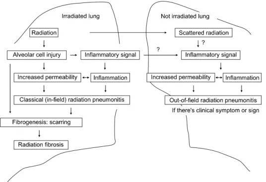
Fig. 1. Schematic mechanism of radiation pneumonitis.
리학적인 기전이 명확하게 밝혀져 있지 않은 임상 증후군 으로 간주되고 있으며, 이를 예측하고 예방할 수 있는 효과 적인 방법이 확립되어 있지 않다. 최근 방사선에 의한 폐 독성에 대한 많은 연구들이 진행되었으며, 이를 검토하여 방사선에 의한 폐 독성의 임상 경과, 기전, 예측 및 예방 방 법 등에 대한 최신 지견을 고찰하고자 한다.
임상 경과
방사선폐렴은 평가하기가 쉽지 않은 임상적인 증후군으 로 방사선치료 후 12주 이내에 주로 발생하며, 길게는 6개 월까지 발생할 수 있다. 일반적으로 마른기침과 호흡곤란 을 주 증상으로 하며 미열이 발생할 수도 있으며, 다양한 호흡기 증상이 발생할 수 있다. 심박동수가 증가할 수 있지 만, 방사선 폐렴의 특징적인 증상은 아니며, 항상 동반되지 도 않는다. 폐 기능이 감소할 수 있으나 방사선 폐렴이 발생 하였다고 항상 폐 기능 검사에서 폐 기능의 감소가 관찰되 는 것은 아니다(6). 흉부단층촬영 소견은 증상의 정도와 항 상 일치하지는 않으며, 컴퓨터 단층촬영을 이용하면 폐에 방사선을 조사 받은 환자의 50∼90%에서 증상의 유무에 관 계없이 방사선폐렴의 소견을 발견할 수 있다(7). 이론적으 로는 일정 용적이상의 정상 폐 조직에 한계 선량 이상의 방사선량이 조사되었다면, 방사선에 의한 정상 폐 조직의 변화는 필연적이며, 적정 시점에서 컴퓨터 단층촬영을 시 행한다면 모든 예에서 방사선폐렴의 소견을 관찰할 수 있
을 것이다.
통상적으로 방사선치료 범위 내에서 발생하는 조사영역 안 방사선폐렴(in-field radiation pneumonitis)과 방사선치료 범위 밖에서 발생하는 조사영역 밖 방사선폐렴(out-of-field radiation pneumonitis)으로 구분하기도 하며, 전자는 방사선 치료 후 쉽게 관찰할 수 있는 전통적 방사선폐렴(classical radiation pneumonitis)으로 부르기도 하며, 후자는 드물게 발 생하는 산발성 방사선폐렴(sporadic radiation pneumonitis)으 로 부르기도 한다. 산발성 폐렴의 개념은 Morgan과 Beit가 림프구가 전체 폐를 침윤하는 림르구과다증으로 처음 제시 하였으며(8), 방사선이 조사된 반대쪽 폐에서는 과민성 반 응으로 림프구가 증가하는 폐렴이 발생한다는 주장도 보고 되어 있다(9). 이러한 과정을 Fig. 1과 같이 요약할 수 있으 며, 임상적으로 방사선조사 범위 밖에서 발생한 방사선폐 렴의 개연성은 인정되지만, 아직까지 모든 사람들이 이 개 념에 동의하고 있지는 않다(10).
최근에는 3차원 입체조형 방사선치료(3-D conformal radi- ation therapy), 세기조절방사선치료(intensity modulated radiation therapy) 등의 방사선량 분포가 복잡하고 정교한 치료 방법 을 사용하기 때문에 2차원적인 방사선치료가 시행된 경우 와 같이 흉부 단층촬영에서 조사면 내인지 아닌지를 판단 하기가 점점 더 어려워지고 있으며, 경우에 따라서는 컴퓨 터 단층촬영 소견에서도 구분하기 쉽지 않다. 컴퓨터 촬영 등의 영상기술의 발전으로 과거에는 모르고 지나갔던 폐 조직의 염증성 병변들이 비교적 쉽게 관찰되기 때문에, 호
흡기 증상이 동반되지 않는 염증 소견도 흔히 볼 수 있다.
거기에 조사영역 밖 방사선폐렴을 인정한다고 하면 방사선 폐렴의 개념은 그 경계가 명확하지 않으며 임상적인 증후 군으로서 명확한 정의를 내리기도 쉽지 않다.
폐 섬유화는 방사선이 조사된 폐 조직에서 수 개월에서 수년에 걸쳐 서서히 진행하며, 통상적으로 1∼2년 후에는 안정화 되는 것으로 알려져 있다(11). 폐 섬유화가 진행된 폐 조직은 동정맥 문합에 의해 산소 교환이 일어나지 않기 때문에 폐 기능의 저하를 초래하게 되며, 그 정도에 따라 다양한 호흡곤란 및 호흡부전 등이 발생한다(10).
방사선 폐 독성의 빈도는 보고에 따라 다른데, 그 이유는 방사선 폐 독성의 정의와 같은 보고 방법 상의 차이, 치료 방법의 차이 등으로부터 기인한다(10). 방사선 폐 독성은 보통 RTOG (Radiation Therapy Oncology Group)의 독성 기 준이나 NCI-CTC (National Cancer Institute-Common Toxicity Criteria)의 독성 기준에 따라 보고되는데, RTOG 독성 기준 에 따르면 증상, 방사선촬영 상의 소견, 스테로이드 등의 치 료방법의 사용 여부 등에 따라 등급을 나누고 있으며, NCI-CTC도 비슷한 기준으로 분류하지만 각 등급의 정의에 는 차이가 있다(12,13). 그 외에도 다양한 독성 분류가 다양 한 연구진에 의해 사용되고 있다(6).
폐에서의 가스 교환을 평가하는 FEV1 (forced expiratory volume in 1 second), FVC (forced vital capacity), DLCO (carbon monoxide diffusing capacity) 검사 등을 방사선폐렴의 평가 를 위해 사용하고자 많은 연구가 진행되었으나, 전술하였 듯이 방사선 폐 독성에서 폐 기능 검사 소견의 변화가 항상 동반되지 않은 다는 점에서 제한적이다. 방사선치료 후의 폐 관류스캔의 변화와의 상관관계에 대한 연구에서도 DLCO나 FEV1 모두 만족할 정도의 상관관계를 보여주지는 못하였다(14,15). 따라서 전술한 모든 방사선 폐 독성에 대 한 독성 기준에서는 폐 기능의 변화를 그 정의에 포함하고 있지 않다(6). 6분 걷기 검사가 일반적인 폐 기능 검사에 비 해 방사선 폐렴을 평가하는데 더 유용하다는 주장도 있다 (16).
이러한 증상과 임상 경과만으로 방사선 폐 독성을 정의 하고 분류하는 것이 널리 사용되고 있지만, 방사선에 의한 폐 독성의 발생과 폐암 자체의 진행을 구별하기는 쉽지 않 으며, 환자가 심폐 기능이 저하되어 있을 때는 방사선 폐렴 에 의한 증상인지를 구별할 수 없어 증상을 기준으로 한 방사선에 의한 폐 독성의 분류도 간단하지만은 않으며, 보 고마다 그 발생 빈도에서 차이를 보이게 된다. 또한 방사선 폐렴 같은 경우는 증상 또는 단순 흉부 촬영 상의 염증소견 이 일시적으로 나타날 수 있기 때문에 얼마나 자주 추적
관찰하는가에 따라서도 그 빈도가 다를 수 있다. 또한 방사 선치료의 조사 기술에 따라 정상 폐 조직에 조사되는 방사 선량이 달라지며, 그에 따라 방사선 폐 독성은 차이를 보일 수 있으며, 총 방사선량, 피폭 폐 용적, 분할조사방법, 항암 화학약품 요법의 병용 여부 등도 방사선 폐렴의 발생에 영 향을 미치는 것으로 알려져 있다(17∼20). 그러나 대부분의 연구에서 증상이 동반되는 중간 정도 또는 심한 정도의 방 사선폐렴의 발생 빈도는 10∼20%로 보고하고 있다(21). 방 사선치료 전의 폐 질환 여부 또는 폐 기능의 정도는 증상을 동반하여 스테로이드 치료가 필요한 방사선 폐렴의 발생을 증가시키며(22), 원인을 정확히 알 수는 없지만 원위부의 폐 조직에서 방사선 폐렴의 발생 위험이 상대적으로 높다 고 보고되고 있다(23).
방사선 폐렴에 대한 치료는 대부분의 경우 일반적인 증 상에 따른 처방으로 충분하며, 기침이 심하거나 호흡곤란 이 발생하는 경우 스테로이드 처방으로 쉽게 증상이 호전 되는 것으로 알려져 있다. 스테로이드 처방은 prednisolone 30∼60 mg/day 또는 1 mg/kg/day의 용량이 일반적으로 사용 되고 있으며, 이보다 적은 용량에서도 효과를 기대할 수 있 다. 또한 조사영역 밖 방사선폐렴(out-of-field radiation pneumonitis)은 특별한 치료 없이도, 부작용 없이 완쾌된다 는 것이 일반적인 정설이다.
그러나 심한 정도의 방사선 폐렴이 발생한 경우 경한 정 도의 방사선폐렴이 발생하거나 방사선폐렴이 발생하지 않 은 경우에 비해 생존율이 감소한다는 보고도 있으며(24), 심각한 방사선 폐렴이 발생하는 경우 사망률이 50%까지 달 한다는 보고도 있다(25).
또한 최근 폐암의 치료에서 널리 사용되고 있는 방사선 치료와 항암약물치료의 병용은 cisplatin, paclitaxel, gemcitabine 등 다양한 약제에서 폐 독성을 증가시킬 수 있는 요인으로 제시되고 있으나(17,26∼29), 그렇지 않다는 연구 결과도 보 고되어 있다(30∼32). 동시에 화학방사선치료를 시행하는 것이 순차적으로 화학방사선치료를 시행하는 것에 비해 방 사선폐렴의 위험성이 높아진다는 보고가 있으나(29), 이 역 시 그렇지 않다는 연구 결과도 있다(32). 유방암 환자를 대 상으로 한 연구에서도 paclitaxel을 병용하면 방사선 폐렴의 빈도가 35%까지 증가하였다고 보고하고 있다(18). 화학방 사선치료가 방사선치료 단독에 비해 방사선폐렴의 발생 빈 도 및 임상경과에 영향을 미칠 가능성이 높고, 최근 폐암의 치료에서 방사선치료 단독보다는 화학방사선치료의 사용 이 증가하는 만큼 화학방사선치료가 방사선치료 단독에 비 해 방사선 폐렴의 발생 양상에 어떤 영향을 미치는지에 대 한 더 많은 연구가 필요할 것이다.
Fig. 2. Histopatholgic findings of radiation-induced lung damage. Histopathologic findings 7 weeks after 54 Gy of radiation. (A) Diffuse alveolar damage and edema, (B) Pneumocyte proliferation after radiation damage.
기 전
일반적으로 방사선폐렴은 방사선치료 후 보통 12주 이내 에 발생하는 염증반응으로 폐포 내 세포 손상에 따른 염증 세포들이 폐포 간극으로 집결하는 양상을 보인다. 이 후 만 성기에 들어가면서 섬유아세포가 증식하며, 교원질을 생성 하며, 정상 폐의 구조가 변형되는 방사선 폐 섬유화라는 비 가역적인 상태에 이르게 된다(8). 이러한 일련의 방사선에 의한 폐 손상의 진행 과정은 주요 과정이 방사선에 의한 폐 조직의 손상에 대한 일반적인 조직의 반응이기 때문에 다른 폐 질환과 비교할 때 그 기전에서 명백한 특이성을 보여주지 않으며, 또한 상당히 다양한 양상으로 진행되기 때문에 그 병리생리학적 기전을 단순하게 설명하기 어렵다.
폐포를 구성하는 상피세포는 1형 폐포세포와 2형 폐포세 포로 구분할 수 있는데, 1형 폐포세포는 폐포의 대부분을 덥고 있으며, 2형 폐포세포는 폐포 내의 표면장력을 유지하 기 위하여 표면활성제를 분비하는 역할을 한다. 방사선이 조사되어 폐 조직의 손상이 발생하면 1형 폐포세포가 우선 적으로 영향을 받아 아포토시스 등을 일으키며, 2형 폐포세 포는 1형 세포의 전구세포로 작용하며 이에 따라 증식이 가속되며, 폐포 내 상피층을 복구한다. 이러한 2형 폐포세 포의 증식은 폐포 손상과 복구를 의미하며, 방사선 조사 후 약 2∼6주 후에 폐포 내에 표면활성제의 생성이 증가되는 것이 알려져 있다(11,33). 1형 폐포세포는 모세혈관과 짝을 이루어 산소 교환을 하게 되는데, 이 모세혈관의 내피세포 또한 방사선에 의해 손상을 받으며, 이에 따라 모세혈관과
폐포 간의 투과성이 증가하게 되어 단백질과 같은 고분자 물질을 포함한 삼출액이 폐포 안으로 분비된다(10).
그와 동시에 폐포의 손상에 따라 활성화된 폐포 내 대식 세포는 다양한 세포활성물질과 염증 전달물질을 분비하여 림프구와 호중구를 불러모으며, 폐포 벽의 섬유아세포 등 에도 작용하여, 염증 반응과 섬유화 과정에 관여한다. 폐포 벽의 섬유아세포는 활성화되어 콜라겐 I, III, IV와 화이브로 넥틴(fibronectin) 등을 생성하여 폐 섬유화가 진행된다(34∼
37).
조직병리학적 소견은 방사선치료 후 폐 조직을 절제하거 나 검시를 시행한 예가 적어 많은 보고가 없으며, 대부분이 동물 실험 결과이다. 수술 전 방사선치료 후 외과적 절제를 시행 받은 환자들의 정상 폐 조직의 방사선에 의한 손상의 조직병리학적인 소견을 살펴보면 폐 조직에 만연된 손상과 부종 소견이 보이며 폐포세포의 증식 소견이 관찰되며, 이 러한 소견은 방사선에 의한 폐 손상의 특징적인 소견이라 기 보다는 폐 조직의 손상에 따른 비 특이적인 소견이라 할 수 있다(Fig. 2) 동물실험에서의 소견을 보면 방사선 조 사 후 초기에는 전체적인 폐포 손상이 관찰되는데, 혈관 투 과율이 증가하여 단백질 등이 폐포 내로 삼출되며 폐포 벽 의 부종이 발생한다(38). 실험모델에서는 염증세포들이 폐 포 내에서 관찰되지만, 수일 후에 급격히 사라지며, 1형 폐 포세포가 소실되며, 동시에 2형 폐포세포가 증식하는 현상 이 관찰된다(39). 섬유아세포가 증식하면서 콜라겐이 폐포 간 조직과 폐포 내에 증가하는 것은 가장 대표적인 조직병 리학적 소견이며, 보통 수주 이내에 폐포 간극이 두꺼워지 며, 폐 섬유화가 명확해진다(11).
또한 방사선 치료 후에는 유전자 발현의 질적, 양적인 변 화가 발생한다(40∼43). 이러한 유전자 변화가 방사선에 의 한 직접적인 변화인지, 세포 손상에 따른 이차적인 반응인 지를 명확하게 구별할 수는 없지만, 이러한 유전자 발현의 변화로 인해 다양한 세포 활성물질과 성장 인자들이 활성 화 되며, 결국 조직병리학적인 변화를 초래하고 임상 증후 군으로 발전하는 것이다.
방사선에 의한 폐 독성에 관여하는 가장 대표적인 세포 활성물질인 TGF-β는 섬유화 과정에 관여하는 주요 물질 로 알려져 있다(44∼51). TGF-β는 콜라겐 합성을 강하게 촉진하는데, 방사선 조사 후 TGF-β 유전자 발현이 증가하 며, 이와 동시에 섬유아세포의 콜라겐과 화이브로넥틴 합 성 유전자가 발현되며 섬유화가 진행된다(52∼54). 그 외에 도 b-FGF, TNF-α, IL-1, IL-6, HIF (hypoxia inducing factor), PDGF, ICAM, i-NOS 등이 관여하는 것으로 보고되고 있다 (34,35,37,55∼61). 이 외에도 더 많은 세포활성물질, 성장 인자 또는 신호전달물질들이 관여할 것이지만, 폐포 손상 후에 나타나는 일반적인 과정과 다르지 않을 것이며, 방사 선에 의한 폐 손상에 특이한 소견은 아니다. 그러나 이들 인자들을 이용하여 폐 손상을 예측하거나 예방 또는 치료 하고자 하는 많은 연구가 시행되고 있다.
예측모델
방사선폐렴 특히 중등도 이상의 심한 방사선 폐렴의 발 생 위험이 높은 환자를 미리 선별할 수 있다면 이들 환자에 서는 정상 폐 조직에 조사되는 방사선량을 상대적으로 최 소화하고, 방사선방어치료의 병합 등을 고려할 수 있을 것 이며, 그에 따라 심각한 방사선폐렴의 빈도를 낮추거나 방 사선량의 증량을 기할 수 있을 것이다. 따라서 방사선 폐렴 의 발생을 예측할 수 있는 인자에 대한 많은 연구가 있어왔 으며, 대표적으로 선량체적히스토그람(dose-volume histogram, DVH), TGF-beta 등에 대한 연구가 있으나(2), 아직까지 명 확한 방법이 정립되어 있지는 않다.
방사선치료 후에 발생하는 방사선 폐 독성은 당연히 정 상 폐 조직에 조사된 방사선량의 분포에 영향을 받는다. 방 사선치료로 인해 정상 폐 조직에 조사되는 방사선량은 지 금까지 알려진 예측인자 중에서 가장 일관되게 방사선 폐 렴을 예측할 수 있는 인자이며, 일정 누적 선량이 조사되는 정상 폐 조직의 %용적으로 표현되는 선량체적히스토그람 으로 정상 폐에 조사되는 방사선량 분포를 표현 할 수 있다.
일정한 누적 방사선량이 이상이 조사된 정상 폐 조직의 용 적으로 정의되는 Vdose 값과 폐 용적당 평균 방사선량의 정
의되는 평균 폐 선량(mean lung dose, MLD) 값과 방사선폐 렴의 발생과의 상관관계에 대한 많은 연구가 있으며(62∼
66), 비소세포성 폐암 환자를 대상으로 3차원 입체조형방사 선치료를 시행한 전향적 연구에서는 V20 (20 Gy의 방사선 량이 조사된 정상 폐 %용적)이 18% 이상, V30 (30 Gy의 방 사선량이 조사된 정상 폐 %용적)이 13% 이상, V40 (40 Gy 의 방사선량이 조사된 정상 폐 %용적)이 10% 이상인 경우 에 방사선폐렴의 발생 위험이 증가한다고 보고하였다(65).
한국 환자를 대상으로 한 Kim 등(66)의 연구에서는 3도 이 상의 심각한 정도의 방사선폐렴의 발생은 단변량 분석상에 서는 평균 폐 선량, V20, V30, V40, V50, NTCP (normal tissue complication probability) 등과 상관관계가 높았으나, 다변량 분석에서는 평균 폐 선량 만이 유의한 상관관계를 보여주 어 평균 폐 선량을 심각한 정도의 방사선폐렴의 발생을 예 측할 수 있는 유의한 인자로 제시하였다. 또한 Lee 등(67)은 3기 비소세포성 폐암 환자에서 동시화학방사선치료를 시 행한 결과에서 방사선폐렴이 29%의 환자에서 발생하였으 며, 이들 환자에서 NTCP 값이 유의한 차이를 보인다고 보 고하였다. NTCP란 방사선량과 조직의 반응을 표현한 S 자 형 곡선에 따라 폐 독성의 발생 가능성을 추정하는 수학적 인 모델의 하나이다(68,69). 몇몇 연구들이 NTCP가 방사선 폐렴을 예측하는데 유용하다고 보고하고 있으나(67,70), 수 학적 모델의 기반이 되는 만큼 근거자료가 얼마나 정확하 냐에 따라 NTCP 값의 신뢰도 및 예측력이 높아질 수 있는 데, 아직까지는 근거 자료가 빈약하고, 임상적인 상황에 적 합한지에 대해서도 논란이 있다.
흉부 방사선치료의 경우 방사선치료가 종료된 후에도 혈 장 TGF-β1 수치가 지속적으로 증가되어 있으며, 이러한 비정상적으로 증가된 정도로 방사선폐렴의 발생 위험도를 예측하는데 활용할 수 있다(47,71). 그러나 폐암의 경우 폐 종양 자체에서 TGF-β1을 생성하는 것으로 알려져 있기 때 문에 방사선치료와 관계 없이 증가되어 있으며(72,73), 따라 서 TGF-β1만으로 방사선폐렴을 예측하는 것은 쉽지 않다.
그 외에도 혈액 내의 IL-6을 측정하여 방사선폐렴을 예측 하려는 시도도 있다(74).
예 방
방사선에 의한 폐 독성을 감소시키기 위한 방안은 크게 방사선치료 기법의 변화와 생물학적인 방사선방어제의 사 용으로 크게 나누어 볼 수 있다.
폐암의 방사선치료에 사용되는 분할조사법은 1.8내지 2 Gy의 방사선량을 하루에 한번 조사하는 통상분할조사법
(conventional fractionation)과 하루에 2회 이상 방사선을 조 사하며 일일 방사선량을 증가시키는 가속과분할조사법 (accelerated hyperfractionation)이 사용된다. 이러한 분할조사 법의 차이는 방사선폐렴의 발생에 영향을 줄 수 있으며, 이 론적으로 분할 조사법의 변화를 통해 방사선폐렴의 발생 빈도를 낮출 수 있다. CHART (continuous hyperfractionated accelerated radiation therapy) 연구에 따르면 가속과분할조사 법이 시행된 환자군에서 통사분할조사법이 시행된 환자군 에 비해 전체적인 방사선폐렴의 발생빈도는 높았지만, 치 료가 필요한 방사선 폐렴의 발생 빈도는 오히려 낮았으며, 영상학적인 변화가 관찰된 예도 CHART가 시행된 환자군 에서 상대적으로 낮았다(75). 그러나 아직까지 연구 결과가 충분하지 않으며, 가속분할조사법을 항암화학약물요법과 병행할 경우 심각한 정도의 방사선 폐렴 등의 폐 독성이 증가할 가능성이 있으므로 근거 중심으로 판단할 때 단정 적으로 권고하기는 어렵다.
방사선치료 기술의 측면에서 보면 3차원입체조형 방사 선치료를 시행함으로써 정상 폐 조직에 조사되는 방사선량 을 줄일 수 있고 그에 따라 방사선폐렴의 발생을 줄일 수 있을 것으로 예측되지만, 임상 연구 결과로는 명확하지 않 으며, 2도 이상의 방사선폐렴의 발생 빈도가 0∼37%로 보 고되고 있다(6). 3차원 입체조형 방사선치료를 시행한 비소 세포성 폐암 환자에서 컴퓨터 단층촬영과 폐기능검사를 통 해 전향적으로 방사선폐렴을 평가한 연구에 따르면 2도 이 상의 방사선 폐렴의 발생빈도가 37%로 기존의 방사선폐렴 의 빈도에 비해 다소 높은 듯이 보이지만(76) 검사 방법, 연 구 설계 등을 고려할 때 그렇게만 결론을 내릴 수는 없을 것이다.
또한 세기조절방사선치료는 3차원 입체조형 방사선치료 에서 발전한 방사선치료 기술로 정상 폐 조직에 조사되는 방사선량을 3차원 입체조형방사선치료에 비해 더 낮출 수 있으며 따라서 방사선 폐렴의 추정 위험도를 낮출 수 있다 는 보고가 있다(77). 이러한 것은 토모쎄라피에서도 마찬가 지로 정상 폐 조직에 조사되는 방사선량을 줄일 수 있다고 보고되고 있다(78). 양성자 치료나 카본이온 방사선치료 또 한 정상 폐 조직에 조사되는 방사선량을 줄일 수 있으며, 따라서 방사선 폐렴의 위험을 낮출 수 있을 것이다(79,80).
따라서 다양한 방사선치료 기술을 이용하여 최대한 방사선 폐렴의 위험도를 낮추기 위해 노력해야 할 것이다. 심폐 기 능의 저하 등으로 수술이 불가능한 초기 폐암 환자를 대상 으로 한 정위 방사선치료에 대한 보고에 따르면 중앙에 위 치한 기관 및 기관지 주변 병변일 경우 심각한 폐 독성이 발생하였다고 보고되어 있기 때문에, 치료 전 폐 기능이 저
하되어 있거나, 분할선량이 큰 경우에는 더욱 정상 폐 조직 에 조사되는 방사선량을 줄이도록 노력하여야 할 것이다 (81).
또 한가지는 생물학적인 약제를 이용하는 방법으로 ami- fostine과 같은 항산화물질을 이용하는 것이 현재 임상에서 사용할 수 있는 가장 대표적인 방법이라 할 수 있다. Ami- fostine은 아미노치올(aminothiol) 합성물로서 자유래디칼을 제거함으로써 그 효과를 나타내며, 임상적으로도 그 효과 를 입증하는 보고가 있다(82∼84). 그러나 3상 비교 임상연 구에서 효과가 없다는 보고가 있는 만큼 그 효과는 미지수 이다(85). Captopril 또한 치올(thiol)기를 포함하고 있는 ACE (angiotensin converting enzyme)약제로 방사선폐렴을 억제할 가능성이 있으나, 증상을 동반한 방사선폐렴의 발생 빈도 를 낮추지는 못하는 것으로 알려지고 있다(86).
TGF-β를 억제하는 소분자 물질을 이용하여 방사선에 의한 폐 손상을 방지하려는 시도도 있으며(87), Pentoxyfillin 의 경우 Smad 경로를 간섭하여 TGF-β를 하향 조절함으로 써 섬유화를 억제하는 효과를 기대할 수 있다(88). 그 외에 도 TGF-β를 억제하는 물질이라면 섬유화를 제어할 수 있 는 목표로서의 가능성이 있을 것이다. 방사선 폐 섬유화가 ICAM-1을 제거한 쥐에서 발생하지 않았다는 보고도 있다 (89).
세포 외 superoxide dismutase를 유전자치료를 통해 과발 현 시킴으로써 방사선에 의한 ROS (reactive oxygen species) 를 감소시켜, 방사선에 의한 직접적인 폐 손상으로부터 폐 를 보호하려는 시도도 있으며(90) 멜라토닌 또한 방사선방 어제로 알려져 있다(91).
아직까지 임상에서 방사선에 의한 폐 독성을 예방할 수 있는 효과적인 생물학적 제제들이 알려져 있지 않지만 분 자생물학의 발전에 힘입어 다양한 치료제가 등장할 것으로 기대된다.
결 론
폐암의 치료에서 방사선치료는 매우 중요한 역할을 담당 하고 있으며, 방사선에 의한 폐 독성은 생존율이나 삶의 질 측면에서 매우 중요하다. 정상 폐 조직에 조사되는 방사선 량을 줄일 수 있는 다양한 방사선치료 기술이 개발되어 사 용되고 있으며, 특히 치료 전 폐 기능이 좋지 않은 경우에는 정상 폐 조직의 방사선량을 최소화할 수 있는 노력이 필요 하다. 방사선 폐 독성에 대한 병리생리학적인 기전을 정확 하게 이해하고, 이에 따라 방사선 치료 전에 폐 독성을 예측 하고 예방할 수 있는 방법들을 모색하는 더욱 많은 연구가
필요하다.
REFERENCES
1. Lingos TI, Recht A, Vicini F, Abner A, Silver B, Harris JR.
Radiation pneumonitis in breast cancer patients treated with conservative surgery and radiation therapy. Int J Radiat Oncol Biol Phys 1991;21:355-360.
2. Marks LB, Munley MT, Bentel GC, et al. Physical and biological predictors of changes in whole-lung function follow- ing thoracic irradiation. Int J Radiat Oncol Biol Phys 1997;
39:563-570.
3. Morgan GW, Freeman AP, McLean RG, Jarvie BH, Giles RW.
Late cardiac, thyroid, and pulmonary sequelae of mantle radiotherapy for Hodgkin's disease. Int J Radiat Oncol Biol Phys 1985;11:1925-1931.
4. Penn CR, Hope-Stone HF. The role of radiotherapy in the management of malignant thymoma. Br J Surg 1972;59:533- 539.
5. Postoperative radiotherapy in non-small-cell lung cancer:
systematic review and meta-analysis of individual patient data from nine randomised controlled trials. PORT Meta-analysis Trialists Group. Lancet 1998;352:257-263.
6. Mehta V. Radiation pneumonitis and pulmonary fibrosis in non-small-cell lung cancer: pulmonary function, prediction, and prevention. Int J Radiat Oncol Biol Phys 2005;63:5-24.
7. Mah K, Van Dyk J, Keane T, Poon PY. Acute radiation- induced pulmonary damage: a clinical study on the response to fractionated radiation therapy. Int J Radiat Oncol Biol Phys 1987;13:179-188.
8. Morgan GW, Breit SN. Radiation and the lung: a reevaluation of the mechanisms mediating pulmonary injury. Int J Radiat Oncol Biol Phys 1995;31:361-369.
9. Martin C, Romero S, Sanchez-Paya J, Massuti B, Arriero JM, Hernandez L. Bilateral lymphocytic alveolitis: a common reac- tion after unilateral thoracic irradiation. Eur Respir J 1999;
13:727-732.
10. Tsoutsou PG, Koukourakis MI. Radiation pneumonitis and fibrosis: mechanisms underlying its pathogenesis and implica- tions for future research. Int J Radiat Oncol Biol Phys 2006;
66:1281-1293.
11. McDonald S, Rubin P, Phillips TL, Marks LB. Injury to the lung from cancer therapy: clinical syndromes, measurable endpoints, and potential scoring systems. Int J Radiat Oncol Biol Phys 1995;31:1187-1203.
12. Cox JD, Stetz J, Pajak TF. Toxicity criteria of the Radiation Therapy Oncology Group (RTOG) and the European Organi- zation for Research and Treatment of Cancer (EORTC). Int J Radiat Oncol Biol Phys 1995;31:1341-1346.
13. Trotti A, Byhardt R, Stetz J, et al. Common toxicity criteria:
version 2.0. an improved reference for grading the acute effects of cancer treatment: impact on radiotherapy. Int J Radiat Oncol Biol Phys 2000;47:13-47.
14. Fan M, Marks LB, Hollis D, et al. Can we predict
radiation-induced changes in pulmonary function based on the sum of predicted regional dysfunction? J Clin Oncol 2001;19:
543-550.
15. Fan M, Marks LB, Lind P, et al. Relating radiation-induced regional lung injury to changes in pulmonary function tests.
Int J Radiat Oncol Biol Phys 2001;51:311-317.
16. Miller KL, Kocak Z, Kahn D, et al. Preliminary report of the 6-minute walk test as a predictor of radiation-induced pulmo- nary toxicity. Int J Radiat Oncol Biol Phys 2005;62:1009- 1013.
17. Reckzeh B, Merte H, Pfluger KH, Pfab R, Wolf M, Havemann K. Severe lymphocytopenia and interstitial pneumonia in patients treated with paclitaxel and simultaneous radiotherapy for non-small-cell lung cancer. J Clin Oncol 1996;14:1071- 1076.
18. Yu TK, Whitman GJ, Thames HD, et al. Clinically relevant pneumonitis after sequential paclitaxel-based chemotherapy and radiotherapy in breast cancer patients. J Natl Cancer Inst 2004;96:1676-1681.
19. Segawa Y, Takigawa N, Kataoka M, Takata I, Fujimoto N, Ueoka H. Risk factors for development of radiation pneu- monitis following radiation therapy with or without chemo- therapy for lung cancer. Int J Radiat Oncol Biol Phys 1997;
39:91-98.
20. Tsujino K, Hirota S, Kotani Y, et al. Radiation pneumonitis following concurrent accelerated hyperfractionated radiotherapy and chemotherapy for limited-stage small-cell lung cancer:
Dose-volume histogram analysis and comparison with conven- tional chemoradiation. Int J Radiat Oncol Biol Phys 2006;64:
1100-1105.
21. Roach M 3rd, Gandara DR, Yuo HS, et al. Radiation pneumonitis following combined modality therapy for lung cancer: analysis of prognostic factors. J Clin Oncol 1995;13:
2606-2612.
22. Lind PA, Marks LB, Hollis D, et al. Receiver operating characteristic curves to assess predictors of radiation-induced symptomatic lung injury. Int J Radiat Oncol Biol Phys 2002;
54:340-347.
23. Seppenwoolde Y, De Jaeger K, Boersma LJ, Belderbos JS, Lebesque JV. Regional differences in lung radiosensitivity after radiotherapy for non-small-cell lung cancer. Int J Radiat Oncol Biol Phys 2004;60:748-758.
24. Inoue A, Kunitoh H, Sekine I, Sumi M, Tokuuye K, Saijo N.
Radiation pneumonitis in lung cancer patients: a retrospective study of risk factors and the long-term prognosis. Int J Radiat Oncol Biol Phys 2001;49:649-655.
25. Wang JY, Chen KY, Wang JT, et al. Outcome and prognostic factors for patients with non-small-cell lung cancer and severe radiation pneumonitis. Int J Radiat Oncol Biol Phys 2002;
54:735-741.
26. Brooks BJ Jr, Seifter EJ, Walsh TE, et al. Pulmonary toxicity with combined modality therapy for limited stage small-cell lung cancer. J Clin Oncol 1986;4:200-209.
27. Lee JS, Scott C, Komaki R, et al. Concurrent chemoradiation therapy with oral etoposide and cisplatin for locally advanced
inoperable non-small-cell lung cancer: radiation therapy oncology group protocol 91-06. J Clin Oncol 1996;14:1055- 1064.
28. Byhardt RW, Scott C, Sause WT, et al. Response, toxicity, failure patterns, and survival in five Radiation Therapy Oncology Group (RTOG) trials of sequential and/or concurrent chemotherapy and radiotherapy for locally advanced non- small-cell carcinoma of the lung. Int J Radiat Oncol Biol Phys 1998;42:469-478.
29. Yamada M, Kudoh S, Hirata K, Nakajima T, Yoshikawa J.
Risk factors of pneumonitis following chemoradiotherapy for lung cancer. Eur J Cancer 1998;34:71-75.
30. Schaake-Koning C, van den Bogaert W, Dalesio O, et al.
Effects of concomitant cisplatin and radiotherapy on inoperable non-small-cell lung cancer. N Engl J Med 1992;326:524-530.
31. Jeremic B, Shibamoto Y, Acimovic L, Milisavljevic S.
Hyperfractionated radiation therapy with or without concurrent low-dose daily carboplatin/etoposide for stage III non-small-cell lung cancer: a randomized study. J Clin Oncol 1996;14:
1065-1070.
32. Robnett TJ, Machtay M, Vines EF, McKenna MG, Algazy KM, McKenna WG. Factors predicting severe radiation pneu- monitis in patients receiving definitive chemoradiation for lung cancer. Int J Radiat Oncol Biol Phys 2000;48:89-94.
33. Rubin P, Siemann DW, Shapiro DL, Finkelstein JN, Penney DP. Surfactant release as an early measure of radiation pneu- monitis. Int J Radiat Oncol Biol Phys 1983;9:1669-1673.
34. Rubin P, Johnston CJ, Williams JP, McDonald S, Finkelstein JN. A perpetual cascade of cytokines postirradiation leads to pulmonary fibrosis. Int J Radiat Oncol Biol Phys 1995;
33:99-109.
35. Chen Y, Williams J, Ding I, et al. Radiation pneumonitis and early circulatory cytokine markers. Semin Radiat Oncol 2002;
12:26-33.
36. Ibuki Y, Goto R. Enhancement of NO production from resident peritoneal macrophages by in vitro gamma-irradiation and its relationship to reactive oxygen intermediates. Free Radic Biol Med 1997;22:1029-1035.
37. Hong JH, Jung SM, Tsao TC, et al. Bronchoalveolar lavage and interstitial cells have different roles in radiation-induced lung injury. Int J Radiat Biol 2003;79:159-167.
38. Gross NJ. Radiation pneumonitis in mice. Some effects of corticosteroids on mortality and pulmonary physiology. J Clin Invest 1980;66:504-510.
39. Abdollahi A, Li M, Ping G, et al. Inhibition of platelet-derived growth factor signaling attenuates pulmonary fibrosis. J Exp Med 2005;201:925-935.
40. Boerma M, van der Wees CG, Vrieling H, et al. Microarray analysis of gene expression profiles of cardiac myocytes and fibroblasts after mechanical stress, ionising or ultraviolet radiation. BMC Genomics 2005;6:6.
41. Boerma M, Wang J, Wondergem J, et al. Influence of mast cells on structural and functional manifestations of radiation- induced heart disease. Cancer Res 2005;65:3100-3107.
42. Christiansen H, Batusic D, Saile B, et al. Identification of
genes responsive to gamma radiation in rat hepatocytes and rat liver by cDNA array gene expression analysis. Radiat Res 2006;165:318-325.
43. Christiansen H, Sheikh N, Saile B, et al. x-Irradiation in rat liver: consequent upregulation of hepcidin and downregulation of hemojuvelin and ferroportin-1 gene expression. Radiology 2007;242:189-197.
44. Anscher MS, Peters WP, Reisenbichler H, Petros WP, Jirtle RL. Transforming growth factor beta as a predictor of liver and lung fibrosis after autologous bone marrow transplantation for advanced breast cancer. N Engl J Med 1993;328:1592- 1598.
45. Anscher MS, Marks LB, Shafman TD, et al. Using plasma transforming growth factor beta-1 during radiotherapy to select patients for dose escalation. J Clin Oncol 2001;19:3758-3765.
46. De Jaeger K, Seppenwoolde Y, Kampinga HH, Boersma LJ, Belderbos JS, Lebesque JV. Significance of plasma transfor- ming growth factor-beta levels in radiotherapy for non- small-cell lung cancer. Int J Radiat Oncol Biol Phys 2004;58:
1378-1387.
47. Anscher MS, Kong FM, Andrews K, et al. Plasma trans- forming growth factor beta1 as a predictor of radiation pneu- monitis. Int J Radiat Oncol Biol Phys 1998;41:1029-1035.
48. Anscher MS, Kong FM, Marks LB, Bentel GC, Jirtle RL.
Changes in plasma transforming growth factor beta during radiotherapy and the risk of symptomatic radiation-induced pneumonitis. Int J Radiat Oncol Biol Phys 1997;37:253-258.
49. Blobe GC, Schiemann WP, Lodish HF. Role of transforming growth factor beta in human disease. N Engl J Med 2000;
342:1350-1358.
50. Burger A, Loffler H, Bamberg M, Rodemann HP. Molecular and cellular basis of radiation fibrosis. Int J Radiat Biol 1998;
73:401-408.
51. Vujaskovic Z, Groen HJ. TGF-beta, radiation-induced pulmo- nary injury and lung cancer. Int J Radiat Biol 2000;76:511- 516.
52. Fine A, Goldstein RH. The effect of transforming growth factor-beta on cell proliferation and collagen formation by lung fibroblasts. J Biol Chem 1987;262:3897-3902.
53. Finkelstein JN, Johnston CJ, Baggs R, Rubin P. Early alterations in extracellular matrix and transforming growth factor beta gene expression in mouse lung indicative of late radiation fibrosis. Int J Radiat Oncol Biol Phys 1994;28:
621-631.
54. Rube CE, Uthe D, Schmid KW, et al. Dose-dependent induction of transforming growth factor beta (TGF-beta) in the lung tissue of fibrosis-prone mice after thoracic irradiation. Int J Radiat Oncol Biol Phys 2000;47:1033-1042.
55. Girinsky T. Effects of ionizing radiation on the blood vessel wall. J Mal Vasc 2000;25:321-324.
56. Gridley DS, Bonnet RB, Bush DA, et al. Time course of serum cytokines in patients receiving proton or combined photon/proton beam radiation for resectable but medically inoperable non-small-cell lung cancer. Int J Radiat Oncol Biol Phys 2004;60:759-766.
57. Vujaskovic Z, Anscher MS, Feng QF, et al. Radiation-induced hypoxia may perpetuate late normal tissue injury. Int J Radiat Oncol Biol Phys 2001;50:851-855.
58. Johnston CJ, Piedboeuf B, Rubin P, Williams JP, Baggs R, Finkelstein JN. Early and persistent alterations in the expression of interleukin-1 alpha, interleukin-1 beta and tumor necrosis factor alpha mRNA levels in fibrosis-resistant and sensitive mice after thoracic irradiation. Radiat Res 1996;145:762-767.
59. Hallahan DE, Virudachalam S. Ionizing radiation mediates expression of cell adhesion molecules in distinct histological patterns within the lung. Cancer Res 1997;57:2096-2099.
60. Nozaki Y, Hasegawa Y, Takeuchi A, et al. Nitric oxide as an inflammatory mediator of radiation pneumonitis in rats. Am J Physiol 1997;272:L651-658.
61. Giaid A, Lehnert SM, Chehayeb B, Chehayeb D, Kaplan I, Shenouda G. Inducible nitric oxide synthase and nitrotyrosine in mice with radiation-induced lung damage. Am J Clin Oncol 2003;26:e67-72.
62. Graham MV, Purdy JA, Emami B, et al. Clinical dose-volume histogram analysis for pneumonitis after 3D treatment for non-small cell lung cancer (NSCLC). Int J Radiat Oncol Biol Phys 1999;45:323-329.
63. Jenkins P, D'Amico K, Benstead K, Elyan S. Radiation pneumonitis following treatment of non-small-cell lung cancer with continuous hyperfractionated accelerated radiotherapy (CHART). Int J Radiat Oncol Biol Phys 2003;56:360-366.
64. Tsujino K, Hirota S, Endo M, et al. Predictive value of dose-volume histogram parameters for predicting radiation pneumonitis after concurrent chemoradiation for lung cancer.
Int J Radiat Oncol Biol Phys 2003;55:110-115.
65. Claude L, Perol D, Ginestet C, et al. A prospective study on radiation pneumonitis following conformal radiation therapy in non-small-cell lung cancer: clinical and dosimetric factors analysis. Radiother Oncol 2004;71:175-181.
66. Kim TH, Cho KH, Pyo HR, et al. Dose-volumetric parameters for predicting severe radiation pneumonitis after three-dimen- sional conformal radiation therapy for lung cancer. Radiology 2005;235:208-215.
67. Lee SW, Choi EK, Lee JS, et al. Phase II study of three-dimensional conformal radiotherapy and concurrent mitomycin-C, vinblastine, and cisplatin chemotherapy for Stage III locally advanced, unresectable, non-small-cell lung cancer. Int J Radiat Oncol Biol Phys 2003;56:996-1004.
68. Kutcher GJ, Burman C. Calculation of complication probability factors for non-uniform normal tissue irradiation: the effective volume method. Int J Radiat Oncol Biol Phys 1989;16:1623- 1630.
69. Lyman JT, Wolbarst AB. Optimization of radiation therapy, III: A method of assessing complication probabilities from dose-volume histograms. Int J Radiat Oncol Biol Phys 1987;
13:103-109.
70. Martel MK, Ten Haken RK, Hazuka MB, Turrisi AT, Fraass BA, Lichter AS. Dose-volume histogram and 3-D treatment planning evaluation of patients with pneumonitis. Int J Radiat
Oncol Biol Phys 1994;28:575-581.
71. Fu XL, Huang H, Bentel G, et al. Predicting the risk of symptomatic radiation-induced lung injury using both the physical and biologic parameters V(30) and transforming growth factor beta. Int J Radiat Oncol Biol Phys 2001;50:
899-908.
72. Kong F, Jirtle RL, Huang DH, Clough RW, Anscher MS.
Plasma transforming growth factor-beta1 level before radio- therapy correlates with long term outcome of patients with lung carcinoma. Cancer 1999;86:1712-1719.
73. Kong FM, Washington MK, Jirtle RL, Anscher MS. Plasma transforming growth factor-beta 1 reflects disease status in patients with lung cancer after radiotherapy: a possible tumor marker. Lung Cancer 1996;16:47-59.
74. Chen Y, Rubin P, Williams J, Hernady E, Smudzin T, Oku- nieff P. Circulating IL-6 as a predictor of radiation pneumo- nitis. Int J Radiat Oncol Biol Phys 2001;49:641-648.
75. Saunders M, Dische S, Barrett A, Harvey A, Gibson D, Parmar M. Continuous hyperfractionated accelerated radiotherapy (CHART) versus conventional radiotherapy in non-small-cell lung cancer: a randomised multicentre trial. CHART Steering Committee. Lancet 1997;350:161-165.
76. Sunyach MP, Falchero L, Pommier P, et al. Prospective evaluation of early lung toxicity following three-dimensional conformal radiation therapy in non-small-cell lung cancer:
preliminary results. Int J Radiat Oncol Biol Phys 2000;48:
459-463.
77. Murshed H, Liu HH, Liao Z, et al. Dose and volume reduction for normal lung using intensity-modulated radiotherapy for advanced-stage non-small-cell lung cancer. Int J Radiat Oncol Biol Phys 2004;58:1258-1267.
78. Scrimger RA, Tome WA, Olivera GH, Reckwerdt PJ, Mehta MP, Fowler JF. Reduction in radiation dose to lung and other normal tissues using helical tomotherapy to treat lung cancer, in comparison to conventional field arrangements. Am J Clin Oncol 2003;26:70-78.
79. Bonnet RB, Bush D, Cheek GA, et al. Effects of proton and combined proton/photon beam radiation on pulmonary function in patients with resectable but medically inoperable non-small cell lung cancer. Chest 2001;120:1803-1810.
80. Kadono K, Homma T, Kamahara K, et al. Effect of heavy-ion radiotherapy on pulmonary function in stage I non-small cell lung cancer patients. Chest 2002;122:1925-1932.
81. Timmerman R, McGarry R, Yiannoutsos C, et al. Excessive toxicity when treating central tumors in a phase II study of stereotactic body radiation therapy for medically inoperable early-stage lung cancer. J Clin Oncol 2006;24:4833-4839.
82. Koukourakis MI. Amifostine in clinical oncology: current use and future applications. Anticancer Drugs 2002;13:181-209.
83. Komaki R, Lee JS, Milas L, et al. Effects of amifostine on acute toxicity from concurrent chemotherapy and radiotherapy for inoperable non-small-cell lung cancer: report of a rando- mized comparative trial. Int J Radiat Oncol Biol Phys 2004;
58:1369-1377.
84. Antonadou D, Petridis A, Synodinou M, et al. Amifostine
reduces radiochemotherapy-induced toxicities in patients with locally advanced non-small cell lung cancer. Semin Oncol 2003;30:2-9.
85. Movsas B, Scott C, Langer C, et al. Randomized trial of amifostine in locally advanced non-small-cell lung cancer patients receiving chemotherapy and hyperfractionated radia- tion: radiation therapy oncology group trial 98-01. J Clin Oncol 2005;23:2145-2154.
86. Wang LW, Fu XL, Clough R, et al. Can angiotensin- converting enzyme inhibitors protect against symptomatic radiation pneumonitis? Radiat Res 2000;153:405-410.
87. Anscher MS, Thrasher B, Zgonjanin L, et al. Small molecular inhibitor of transforming growth factor-beta protects against development of radiation-induced lung injury. Int J Radiat Oncol Biol Phys 2008;71:829-837.
88. Ozturk B, Egehan I, Atavci S, Kitapci M. Pentoxifylline in prevention of radiation-induced lung toxicity in patients with breast and lung cancer: a double-blind randomized trial. Int J Radiat Oncol Biol Phys 2004;58:213-219.
89. Hallahan DE, Geng L, Shyr Y. Effects of intercellular adhesion molecule 1 (ICAM-1) null mutation on radiation- induced pulmonary fibrosis and respiratory insufficiency in mice. J Natl Cancer Inst 2002;94:733-741.
90. Kang SK, Rabbani ZN, Folz RJ, et al. Overexpression of extracellular superoxide dismutase protects mice from radiation- induced lung injury. Int J Radiat Oncol Biol Phys 2003;57:
1056-1066.
91. Vijayalaxmi, Reiter RJ, Tan DX, Herman TS, Thomas CR Jr.
Melatonin as a radioprotective agent: a review. Int J Radiat Oncol Biol Phys 2004;59:639-653.