Received:February 21, 2017, Revised:April 6, 2017, Accepted:May 9, 2017
Corresponding to:Won-Seok Lee, Division of Rheumatology, Department of Internal Medicine, Chonbuk National University Medical School and Research Institute of Clinical Medicine of Chonbuk National University Hospital, 20 Geonji-ro, Deokjin-gu, Jeonju 54907, Korea.
E-mail:[email protected]
Copyright ⓒ 2017 by The Korean College of Rheumatology. All rights reserved.
This is a Open Access article, which permits unrestricted non-commerical use, distribution, and reproduction in any medium, provided the original work is properly cited.
Neutrophil-to-lymphocyte Ratio in Diagnosis of Systemic Sclerosis for Prediction of Interstitial Lung Disease
Ji-Hyeon Jung, Yu-Mi Lee, Eun-Gyeong Lee, Wan-Hee Yoo, Won-Seok Lee
Division of Rheumatology, Department of Internal Medicine, Chonbuk National University Medical School and Research Institute of Clinical Medicine of Chonbuk National University Hospital, Jeonju, Korea
Objective. The neutrophil-to-lymphocyte ratio (NLR) is elevated in inflammatory diseases, but its clinical significance in sys- temic sclerosis (SSc) is unclear. This study evaluated NLR in diagnosing SSc and in predicting lung involvement such as inter- stitial lung disease (ILD). Methods. The medical records of 88 patients with SSc and 50 healthy controls were reviewed.
Exclusion criteria included active infection or the presence of any hematological, cardiovascular, or metabolic disorder. The NLR was compared between patients with SSc and healthy controls, and associations between NLR and lung involvement were analyzed. Results. The NLR was significantly higher in patients with SSc compared to healthy controls (NLR, 3.95±6.59 vs.
2.00±1.07, p<0.01). Patients with SSc and ILD had higher NLR levels than those without ILD (p<0.01, p<0.05). NLR was negatively associated with forced vital capacity (r=−0.341, p<0.01), but not with diffusing capacity for carbon monoxide.
Receiver-operating characteristics analysis of NLR to predict ILD in patients with SSc showed that the area under the curve was 0.763. The cut-off NLR value for prediction of lung involvement was determined to be 2.59 (sensitivity, 0.700; specificity, 0.729; p<0.01). Conclusion. NLR may be a promising marker that reflects ILD in patients with SSc, and values greater than 2.59 were useful in predicting ILD. (J Rheum Dis 2017;24:138-142)
Key Words. Neutrophils, Lymphocytes, Blood platelets, Systemic sclerosis, Interstitial lung disease
INTRODUCTION
Systemic sclerosis (SSc) is an acquired chronic con- nective tissue disease characterized by progressive fib- rosis and thickening of the skin, vascular damage, and au- toantibodies with variable involvement of major organs such as the lungs. From a clinical standpoint, SSc is sub- divided into limited versus diffuse cutaneous disease based on the presence and extent of skin involvement [1].
Pulmonary disease is currently the leading cause of death in patients with SSc [2]. Pulmonary disease in pa- tients with SSc is divided into two broad categories: inter- stitial lung disease (ILD) and pulmonary hypertension.
ILD is one of the most common and important complica- tions of SSc. Moderate-to-severe pulmonary fibrosis is found in approximately 25% of SSc cases. These patients
may remain asymptomatic despite the presence of phys- ical findings such as crackles on auscultation or inter- stitial thickening on chest radiography.
Complete blood count is a standard part of modern labo- ratory evaluation in many clinical settings. Leukocyte counts and their classification are reported as inflammatory markers in chronic inflammatory disease [3,4]. NLR is the ratio of neutrophil-to-lymphocytes. Its stability is less influenced by physiological, pathological, and physical factors [5,6]. NLR also has a close correlation with other inflammation markers in patients with connective tissue disease, such as systemic lupus erythematosus (SLE) or Sjögren’s syndrome [7-10].
However, there is a lack of data in the literature about the association of NLR and platelet to lymphocyte ratio (PLR) with SSc. In this retrospective study, we analyzed
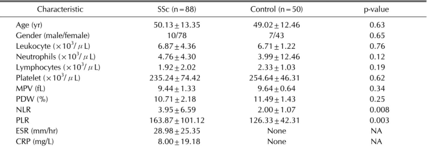
Table 1. Demographic and baseline clinical characteristics of patients with systemic sclerosis (SSc) and healthy groups
Characteristic SSc (n=88) Control (n=50) p-value
Age (yr) 50.13±13.35 49.02±12.46 0.63
Gender (male/female) 10/78 7/43 0.65
Leukocyte (×103/μL) 6.87±4.36 6.71±1.22 0.76
Neutrophils (×103/μL) 4.76±4.30 3.99±12.46 0.12
Lymphocytes (×103/μL) 1.92±2.02 2.33±1.03 0.19
Platelet (×103/μL) 235.24±74.42 254.64±46.31 0.62
MPV (fL) 9.44±1.33 9.64±0.64 0.34
PDW (%) 10.71±2.18 11.49±1.43 0.25
NLR 3.95±6.59 2.00±1.07 0.008
PLR 163.87±101.12 126.33±42.31 0.003
ESR (mm/hr) 28.98±25.35 None NA
CRP (mg/L) 8.00±19.18 None NA
Values are presented as the mean±standard deviation or number. MPV: mean platelet volume, PDW: platelet distribution width, NLR: neutrophil/lymphocyte ratio, PLR: platelet/lymphocyte ratio, ESR: erythrocyte sedimentation rate, CRP: C-reactive protein, NA: not available.
the medical records of 88 patients with SSc and 50 healthy individuals in an attempt to define a possible association between NLR, PLR, and SSc-ILD involvement.
MATERIALS AND METHODS
Participants
We retrospectively reviewed the medical records of all patients with SSc at our institution between February 2010 and May 2016. We diagnosed SSc according to the American College of Rheumatology 1980 classification for SSc or the presence of three out of five CREST criteria (calcinosis cutis, Raynaud phenomenon, esophageal dys- motility, sclerodactyly, and telangiectasias) [11]. A retro- spective chart review was conducted to determine the number of subjects that fulfilled the new 2013 SSc criteria [12]. We included only samples collected when SSc was diagnosed for the first time before any therapies were initiated. Significant ILD was defined as a forced vital ca- pacity (FVC) ≤80 and evidence of ILD on thoracic high-resolution computed tomography (HRCT) images.
Patients were excluded if they also had one of the follow- ing diseases/situations: 1) other autoimmune diseases such as rheumatoid arthritis, systemic lupus eryth- ematosus, or Sjögren’s syndrome; 2) malignancies; 3) end-stage renal disease; 4) hematology disease or admin- istration of blood transfusion during the past 4 months before enrollment; 5) infections within 4 weeks before enrollment; or 6) anemia.
Fifty healthy age- and gender-matched individuals who
underwent routine physical examination between February 2014 and March 2015 were enrolled as controls.
No significant underlying disease, including autoimmune disease, liver disease, diabetes, hypertension, hemato- logic disease, or malignancies, was detected on physical examination or laboratory tests including electrocardio- graphy, liver and renal function tests, lipid profiles, and serum glucose tests. Individuals who contracted an in- fectious disease, such as pneumonia or urinary tract in- fection, within two months before enrollment were excluded.
This study was conducted in accordance with the Declaration of Helsinki and approved by the ethics board of Chonbuk National University Hospital (IRB No.
2016-12-002).
Statistical analysis
All statistical analyses were performed using SPSS stat- istical software (IBM SPSS Statistics 19.0; IBM Co., Armonk, NY, USA). Continuous variables are presented as mean±standard deviation, and categorical variables are expressed as percentages. The Student’s t-test or Mann-Whiney U test was used to compare the two in- dependent groups according to distribution state. The chi-square test was used to compare proportions in differ- ent groups. Logistic regression analysis was also con- ducted to assess relationships. To evaluate the discrim- ination value of NLR for SSc and SSc-ILD, we evaluated the area under the curve using the receiver operating characteristic (ROC) curve. The p-values less than 0.05
Figure 1. Receiver operating characteristic (ROC) curve for the neutrophil/lymphocyte ratio (NLR) to predict interstitial lung disease (ILD) involvement. Receiver-operating character- istics analysis of NLR to predict ILD showed that the area under the curve was 0.763. The cutoff value using the ROC curve was 2.59 (sensitivity, 0.700, specificity, 0.729; p<0.001).
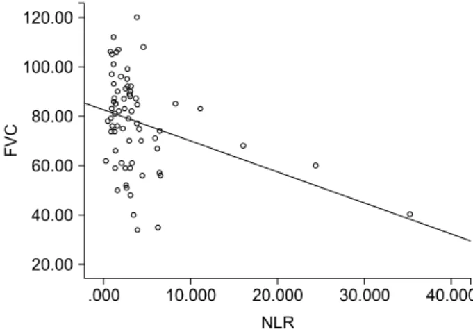
Figure 2. Correlation between neutrophil/lymphocyte ratio (NLR) and forced vital capacity (FVC). The NLR was negatively correlated with FVC, with a correlation coefficient of −0.341 (p=0.004).
Table 2. Comparison of the NLR and PLR in SSc-ILD and SSc without ILD
Variable SSc-ILD (n=40) SSc-non ILD (n=48) p-value
NLR 6.13±9.18 2.12±1.71 <0.005
PLR 189.57±126.86 142.46±67.40 0.048 Values are presented as the mean±standard deviation. NLR:
neutrophil/lymphocyte ratio, PLR: platelet/lymphocyte ratio, SSc: systemic sclerosis, ILD: interstitial lung disease.
were considered statistically significant.
RESULTS
Patient characteristics
Patient and control group demographics and clinical and laboratory characteristics are summarized in Table 1.
During the study period, there were 88 patients with SSc and 50 healthy subjects. The 88 patients with SSc in- cluded 78 women and 10 men whose mean age was 50.13±13.35 years. There was no significant difference between the two groups regarding age and sex distribution.
There was no significant difference in leukocytes, abso- lute neutrophil count, or lymphocytes.
NLR in patients with SSc vs. healthy individuals
NLR was significantly higher in patients with SSc than in healthy subjects (mean, 3.95±6.59 vs. 2.00±1.07) (Table 1). PLR also was significantly higher in patients with SScthan in healthy subjects (mean, 163.87±101.12 vs.
126.33±42.31).
Correlations of NLR with clinical characteristics in patients with SSc
At the first diagnosis, 30 patients with SSc also had ILD, and another 10 patients developed ILD during the fol- low-up period. Therefore, of the 88 patients with SSc, 40 had ILD at the time of data analysis. This study found that patients with SSc-ILD had a higher NLR than patients without ILD (mean, 6.13±9.18 vs. 2.12±1.71) (Table 2).
PLR was found to be statistically meaningful, but the stat- istical significance was low.
Predictability of NLR in the development of SSc with ILD
ROC analysis of NLR to predict SSc-ILD was performed.
The area under the ROC curve for NLR was 0.763 (95%
confidence interval), with an optimal cut-off value of 2.59, sensitivity of 70%, and specificity of 73% in differ- entiating between SSc-ILD and SSc without ILD (Figure 1). NLR was negatively correlated with FVC with a corre- lation coefficient of −0.341 (p=0.004) (Figure 2).
DISCUSSION
This retrospective study evaluated the predictive value of NLR in patients with SSc without ILD and patients with SSc-ILD. The present study, to our knowledge, is the first Korean report on the clinical significance of NLR in SSc. According to the data we obtained from this study,
NLR may be a predictive marker of ILD in patients with SSc, and values greater than 2.59 were useful in the pre- diction of ILD.
Markers of inflammation, such as NLR, and their clinical significance in patients with SSc are still under evaluation.
NLR is cost-effective and can be easily calculated, so NLR has been used frequently to predict outcomes in patients with cancer and systemic inflammatory disease such as SLE [7,8], Sjögren’s syndrome [9,10] and rheumatoid ar- thritis [13]. Recently, Atilla et al. [14] showed that the NLR level may serve as a marker of lung involvement in the presence of ILD in patients with SSc in Turkey.
However, this study was conducted at a single center in Turkey, and there has been no research on the role of NLR in SSc in Korea.
Our results showed that patients with SSc had a higher NLR index compared to healthy subjects. This is similar to other systemic inflammatory diseases. For example, Oehadian et al. [15] conducted a study in a small cohort of 21 patients with SLE and 30 controls and found that NLR was significantly higher in patients with SLE.
SSc-ILD is characterized by interstitial and alveolar in- flammation and fibrosis, typically manifested on radio- graphic imaging and histology as a nonspecific interstitial pneumonia pattern [16]. SSc-ILD mainly occurs in the first years after disease onset and may progress to respira- tory failure and death. The pathogenesis of SSc-ILD is not well understood. It is presumed to be related to abnormal interactions between endothelial cells, lymphocyte/mono- cytes, and fibroblasts in the setting of tissue hypoxia and vascular hyperreactivity [17]. The authors wondered whether the NLR index of patients with SSc could be use- ful in predicting the invasion of major organs, especially lung involvement. The result of this study showed that patients with SSc-ILD have a higher NLR than patients without ILD. This is similar to the results of a previous study in Turkey [14]. We could not confirm the relation- ship with pulmonary arterial hypertension, another as- pect of pulmonary involvement, but this was a limitation of the retrospective nature of the study. Not all patients underwent echocardiography, but statistical analysis re- vealed no statistical significance (data not shown).
NLR in patients with SSc-ILD had a negative correlation with FVC, indicating that the severity of lung function re- duction may also be associated with SSc-ILD. Of course, further studies are needed, but increased NLR on clinical follow-up may be helpful in predicting decreased lung function if there is no evidence of other infections.
The cornerstone for detection of lung fibrosis in SSc is HRCT. Nevertheless, the value of serial CT assessment over longitudinal measurement of lung function together with baseline HRCT has not been established [18]. Even if chest X-rays are frequently performed, it is difficult to detect a small degree of invasion. However, it is not possi- ble to perform HRCT every year. As our study confirmed that NLR is associated with ILD, we recommend that HRCT should be performed only in patients with SSc who have a significantly increased NLR rather than in all pa- tients diagnosed with SSc.
Several limitations of this study are noteworthy. One limitation is its sample size, which was relatively small for generalizing our findings. Another limitation was its ret- rospective design, which is susceptible to bias in data se- lection and analysis. Thus, prospective studies with large sample sizes should be designed to achieve a cut-off value or prediction rule based on several clinical and laboratory parameters. The third limitation is that the NLR value was measured only once in each patient (i.e., there was no longitudinal follow-up). NLR differs among individuals and can fluctuate because of medications. These factors cannot be fully accounted for in this study.
Despite the aforementioned limitations, this is the first Korean study showing the promising role of NLR in dif- ferentiating whether or not patients with SSc have ILD.
Performing HRCT in all patients with SSc is both difficult and inefficient. However, NLR is independently asso- ciated with both SSc and SSc-ILD. In patients with high NLR levels, HRCT may be helpful in making an early di- agnosis of ILD, even if normal findings are seen on chest X-ray.
CONCLUSION
The present study showed that NLR was significantly higher in patients with SSc-ILD compared to patients with SSC without ILD. Prospective studies should be car- ried out that take into account the clinical presentation as well as several laboratory markers to aid in identifying predictive markers that reflect ILD in patients with SSc.
ACKNOWLEDGMENTS
This paper was supported by the Fund of Biomedical Research Institute, Chonbuk National University Hospital.
CONFLICT OF INTEREST
No potential conflict of interest relevant to this article was reported.
REFERENCES
1. Merkel PA, Clements PJ, Reveille JD, Suarez-Almazor ME, Valentini G, Furst DE; OMERACT 6. Current status of out- come measure development for clinical trials in systemic sclerosis. Report from OMERACT 6. J Rheumatol 2003;
30:1630-47.
2. Steen VD, Conte C, Owens GR, Medsger TA Jr. Severe re- strictive lung disease in systemic sclerosis. Arthritis Rheum 1994;37:1283-9.
3. Zahorec R. Ratio of neutrophil to lymphocyte counts--rapid and simple parameter of systemic inflammation and stress in critically ill. Bratisl Lek Listy 2001;102:5-14.
4. Ahsen A, Ulu MS, Yuksel S, Demir K, Uysal M, Erdogan M, et al. As a new inflammatory marker for familial Mediterranean fever: neutrophil-to-lymphocyte ratio. Inflammation 2013;
36:1357-62.
5. Núñez J, Núñez E, Bodí V, Sanchis J, Miñana G, Mainar L, et al. Usefulness of the neutrophil to lymphocyte ratio in pre- dicting long-term mortality in ST segment elevation my- ocardial infarction. Am J Cardiol 2008;101:747-52.
6. Gibson PH, Croal BL, Cuthbertson BH, Small GR, Ifezulike AI, Gibson G, et al. Preoperative neutrophil-lymphocyte ra- tio and outcome from coronary artery bypass grafting. Am Heart J 2007;154:995-1002.
7. Li L, Xia Y, Chen C, Cheng P, Peng C. Neutrophil-lympho- cyte ratio in systemic lupus erythematosus disease: a retro- spective study. Int J Clin Exp Med 2015;8:11026-31.
8. Qin B, Ma N, Tang Q, Wei T, Yang M, Fu H, et al. Neutrophil to lymphocyte ratio (NLR) and platelet to lymphocyte ratio (PLR) were useful markers in assessment of inflammatory response and disease activity in SLE patients. Mod Rheumatol 2016;26:372-6.
9. Sekeryapan B, Uzun F, Buyuktarakci S, Bulut A, Oner V.
Neutrophil-to-lymphocyte ratio increases in patients with dry eye. Cornea 2016;35:983-6.
10. Hu ZD, Sun Y, Guo J, Huang YL, Qin BD, Gao Q, et al. Red blood cell distribution width and neutrophil/lymphocyte ra- tio are positively correlated with disease activity in primary Sjögren's syndrome. Clin Biochem 2014;47:287-90.
11. Preliminary criteria for the classification of systemic scle- rosis (scleroderma). Subcommittee for scleroderma criteria of the American Rheumatism Association Diagnostic and Therapeutic Criteria Committee. Arthritis Rheum 1980;23:
581-90.
12. van den Hoogen F, Khanna D, Fransen J, Johnson SR, Baron M, Tyndall A, et al. 2013 classification criteria for systemic sclerosis: an American College of Rheumatology/European League against Rheumatism collaborative initiative.
Arthritis Rheum 2013;65:2737-47.
13. Ghang B, Kwon O, Hong S, Lee CK, Yoo B, Kim YG.
Neutrophil-to-lymphocyte ratio is a reliable marker of treat- ment response in rheumatoid arthritis patients during toci- lizumab therapy. Mod Rheumatol 2017;27:405-10.
14. Atilla N, Yıldırım Çetin G, Balkarlı A. Association of neu- trophil/lymphocyte ratio with the degree ofinterstitial lung disease in systemic sclerosis. Turk J Med Sci 2016;46:
1871-4.
15. Oehadian A, Suryadinata H, Dewi S, Pramudyo R, Alisjahbana B. The role of neutrophyl lymphocyte count ra- tio as an inflammatory marker in systemic lupus erythe- matosus. Acta Med Indones 2013;45:170-4.
16. Herzog EL, Mathur A, Tager AM, Feghali-Bostwick C, Schneider F, Varga J. Review: interstitial lung disease asso- ciated with systemic sclerosis and idiopathic pulmonary fib- rosis: how similar and distinct? Arthritis Rheumatol 2014;
66:1967-78.
17. Tamby MC, Chanseaud Y, Guillevin L, Mouthon L. New in- sights into the pathogenesis of systemic sclerosis. Autoim- mun Rev 2003;2:152-7.
18. Todd NW, Lavania S, Park MH, Iacono AT, Franks TJ, Galvin JR, et al. Variable prevalence of pulmonary hyper- tension in patients with advanced interstitial pneumonia. J Heart Lung Transplant 2010;29:188-94.