PGHN
Original Article
The Importance of Esophageal and Gastric Diseases as Causes of Chest Pain
Yong Joo Kim, Eun Jung Shin, Nam Su Kim, Young Ho Lee, and Eun Woo Nam
Department of Pediatrics, Hanyang University College of Medicine, Seoul, Korea
Purpose: Pediatric chest pain is considered to be idiopathic or caused by benign diseases. This study was to find out how much upper gastrointestinal (UGI) diseases are major causes of chest pain in pediatric patients.
Methods: The records of 75 children (42 boys and 33 girls, aged 3-17 years old) who have presented with mainly chest pain from January 1995 to March 2015 were retrospectively reviewed. Chest X-ray and electrocardiography (ECG) were performed in all aptients. Further cardiologic and gastrointestinal (GI) evaluations were performed in indicated patients.
Results: Chest pain was most common in the children of 6 and 9 to 14 years old. Esopha-gogastric diseases were unexpectedly the most common direct causes of the chest pain, the next are idiopathic, cardiac diseases, chest trauma, respiratory disease, and psychosomatic disease. Even though 21 showed abnormal ECG findings and 7 showed abnormalities on echocardiography, cardiac diseases were determined to be the direct causes only in 9.
UGI endoscopy was performed in 57 cases, and esophago-gastric diseases which thereafter were thought to be causative diseases were 48 cases. The mean age of the children with esophago-gastric diseases were different with marginal significance from that of the other children with chest pain not related with esophago-gastric diseases.
All the 48 children diagnosed with treated with GI medicines based on the diagnosis, and 37 cases (77.1%) sub- sequently showed clinical improvement.
Conclusion: Diagnostic approaches to find out esophageal and gastric diseases in children with chest pain are im- portant as well as cardiac and respiratory investigations.
Key Words: Chest pain, Child, Esophageal diseases, Stomach diseases, Endoscopy
Received:August 27, 2015, Revised:September 18, 2015, Accepted:September 30, 2015
Corresponding author: Yong Joo Kim, Department of Pediatrics, Hanyang University Seoul Hospital, 222 Wangsimni-ro, Seongdong-gu, Seoul 04763, Korea. Tel: +82-2-2290-8390, Fax: +82-2-2290-8380, E-mail: [email protected]
Copyright ⓒ 2015 by The Korean Society of Pediatric Gastroenterology, Hepatology and Nutrition
This is an openaccess article distributed under the terms of the Creative Commons Attribution NonCommercial License (http://creativecommons.org/licenses/by-nc/4.0/) which permits unrestricted noncommercial use, distribution, and reproduction in any medium, provided the original work is properly cited.
INTRODUCTION
Chest pain in the pediatric population is not rare and is mostly a benign symptom. The causes of chest
pain are diverse, and formulating a differential diag- nosis is not easy. In most cases pediatric chest pain is either idiopathic or is caused by benign diseases. This is not the case for adults, in whom chest pain is usu-
Fig. 1. Age and sex distribution of the 75 children with chest pain.
ally the result of more aggressive diseases [1-3]. But, few clinical studies have addressed the causes and clinical characteristics of pediatric chest pain. The most common causes are musculoskeletal diseases, pulmonary diseases, and idiopathic cases. Less com- mon cause are gastrointestinal diseases and cardiac diseases [4]. The situation in Korea is similar [5].
Sabri et al. [6] suggested that there is a higher pro- portion of upper gastrointestinal (UGI) disease among children and teenagers with chest pain, and recommended including a clinical examination fo- cusing on GI disease to determine the true source of chest pain.
The aim of this study is to evaluate the causes of chest pain in children and to determine the useful- ness of several diagnostic examinations used in for- mulating the differential diagnosis of pediatric chest pain.
MATERIALS AND METHODS
We retrospectively surveyed 75 children (42 males, 56.0%; 33 females, 44.0%) mainly presenting with chest pain who visited the pediatric clinic of Hanyang University Hospital (Seoul, Korea) from January 1995 to March, 2015 Children 9-14 years of age (57.3%) were most common, followed by 6-year-olds (14.7%) (Fig. 1).
Data on clinical history, physical examination, chest X-ray, and electrocardiography (ECG) were re- viewed for each patient using outpatient records.
Thorough cardiovascular examinations (including 24-hour Holter monitor recording, echocardiog- raphy, and exercise stress test), and UGI examina- tions (endoscopy, gastroesophageal scan, 24-hour esophageal pH monitoring) were performed in select patients depending on specific history and clinical status.
We investigated what diseases and organ diseases were the primary causes of chest pain in these chil- dren, and how valuable the diagnostic procedures were in terms of differential diagnoses. A simple ab- normality in a clinical examination without true cor- relation was not regarded as the primary cause of the symptom. We made the final diagnoses in the pa- tients according to the symptomatic improvement in 6 months upon specific treatment and whether the symptoms recurred or not afterwards. This study was approved from the institutional review board of Hanyang University Hospital (IRB No. HYUH 2015-07-029-001).
Statistical analysis
General descriptive statistics of patients are pre- sented as frequencies and percentages (%). The prev- alence and the proportion were evaluated by the ex- act or normal approximation test to test the hypoth- esis of a single population proportion, and the com- parison of means was conducted using the Wilcoxon rank-sum test. All the tests are bilateral at a sig- nificance level of α=0.05. All statistical analyses were performed using SAS ver. 9.3 (SAS Institute Inc., Cary, NC, USA).
RESULTS
Gastrointestinal endoscopy was performed in 57 patients. Of these, 48 patients showed obvious mu- cosal lesions. These patients subsequently under- went further GI examinations, gastroesophageal scan, and 24-hour esophageal pH monitoring. The mucosal lesion sites of these patients were mainly
Table 1.Endoscopic Findings Diagnosed as the Main Cause of Chest Pain (n=75)
Endoscopic finding
Diagnosed as the main cause of
chest pain
Cases with positive H. pylori Reflux esophagitis 27 (36.0) 1 (1.3) Erosive esophagitis 8 (10.7) 1 (1.3)
Esophageal ulcer 7 (9.3) 1 (1.3)
Nodular gastritis 3 (4.0) 4 (5.3)
Gastric ulcer 3 (4.0) 1 (1.3)
Total 48 (64.0) 8 (10.7)*
Values are presented as number (%).
*The prevalence rate of Helicobacter pylori in these children showed statistical significance (p<0.001) compared with that in the children with chronic abdominal pain (6.7%, 2011-2014) and those in general population children (5.8%, 2011-2014) [7].
Accompanied endoscopic findings in addition with the above (number of cases): chronic superficial gastritis, 13 duodenogastric reflux, 12 chronic duodenitis, 10 and duodenal ulcer, 1.
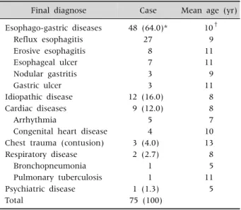
Table 2.Final Diagnoses of the Children Presenting with Chest Pain
Final diagnose Case Mean age (yr)
Esophago-gastric diseases 48 (64.0)* 10†
Reflux esophagitis 27 9
Erosive esophagitis 8 11
Esophageal ulcer 7 11
Nodular gastritis 3 9
Gastric ulcer 3 11
Idiopathic disease 12 (16.0) 8
Cardiac diseases 9 (12.0) 8
Arrhythmia 5 7
Congenital heart disease 4 10
Chest trauma (contusion) 3 (4.0) 13
Respiratory disease 2 (2.7) 8
Bronchopneumonia 1 5
Pulmonary tuberculosis 1 11
Psychiatric disease 1 (1.3) 5
Total 75 (100)
Values are presented as number (%) or number only.
*Among the children presenting with chest pain, the proportion of children diagnosed as esophago-gastric diseases finally is significantly higher than half (p=0.015). †The mean age of the children with esophago-gastric diseases was higher than that of the children with other diseases, but not statistically significant (p=0.056).
esophageal: reflux esophagitis (n=27), acute erosive esophagitis (n=8), esophageal ulcer (n=7), nodular gastritis (n=3), and gastric ulcer (n=3). Eight (16.7%) among these 48 patients also showed pos- itive Helicobacter pylori infections. Accompanied endo- scopic findings in addition with the above were chronic superficial gastritis in 13 cases, duodenogas- tric reflux in 12, chronic duodenitis in 10, and duode- nal ulcer in one.
The prevalence rate of H. pylori in these children showed statistical significance (p<0.001) compared with the rate in children with chronic abdominal pain (6.7%, 2011-2014) and in the general pop- ulation of children (5.8%, 2011-2014) (Table 1) [7].
The most common cause of the chest pain was esophago-gastric diseases (64.0%; esophageal 56.0%, gastric 8.0%), followed by idiopathic cases (16.0%), cardiac diseases (12.0%), chest trauma (4.0%), respi- ratory diseases (2.7%), and psychiatric disease (1.3%).
Among the children presenting with chest pain, the proportion finally diagnosed as esophago-gastric dis- eases was significantly higher than half (p=0.015).
The mean age of the children with esophago-gastric diseases was higher than that of the children with other diseases, but not statistically significant (p=0.056) (Table 2).
ECG was performed in all patients, of whom 21 (28.0%) showed abnormalities. Sinus arrhythmia was most commonly noted, right bundle branch block (RBBB) in three, with one patients each dis- playing ST elevation, left axis deviation, PR prolonga- tion, left ventricular hypertrophy, and right axis deviation. Only two cases of sinus arrhythmia showed the same abnormality in 24-hour Holter monitoring; one case with RBBB and one case with PR prolongation were in ventricular septal defect (VSD) status with normal findings in echocardio- graphy. Echocardiography performed in 34 children revealed nine abnormalities including three VSD, one aortic regurgitation, one atrial septal defect, one septal hypertrophy, and one high overload. Twenty four-hour Holter monitoring was performed in four, of whom two showed sinus arrhythmia (Table 3).
Chest X-ray performed in all patients revealed ab- normalities in three, consisting of one each of en- largement of pulmonary conus, bronchopneumonia, and cardiomegaly due to VSD. The patient with en-
Table 4.Chest Radiologic Findings and the Related Cardiologic Problem of the Children
Chest Radiologic Findings Case (n) Cardiologic problem Enlargement of left pulmonary
conus (pulmonary tuberculosis)
1 Aortic regurgitation
Bronchopneumonia 1 None
Cardiomegaly 1 VSD
VSD: ventricular septal defect.
Table 5. Symptoms Accompanying Chest Pain Accompanied
symptom
UGI diseases as causes
(n=48)
Non-UGI diseases as causes
(n=27)
General symptoms 2 (2.7) 3 (4)
Dizziness and/or fatigue 0 2
Headache 1 1
Anorexia 1 0
Gastrointestinal symptoms 15 (20.0) 2 (2)
Vomiting 8 0
Epigastric pain 6 1
Chronic abdominal pain 1 1
Respiratory symptoms 4 (5.3) 5 (6.7)
Dyspnea 4 3
Cough 0 2
Total 21 10
Values are presented as number (percentage in 75 patients) or number only.
UGI, upper gastrointestinal.
Table 3.Cardiologic Evaluation Findings of the Children with Chest Pain
Variable Data
Electrocardiography
Sinus arrhythmia 12 (16.0)*
RBBB† 3 (4.0)
ST-elevation 1 (1.3)
LAD 1 (1.3)
PR prolongation‡ 1 (1.3)
LVH 1 (1.3)
RAD 1 (1.3)
Sinus bradyarrhythmia 1 (1.3)
Total 21 (28.0)
Echocardiography
VSD 3 (4.0)
ASD 1 (1.3)
AR 1 (1.3)
Septal hypertrophy 1 (1.3)
High overload 1 (1.3)
Total 7 (9.3)
24-hr Holter monitoring
Sinus arrhythmia 2 (2.7)
Total 2 (2.7)
Values are presented as number (%).
RBBB: right bundle branch block, LAD: left axis deviation, LVH:
left ventricular hypertrophy, RAD: right axis deviation, VSD:
ventricular septal defect, ASD: atrial septal defect, AR: aortic regurgitation, ECG: electrocardiography.
*Case number 12 and 25 showed sinus arrhythmia both in ECG and in 24-hr Holter monitoring. †Case number 33 showed RBBB in ECG and postop status of VSD in echocardiography.
‡Case number 7 showed PR prolongation in ECG and postop status of VSD.
largement of pulmonary conus was diagnosed as pulmonary tuberculosis and in the echocardiography.
Aortic regurgitation was also noted, and prompted a clinic with complaint of chest pain 5 years later (Table 4).
The patients whose primary diseases were esoph- ageal or gastric had accompanying symptoms that most often were nausea (n=8) and vomiting (n=6).
Patients whose primary diseases were not UGI dis- eases showed few GI symptoms. There was no dis- tinct difference concerning general symptoms and thoracic symptoms between children with and with- out primary UGI diseases (Table 5).
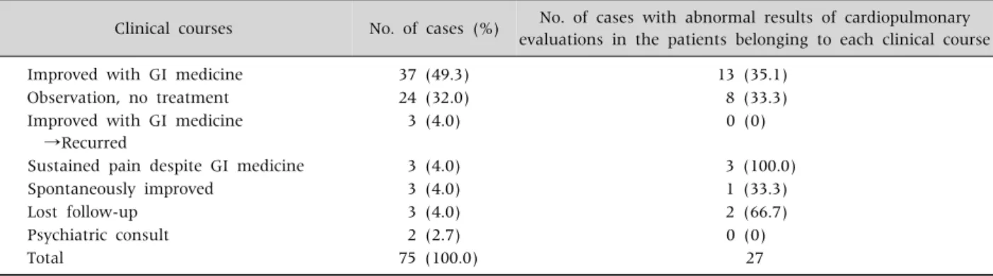
After the final diagnoses of chest pain, patients were followed-up for at least 6 months to assess
whether symptoms disappeared with treatment, persisted, or recurred after initial improvement. Chest pain was improved with GI medicines in 37 patients (49.3%). Of these 37, 13 (35.1%) showed abnormal- ities in their cardiac examinations, which eventually showed little clinical correlation as a cause of chest pain. We continued to observe 24 patients (32.0%) who were not treated and did not display symptom recurrence; eight showed abnormal findings in their cardiac examination but did not require specific treatment. Three improved but the symptoms re- curred, three (4.0%) suffered from sustained chest pain despite GI medicine, in whom abnormal cardiac examination results were already reported and might be the main reason for the persistence of
Table 6.Post-diagnosis Clinical Courses of the Changes of Chest Pain of the Children
Clinical courses No. of cases (%) No. of cases with abnormal results of cardiopulmonary evaluations in the patients belonging to each clinical course
Improved with GI medicine 37 (49.3) 13 (35.1)
Observation, no treatment 24 (32.0) 8 (33.3)
Improved with GI medicine →Recurred
3 (4.0) 0 (0)
Sustained pain despite GI medicine 3 (4.0) 3 (100.0)
Spontaneously improved 3 (4.0) 1 (33.3)
Lost follow-up 3 (4.0) 2 (66.7)
Psychiatric consult 2 (2.7) 0 (0)
Total 75 (100.0) 27
GI: gastrointestinal.
symptoms. In three patients chest pain sponta- neously resolved without any treatment (Table 6).
DISCUSSION
Chest pain in children is not a frequent symptom, as in adults. Pediatric chest pain usually present around 10-years-of-age [1,8-10], and females pres- ent more commonly than males. In this study, the majority of patients were 9 to 14-years-of-age (57.3%) and 6-years-of-age (14.7%). The male and female ra- tio was about 4:3.
Children with chest pain are usually referred to pe- diatric cardiologists, but chest pain is not an im- portant or frequent symptom in children with car- diac diseases [4,11,12]. Careful history-taking, phys- ical examination, and diverse laboratory inves- tigations are required to determine the etiology of chest pain [13-15]. Musculoskeletal diseases (trauma, severe muscle sprain), respiratory diseases (pneu- monia, pleuritis, asthma, chronic cough), UGI dis- eases (esophagitis, gastrointestinal reflux disease, foreign body, esophageal spasm, peptic ulcer dis- ease), cardiovascular diseases (cardiomyopathy, en- docarditis, valvular diseases, myocardial ischemia), and idiopathic causes are included in the differential diagnosis [4]. Idiopathic chest pain is most common in girls around 10-years-of-age [16].
Rowe et al. [17] studied clinical characteristics and causes of chest pain in 325 children visiting the
emergency room. The most common cause was idio- pathic (33%), followed by chest wall pain (28%), res- piratory disease (19%), trauma (15%), and psycho- genic (5%). Very few patients (2%) were hospitalized and there were no patients with cardiovascular disease. There have been several studies regarding the causes of pediatric chest pain; in most of these UGI endoscopic examinations were seldom done so detailed explanations concerning the primary prob- lems are lacking.
Chest pain caused by cardiovascular disease is not common in children with an incidence of 4% to 6%
[18]. Mitral regurgitation is a major cardiac cause of chest pain in children, and is accompanied by verti- go, dyspnea, anxiety, and palpitations [19]. In this study, diverse examinations were performed to in- vestigate cardiovascular disease, but clinically patho- logic cardiac disease was not a major cause.
Several recent studies have suggested that UGI disease should be included in the differential diag- nosis of chest pain [6,20-23]. Hsia et al. [21] per- formed UGI endoscopy and esophageal manometry in 100 patients with non-cardiac chest pain and di- agnosed 24 cases of esophagitis, 18 cases of gastritis or duodenitis, 14 cases of esophageal hernia, and six cases of peptic ulcer disease, and recommended spe- cific examinations for UGI disease in non-cardiac chest pain. Berezin et al. [22] diagnosed UGI dis- eases in 78% of 27 patients whose causes were specif- ic; these patients showed clinical improvement after
the treatment of the UGI diseases. In this study, UGI disease was suspected in 18 children, and evalua- tions specific for the UGI diseases were performed.
Specific UGI diseases were diagnosed in all these children and the symptoms improved after proper treatment. Lipsitz et al. [24] reported that psychoso- matic anxiety disorders could be major causes of non-cardiac chest pain in children; however, further evaluation for UGI disease was not done. The au- thors reported the diagnostic efficacies of several ex- aminations for pediatric chest pains in 2003 [25]. In the former study, the evaluation of UGI diseases were performed only in 18% of patients of the overall patient study group, and idiopathic cases were most common. Performing more examinations to de- termine whether UGI disease is a cause of cardiac pain, as UGI diseases were shown to be major causes of chest pain in children in this study. That is, the idi- opathic causes of chest pain were less than 20%, while UGI disease caused chest pain in 40% of all cas- es in this study.
In our study, the incidence of H. pylori among the children with UGI diseases was higher than that among those with chronic abdominal pain in Korean children. We could not find our similar study per- formed in Korean children, but it was already re- ported that H. pylori infection have a important role in non-cardiac chest pain from Asia-Pacific survey [26].
Study limitations include the retrospectively de- sign, lack of complete laboratory examinations in some patients, and failure of a specific treatment for certain organ disease to alleviate patient suffering.
More differential diagnostic plans are needed, and should be investigated using a well-designed protocol.
In conclusion, careful history taking, physical ex- amination, and proper clinical interventions are usu- ally required to determine the diverse causes of chest pain. GI evaluation, including UGI endoscopic ex- amination, is recommended to diagnose esophageal and gastric diseases in patients with non-cardiac chest pain.
REFERENCES
1. Driscoll DJ, Glicklich LB, Gallen WJ. Chest pain in chil- dren: a prospective study. Pediatrics 1976;57:648-51.
2. Fyfe DA, Moodie DS. Chest pain in pediatric patients presenting to a cardiac clinic. Clin Pediatr (Phila) 1984;23:321-4.
3. Pantell RH, Goodman BW Jr. Adolescent chest pain: a prospective study. Pediatrics 1983;71:881-7.
4. Bernstein D. History and physical examination. In:
Kliegman RM, Stanton BF, St Geme JW, Schor NF, eds.
Nelson textbook of pediatrics. 20th ed. Philadelphia:
Elsevier Saunders, 2015:2163-70.
5. Yoon KL. Chest pain in children and adolescents. J Korean Med Assoc 2010;53:407-14.
6. Sabri MR, Ghavanini AA, Haghighat M, Imanieh MH.
Chest pain in children and adolescents: epigastric ten- derness as a guide to reduce unnecessary work-up.
Pediatr Cardiol 2003;24:3-5.
7. Jang KM, Choe BH, Choe JY, Hong SJ, Park HJ, Chu MA, et al. Changing prevalence of helicobacter pylori infections in korean children with recurrent abdominal pain. Pediatr Gastroenterol Hepatol Nutr 2015;18:
10-6.
8. Asnes RS, Santulli R, Bemporad JR. Psychogenic chest pain in children. Clin Pediatr (Phila) 1981;20:788-91.
9. Raiola G, Galati MC, De Sanctis V, Salerno D, Arcuri VM, Mussari A. Chest pain in adolescents. Minerva Pediatr 2002;54:623-30.
10. Selbst SM. Chest pain in children. Pediatrics 1985;
75:1068-70.
11. Swenson JM, Fischer DR, Miller SA, Boyle GJ, Ettedgui JA, Beerman LB. Are chest radiographs and electro- cardiograms still valuable in evaluating new pediatric patients with heart murmurs or chest pain? Pediatrics 1997;99:1-3.
12. Kim JH, Moon HK, Jun JG. Clinical study of chest pain in children. J Korean Pediatr Soc 1990;33:1526-32.
13. Leung AK, Robson WL, Cho H. Chest pain in children.
Can Fam Physician 1996;42:1156-60, 1163-4.
14. Selbst SM. Consultation with the specialist. Chest pain in children. Pediatr Rev 1997;18:169-73.
15. Evangelista JA, Parsons M, Renneburg AK. Chest pain in children: diagnosis through history and physical examination. J Pediatr Health Care 2000;14:3-8.
16. Selbst SM, Ruddy RM, Clark BJ, Henretig FM, Santulli T Jr. Pediatric chest pain: a prospective study. Pedia- trics 1988;82:319-23.
17. Rowe BH, Dulberg CS, Peterson RG, Vlad P, Li MM.
Characteristics of children presenting with chest pain
to a pediatric emergency department. CMAJ 1990;143:
388-94.
18. Kocis KC. Chest pain in pediatrics. Pediatr Clin North Am 1999;46:189-203.
19. Woolf PK, Gewitz MH, Berezin S, Medow MS, Stewart JM, Fish BG, et al. Noncardiac chest pain in adolescents and children with mitral valve prolapse. J Adolesc Health 1991;12:247-50.
20. Berezin S, Medow MS, Glassman MS, Newman LJ.
Esophageal chest pain in children with asthma. J Pediatr Gastroenterol Nutr 1991;12:52-5.
21. Hsia PC, Maher KA, Lewis JH, Cattau EL Jr, Fleischer DE, Benjamin SB. Utility of upper endoscopy in the evaluation of noncardiac chest pain. Gastrointest Endosc 1991;37:22-6.
22. Berezin S, Medow MS, Glassman MS, Newman LJ.
Chest pain of gastrointestinal origin. Arch Dis Child 1988;63:1457-60.
23. Katz PO. Approach to the patient with unexplained chest pain. Semin Gastrointest Dis 2001;12:38-45.
24. Lipsitz JD, Masia C, Apfel H, Marans Z, Gur M, Dent H, et al. Noncardiac chest pain and psychopathology in children and adolescents. J Psychosom Res 2005;59:
185-8.
25. Shin SA, Kim YJ, Lee JW, Kim NS, Moon SJ. Clinical evaluation and diagnosis of children with chest pain. J Korean Pediatr Soc 2003;46:1248-52.
26. Cheung TK, Lim PW, Wong BC. Noncardiac chest pain--an Asia-Pacific survey on the views of primary care physicians. Dig Dis Sci 2007;52:3043-8.