INTRODUCTION
Incontinentia pigmenti (IP) is a rare X-linked genoderma- tosis and is also called ‘Bloch-Sulzberger syndrome’ (1, 2). Mu- tation of NEMO/IKKr gene in Xq28 is believed to play a role in pathogenesis and it occurs mostly in female infants due to its fatality in male in utero (1, 2). Skin manifestation is the earliest symptom and is classified as 3 or 4 stages such as vesicular, verrucous, hyperpigmented, and sometimes hy- popigmented stage (2, 3). Other accompanying diseases are dental, ocular, auricular, central nervous system (CNS), mus- culoskeletal, and cardiovascular anomalies of different severi- ties (2-4).
We investigated clinical symptoms, disease onset, and ac- companying disorders in 40 cases of IP by reviewing their medical records, laboratory data, clinical photographs, and telephone survey.
MATERIALS AND METHODS Subjects
We have reviewed 64 cases of IP who visited or were re- ferred to dermatologic clinic of Seoul National University Hospital or Chung-Ang University Hospital from June 1995
to May 2005. Total 24 cases were excluded by their incom- patibility or lack of evidences supporting diagnostic criteria of IP set by Landy and Donnai (3). Therefore, 40 cases were included in this study. The study was approved by the Insti- tutional Review Board of the Seoul National University Hos- pital.
Data collection
Medical records of neonatal data as well as inpatient and outpatient charts, clinical photographs, biopsied sample slides, laboratory results, skeletal radiographs, and telephone survey of all affected patients were reviewed by two experienced der- matologists. Information included sex, disease onset, skin stage at the time of diagnosis, distribution of skin lesions, eosino- philic count in peripheral blood analysis, and whether to have neurologic anomalies, dental defects, oculopathies, and other accompanied disorders.
Clinical stages
From the medical records and clinical photographs of the skin lesions, clinical stages were checked as 1st vesicular, 2nd verrucous, 3rd hyperpigmented, and 4th hypopigmented stage to identify whether their time of initial diagnosis were being made early or not (Fig. 1).
Beom Joon Kim, Hyo Seung Shin, Chong Hyun Won, Jong Hee Lee, Kyu Han Kim, Myeung Nam Kim*, Byung In Ro*, Oh Sang Kwon
Department of Dermatology, Seoul National University College of Medicine, Seoul; Department of Dermatology*, College of Medicine, Chung-Ang University, Seoul, Korea
Address for correspondence Oh Sang Kwon, M.D.
Department of Dermatology, Seoul National University College of Medicine, 28 Yongon-dong, Jongno-gu, Seoul 110-744, Korea
Tel : +82.2-2072-1996, Fax : +82.2-742-7344 E-mail : [email protected]
*Not related with any kind of research grants or con- flict of interests.
474 J Korean Med Sci 2006; 21: 474-7
ISSN 1011-8934
Copyright � The Korean Academy of Medical Sciences
Incontinentia Pigmenti: Clinical Observation of 40 Korean Cases
Incontinentia pigmenti (IP) is an uncommon genodermatosis that usually occurs in female infants. It is characterized by ectodermal, mesodermal, neurological, ocular, and dental manifestations. The aim of this study was to clarify clinical symptoms, accompanying diseases, and complications of IP. Forty cases of IP have been re- viewed by their medical records, laboratory data, clinical photographs, and telephone survey. Male-to-female ratio was 1 to 19 and their onsets were mostly in utero. They were usually diagnosed during the neonatal period owing to their early expression of skin manifestation. Central nervous system anomalies were found in 46.7%. Ocu- lar disorders and dental defects were detected in 66.7% and 72.7% respectively.
The most commonly diagnosed anomalies were hypodontia, retinopathy, and sei- zure. For better understanding of IP, long term and close cooperation between der- matologists, pediatricians, neuroscientists, genentic counselors, and even dentists is crucial.
Key Words : Incontinentia Pigmenti; Skin Diseases, Genetic; Genodermatosis; Seizures; Retinal Diseases
Received : 17 August 2005 Accepted : 10 November 2005
Incontinentia Pigmenti in Korea 475
RESULTS Sex ratio and disease onset
Among 40 cases of IP, only 2 cases (5%) were males and the others were females. The onset of disease has been classi- fied as inborn, neonatal, infant, and early childhood. Disease of onset has been marked as ‘since born’ when patient showed skin symptom inborn, ‘neonatal’ within 4 weeks since birth,
‘infant’ 1 month to 12 months, and ‘early childhood’ from 1 yr to 6 yr old. The most common period of disease onset was inborn 67.5% (27 cases), and neonatal period 17.5% (7 cases), infant 10% (4 cases), early childhood 5% (2 cases) in order.
Clinical stages
Clinical stage at the time of initial visit has been analyzed.
Vesicular stages were 52.5% (21 cases), verrucous stages 30%
(12 cases), hyperpigmented stages 15% (6 cases) and hypopig- mented stages 2.5% (1 case), respectively. And there was no patient whose initial visit was at stage 4. Therefore, initial visit of the patients were made relatively early clinical stages in more than half of cases (Table 1).
Distribution of skin lesions
Skin lesions were widely distributed and involved the whole body in 20% (8 cases). In 37.5% (15 cases), skin lesions were found in whole body except face. Involvement of only trunk and lower extremities was 17.5% (7 cases). In only 1 case, skin lesions were limited only in trunk.
Distribution of the skin lesions were also evaluated by ana- tomic area. Lower extremities (97.5%, 39 cases) were the most common site of involvement and trunk (77.5%, 31 cases), upper extremities (57.5%, 23 cases), face (20%, 8 cases) in order.
Eosinophilia
Among 40 cases, blood eosinophilia was not checked in 17 cases. In 23 evaluated cases, blood eosinophilia has been de- tected in 47.8% (11 cases) at least one of serial checks during follow-up periods.
Accompanying diseases
Thirty cases were evaluated for their CNS anomalies by sei- zure history, brain CT, brain MRI, and electroencephalogram.
The other 10 cases free from objective symptom were not checked for neurologic anomalies. CNS anomalies of various severities were detected in 46.7% (14 cases). Seizure (57.1%, 8 of 14 cases) was the most common CNS related symptom and followed by ataxia and sphingoencephalitis.
Ocular anomalies were evaluated from medical chart records, fundoscopy, and slip lamp examination. Twenty four cases were evaluated for their oculopathies and 66.7% (16 cases) of them presented anomalies or ocular disorders. Retinopa- thy (56.3%, 9 cases) was the most common oculopathy and the next was strabismus (31.3%, 5 cases).
Twenty two cases were examined for their dental anoma- lies and 72.7% (16 cases) showed defects or anomalies in den- tal system. Hypodontia (43.8%, 7 cases) was the most com- mon dental anomaly and the next was delayed eruption of teeth (37.5%, 6 cases).
DISCUSSION
IP is a rare genodermatosis and its prevalence in general is about 1 per 40,000 (3). More than 95% of the patients are female infants because it may be fatal when it occurs in male fetus (4, 5). In this study, affected male infants were also found in two cases. Affected male infant who survived may have a
Fig. 1.Clinical stages of incontinentia pigmenti. (A) Linear vesicles and bullae of stage 1. (B) Dark brown colored verrucous papules and plaques of stage 2. (C) Light brown colored swirling hyperpigmentation of stage 3. (D) White atrophic patches of stage 4.
A B C D
476 B.J. Kim, H.S. Shin, C.H. Won, et al.
potential possibility of Klinefelter syndrome (1, 6, 7). Unfor- tunately, we were not able to perform chromosomal studies due to refusal of further genetic studies by parents of the male patients. Skin lesions may follow Blaschko lines and their melanoblasts start to transform into melanocytes after birth (8, 9). Initial appearance of skin manifestations can be obser- ved at birth or within neonatal period. Therefore, the role of dermatologists and pediatrician is crucial in early diagnosis of IP. Skin manifestation of stage 1 usually presents within 2 weeks after birth and it may disappear within 4 months
in 90% of cases (5). Skin lesions of stage 2 can be identified within 2 months and disappear within 6 months after birth (3, 5). In this study, 82.5% of the cases presented skin lesions of stage 1 or 2 at birth or within 1 month after birth, which was similar to the results of the studies by Carney (70-90%) (5) and Hadj-Rabia et al. (80-92%) (10). So, diagnosis of IP in Korea is considered to be made as soon as skin manifesta- tion presents. But, hypopigmented patches of stage 4, which are important clues of diagnosis of IP (11), were not found in this study due to young ages of the cases.
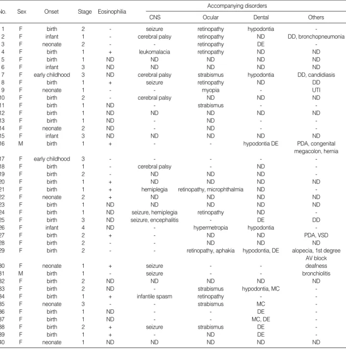
No. Sex Stage Eosinophilia
CNS Ocular
Accompanying disorders
Dental Others
Onset
1 F birth 2 - seizure retinopathy hypodontia -
2 F infant 1 - cerebral palsy retinopathy ND DD, bronchopneumonia
3 F neonate 2 - - retinopathy DE -
4 F birth 1 + leukomalacia retinopathy ND ND
5 F birth 1 ND ND ND ND ND
6 F infant 3 ND ND ND ND ND
7 F early childhood 3 ND cerebral palsy strabismus hypodontia DD, candidiasis
8 F birth 1 + seizure retinopathy ND DD
9 F neonate 1 - - myopia - UTI
10 F birth 2 - cerebral palsy ND ND ND
11 F birth 1 ND - strabismus - -
12 F birth 1 ND ND ND ND ND
13 F birth 1 ND - ND - -
14 F neonate 2 ND - ND - -
15 F infant 3 ND ND ND ND ND
16 M birth 1 + - - hypodontia DE PDA, congenital
megacolon, hernia
17 F early childhood 3 - - - - -
18 F birth 1 - cerebral palsy - ND -
19 F birth 2 - ND ND ND -
20 F birth 1 + ND ND ND ND
21 F birth 1 + hemiplegia retinopathy, microphthalmia ND -
22 F neonate 2 + ND ND ND ND
23 F birth 1 ND ND ND ND ND
24 F birth 1 ND seizure, hemiplegia retinopathy ND -
25 F birth 3 ND seizure, encephalitis - DE DD
26 F infant 4 ND - hypermetropia hypodontia -
27 F birth 2 + - ND ND PDA, VSD
28 F birth 2 - - ND ND ND
29 F birth 2 - - retinopathy, aphakia hypodontia, DE alopecia, 1st degree
AV block
30 F neonate 1 + seizure - - deafness
31 M birth 1 - seizure - - bronchiolitis
32 F birth 2 ND ND ND ND ND
33 F birth 2 ND - strabismus hypodontia, MC -
34 F birth 1 + infantile spasm retinopathy - -
35 F neonate 3 - - strabismus MC -
36 F birth 1 ND - - DE -
37 F birth 1 ND - - MC, DE -
38 F birth 2 + seizure strabismus DE -
39 F birth 1 + - ND DE -
40 F neonate 1 ND ND ND ND ND
Table 1.Summary of patients with Incontinentia pigmenti
W, whole body; H, head and neck; T, trunk; UE, upper extremities; LE, lower extremities; DD, developmental delay; DE, delayed eruption of teeth; UTI, urinary tract infection; PDA, patent ductus arteriosus; VSD, ventricular septal defect; MC, malformed crowns; ND, not done.
Incontinentia Pigmenti in Korea 477
Existence of blood eosinophilia is also a diagnostic clue espe- cially in neonatal period. Blood count of eosinophils gradually decreases without invading internal organs as the patient grows up (12). In addition, severity of skin lesion has no significant relation with blood count of eosinophils (3, 13). In this study, presence of eosinophilia was identified only in 47.8% of 23 cases who had serial blood checks during neonatal periods.
Anomalies of other organs especially CNS, dental, and ocu- lar systems should be also evaluated. Oculopathies of variable severities that can be identified in more than 1/3 of the IP cases include speckled diffuse hypopigmentation in the reti- na, which is the pathognomonic finding of IP, microphthal- mia, lenticular hemorrhage, retrolental fibroplasia, cataract, and atrophy of the optic nerve (1, 5). Dental anomalies are found in 65-90% of IP cases and they are usually delayed eruption of teeth, changes in dental contour (circular or coni- cal shape), and hypodontia (1, 4, 5). CNS related disorders such as seizure, microcephaly, mental retardation, and spas- tic paralysis may be detected in 10-40% of the cases (1, 2, 5).
In this study, percentages of oculopathies (66.7%) and CNS anomalies (46.7%) were relatively higher than those of pre- vious Western reports (1-5). The percentages of dental defects were slightly lower when it was compared with previous We- stern reports (1-5). We do not know the reason of different rates in ocular, dental, and CNS diseases between IP cases of Western and Korean studies. However, accessibility of den- tal care by this study population may partially explain these differences. Dental hospitals or dental clinics are not easily accessible by patient referral system which is requested by dermatologists or pediatricians. Another possible explanation is that dentists are not so much familiar with IP as physicians so that they may ignore minor anomalies and do not record them in medical records.
Compared with previous Korean IP study report by Chun et al. (14), accompanying anomalies were markedly increased and more specified in our study. These diverse anomalies might result from the growth of social-economic status, increased familiarity of physicians due to studies and researches related with genetic diseases, and long term cooperation between physicians of many different specialties such as dermatology, pediatrics, dentistry, neurology, and genetics. Anomalies may not appear at the initial evaluation but continuing work-up and long term precise follow-up using referring system may increase the detection rate of potential anomalies which may occur in early childhood or later. Therefore, long term and close cooperation between dermatologists, pediatricians, neu- rologists, genetic counselors, and even dentists is crucial for better understanding of IP and prediction of occurrence of
potential anomalies in the later life.
REFERENCES
1. Berlin AL, Paller AS, Chan LS. Incontinentia pigmenti: a review and update on the molecular basis of pathophysiology. J Am Acad Dermatol 2002; 47: 169-87.
2. Cohen BA. Incontinentia pigmenti. Neurol Clin 1987; 5: 361-77.
3. Landy SJ, Donnai D. Incontinentia pigmenti (Bloch-Sulzberger syn- drome). J Med Genet 1993; 30: 53-9.
4. Macey-Dare LV, Goodman JR. Incontinentia pigmenti: seven cases with dental manifestations. Int J Paediatr Dent 1999; 9: 293-7.
5. Carney RG. Incontinentia pigmenti. A world statistical analysis.
Arch Dermatol 1976; 112: 535-42.
6. Kenwrick S, Woffendin H, Jakins T, Shuttleworth SG, Mayer E, Gre- enhalgh L, Whittaker J, Rugolotto S, Bardaro T, Esposito T, D’Urso M, Soli F, Turco A, Smahi A, Hamel-Teillac D, Lyonnet S, Bonne- font JP, Munnich A, Aradhya S, Kashork CD, Shaffer LG, Nelson DL, Levy M, Lewis RA; International IP Consortium. Survival of male patients with incontinentia pigmenti carrying a lethal mutation can be explained by somatic mosaicism or Klinefelter syndrome. Am J Hum Genet 2001; 69: 1210-7.
7. Scheuerle AE. Male cases of incontinentia pigmenti: case report and review. Am J Med Genet 1998; 77: 201-18.
8. Moss C. Cytogenetic and molecular evidence for cutaneous mosai- cism: the ectodermal origin of Blaschko lines. Am J Med Genet 1999; 85: 330-3.
9. Cohen PR. Incontinentia pigmenti: clinicopathologic characteris- tics and differential diagnosis. Cutis 1994; 54: 161-6.
10. Hadj-Rabia S, Froidevaux D, Bodak N, Hamel-Teilloc D, Smahi A, Touil Y, Fraitag S, de Prost Y, Bodemer C. Clinical study of 40 cases of incontinentia pigmenti. Arch Dermatol 2003; 139: 1163- 70.
11. Phan TA, Wargon O, Turner AM. Incontinentia pigmenti case series:
clinical spectrum of incontinentia pigmenti in 53 female patients and their relatives. Clin Exp Dermatol 2005; 30: 474-80.
12. el-Benhawi MO, George WM. Incontinentia pigmenti. Cutis 1988;
41: 259-62.
13. Berretty PJ, Cormane RH. Eosinophilic granulocytes and skin dis- orders. Int J Dermatol 1981; 20: 531-40.
14. Chun IK, Chung TB, Kim YP. Clinical observation of incontinentia pigmenti. Korean J Dermatol 1985; 23: 171-6.
15. Patrizi A, Neri I, Guareschi E, Cocchi G. Bullous recurrent eruption of incontinentia pigmenti. Pediatr Dermatol 2004; 21: 613-4.
16. Wiederholt T, Poblete-Gutierrez P, Ott H, Lehmann S, Grussendorf- Conen EI, Beermann T, Frank J. Incontinentia pigmenti in a five-week- old girl. Hautarzt 2004; 55: 999-1001.