INTRODUCTION
Basaloid squamous carcinoma of the uterine cervix is a rare tumor type characterized by an ulcerated, infiltrating growth pattern; nests or cords of small basaloid cells; prominent peri- pheral palisading of cells in the tumor cell nests; and the ab- sence of significant stromal reaction (1). These tumors can arise from various anatomic sites, including the hypopharynx, base of the tongue, salivary glands, esophagus, anal canal, pro- state, thymus, vulva, and urinary bladder (2-12), but origin of uterine cervix is rare. Basaloid squamous carcinoma of the uterine cervix is neither recognized nor included as a specific histologic subtype in the current World Health Organiza- tion (WHO) classification of cervical tumors. Since basaloid squamous carcinomas are thought to behave aggressively (13) but the evidence of supprting this behavior is not powerful, accurate diagnosis and accumulated data of this tumor are important for their clinical management and prognosis.
CASE REPORT
A 70-yr-old woman, gravida 8 para 3, was referred to a ter- tiary medical center from a local hospital for vaginal bleeding
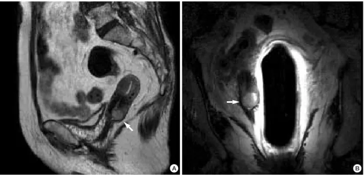
of 3 weeks’ duration. She had undergone a punch biopsy at the local hospital, which resulted in a preliminary diagnosis of adenoid cystic carcinoma or carcinoid tumor. Her medical history included diabetes mellitus (DM) for 10 yr which had been controlled by medication; in addition, her mother died of uterine cervical cancer. Physical examination showed uter- ine cervical erosion. The biopsy specimen taken at the local hospital showed pathologic evidence of a high grade malig- nant epithelial tumor, with features unusual for a cervical tumor. She therefore underwent a loop electrosurgical exci- sion procedure (LEEP) cone biopsy, which revealed a basaloid squamous cell carcinoma. A colonoscopy, intravenous pyelo- gram, and cystoscopy showed no evidence of metastatic dis- ease. Magnetic resonance imaging showed a 2 cm sized can- cerous mass confined to the cervix, with no evidence of inva- sion of the vagina or fornix and no evidence of pelvic lym- phadenopathy (Fig. 1). The tumor was classified as clinical stage Ib1. A radical hysterectomy was performed, along with bilateral salpingo-oophorectomy, pelvic lymph node dissec- tion, and paraaortic lymph node sampling. The pathologic diagnosis was basaloid squamous cell carcinoma. The depth of invasion was 5/7 mm full thickness of the cervical wall.
There was no evidence of tumor in sections taken from 26 lymph nodes. The resection margin of the vaginal cuff was
542
Yong Soon Kwon1,*, Yong Man Kim1, Ga Won Choi2, Young Tak Kim1, and Joo-Hyun Nam1
Departments of Obstetrics and Gynecology1, and Pathology2, College of Medicine, University of Ulsan, Asan Medical Center, Seoul, Korea
*Current address: Department of Obstetrics and Gynecology, College of Medicine, Kwandong University, Cheil General Hospital and Women’s Healthcare Center, Seoul, Korea
Address for correspondence Yong-Man Kim, M.D.
Department of Obstetrics and Gynecology, College of Medicine, University of Ulsan, Asan Medical Center, 388-1 Pungnap-2-dong, Songpa-gu, Seoul 138-736, Korea
Tel: +82.2-3010-3640, Fax: +82.2-3010-6944 E-mail: [email protected]
J Korean Med Sci 2009; 24: 542-5 ISSN 1011-8934
DOI: 10.3346/jkms.2009.24.3.542
Copyright � The Korean Academy of Medical Sciences
Pure Basaloid Squamous Cell Carcinoma of the Uterine Cervix:
A Case Report
Basaloid squamous cell carcinoma of the uterine cervix is an extremely rare malig- nancy of the female genital tract with a poorer clinical outcome than squamous cell carcinoma of the uterine cervix. We report a case of pure basaloid squamous cell carcinoma of the uterine cervix. A 70-yr-old woman with vaginal bleeding was referred to our institute. A basaloid squamous cell carcinoma of the uterine cervix, of Interna- tional Federation of Gynecology and Obstetrics (FIGO) stage Ib1, was diagnosed by a loop electrosurgical excision procedure cone biopsy. A radical hysterectomy was performed, along with bilateral salpingo-oophorectomy, pelvic lymph node dis- section, and para-aortic lymph node sampling. Pathologic findings were consistent with a basaloid squamous cell carcinoma confined to the cervix without an extrac- ervical tumor. No further treatment was administered and there was no clinical evi- dence of recurrence during the 12 months of follow-up. Follow-up for the patient is ongoing. Although basaloid squamous cell carcinoma of the uterine cervix is thought to behave aggressively, accumulation of data on these rare tumors is necessary to determine whether their behavior differs significantly from that of conventional cer- vical squamous cell carcinoma of similar clinical stage. These data would be use- ful for defining the best diagnosis and treatment for these rare tumors.
Key Words : Basaloid; Carcinoma, Squamous Cell; Cervix Uteri
Received : 5 December 2007 Accepted : 7 July 2008
Pure Basaloid Squamous Cell Carcinoma of the Uterine Cervix 543
clear. No adjuvant treatment was administered, and the pa- tient was discharged. In the 12 months since discharge, she has shown no evidence of recurrent or metastatic disease. Fol- low-up is ongoing at Asan Medical Center.
Pathologic findings
A well-defined, fungating firm mass (1.5×1.0×0.6 cm) was present in the posterior wall of the cervix and invaded 5 mm into the cervical wall (full thickness, 7 mm). The parame- tria and vaginal cuff showed no tumor invasion. The cut sur- face of the mass was gray and granular. The tumor cells were immunopositive for p63 and immunonegative for S-100 pro-
tein. The cells appeared basaloid with small hyperchromatic nuclei, distinct nucleoli and scanty cytoplasm. There was also peripheral palisading, supporting the above diagnosis (Fig. 2).
DISCUSSION
Cervical basaloid carcinoma has recently been classified as a specific histologic subtype, with “pure” basaloid carcinomas being extremely rare (14). Often few incidence of this diag- nosis can cause clinical and pathological misinterpretation.
In our case, based on a punch biopsy specimen, our patient was initially diagnosed at the local hospital with adenoid cys-
Fig. 1.MRI findings in this patient. (A) T2 weighted image (B) T2 weighted image using an endorectal coil. The white arrow indicates a 2.0×1.6 cm cervical mass, comparable with cervical cancer.
A B
Fig. 2.Histopathologic findings. The tumor is mainly composed of immature basaloid squamous cells with scanty cytoplasm and periph- eral palisading (arrow). Keratinization foci are seen in the center of the nests (arrowhead). Both tumor components show p63 immunoposi- tivity (A: H&E, ×200; B: p63, ×200).
A B
544 Y.S. Kwon, Y.M. Kim, G.W. Choi, et al.
tic carcinoma or carcinoid tumor, which was not an accurate diagnosis. To obtain an accurate diagnosis, an LEEP cone biop- sy, which yields a larger tissue sample, was performed and resulted in a diagnosis of basaloid carcinoma of the uterine cervix, indicating that diagnostic difficulties can be avoided by obtaining larger specimens for pathologic diagnosis (15).
The term “basaloid carcinoma of the uterine cervix” refers to any neoplastic lesion of the uterine cervix analogous to a cutaneous basal cell carcinoma (BCC), and may include some of the recognized morphologic variants of BCC. Among the morphologic characteristics of basaloid carcinomas of the uter- ine cervix are nests or islands of small basaloid cells, hyper- chromatic epithelial cells with high nucleus-to-cytoplasm ratios, and a tendency to palisade at the periphery of the tumor islands. A basaloid carcinoma of the cervix may be associat- ed with squamous dysplasia, in situ SCC, or invasive SCC (16, 17).
Immunocytochemistry can occasionally be helpful in iden- tifying the epithelial origin of a basaloid carcinoma of the cervix. Some of these tumors have been shown to be strong- ly positive for high molecular weight cytokeratin (18, 19), whereas others have shown little or no expression of cytok- eratins (17, 20). Most tumors are positive for epithelial mem- brane antigen (EMA), but show little or no expression of vimentin, smooth muscle actin, desmin or neuroendocrine markers (2, 18, 19).
The major differential diagnosis of basaloid SCC includes the solid variant of adenoid cystic carcinoma (ACC), small cell carcinoma, and large cell neuroendocrine carcinoma (LC NEC) of the cervix. Solid ACC is distinguished from basa- loid SCC by the focal presence of basement membrane mate- rial enveloped by basaloid neoplastic cells; in addition, the solid variant of ACC may show malignant squamous differ- entiation. Small cell carcinoma may be composed of variably sized nests of relatively small, hyperchromatic tumor cells, which may mimic adenoid basal carcinoma (ABC) and small- er ABC-like nests. Rare cases may present with large neoplas- tic islands having peripheral palisading of tumor cells, which may be confused with the solid variant of ACC (21). Small cell neuroendocrine carcinomas may be diagnosed by immuno- histochemical or ultrastructural demonstration of neuroen- docrine differentiation. Cervical LCNEC, another neoplasm omitted from the current WHO classification of uterine cer- vical neoplasms, is characterized by large cells with vesicu- lar nuclei and prominent nucleoli, a mitotic index in excess of 10 per 10 high-power fields, geographic areas of tumor necrosis, and positive staining with appropriate immunohis- tochemical markers of neuroendocrine differentiation (22, 23). Cervical LCNEC composed of large basaloid islands may mimic basaloid SCC, and may also show a considerable degree of morphologic overlap with solid ACC, a lesion that may contain trabecular structures composed of basaloid cells that palisade at the periphery (22). Accurate diagnosis is of prog- nostic importance because of the biologically aggressive na-
ture of this uncommon type of cervical cancer.
The lack of standard diagnostic criteria for pure basaloid squamous cell carcinoma of the uterine cervix has made it dif- ficult to predict their precise biologic behavior and to design optimal management strategies. Accumulation of data on these rare tumors is therefore necessary to determine whether their behavior differs significantly from that of conventional cervical SCCs of similar clinical stage. Long-term follow-up of this patient and other such patients is therefore important.
REFERENCES
1. Daroca PJ Jr, Dhurandhar HN. Basaloid carcinoma of uterine cervix.
Am J Surg Pathol 1980; 4: 235-9.
2. Banks ER, Frierson HF Jr, Mills SE, George E, Zarbo RJ, Swanson PE. Basaloid squamous cell carcinoma of the head and neck: a clini- copathologic and immunohistochemical study of 40 cases. Am J Surg Pathol 1992; 16: 939-46.
3. Wain SL, Kier R, Vollmer RT, Bossen EH. Basaloid-squamous car- cinoma of the tongue, hypopharynx and larynx: report of 10 cases.
Hum Pathol 1986; 17: 1158-66.
4. Batsakis JG, el Naggar A. Basaloid-squamous carcinoma of the upper aerodigestive tracts. Ann Otol Rhinol Laryngol 1989; 98: 919-20.
5. Coppola D, Catalano E, Tang CK, Elfenbein IB, Harwick R, Mohr R. Basaloid squamous cell carcinoma of floor of mouth. Cancer 1993; 72: 2299-305.
6. Gallimore AP, Spraggs PD, Allen JP, Hobsley M. Basaloid carci- nomas of salivary glands. Histopathology 1994; 24: 139-44.
7. Abe K, Sasano H, Itakura Y, Nishihira T, Mori S, Nagura H. Basa- loid-squamous carcinoma of the esophagus: a clinicopathologic, DNA ploidy, and immunohistochemical study of seven cases. Am J Surg Pathol 1996; 20: 453-61.
8. Kurman RJ, Toki T, Schiffman MH. Basaloid and warty carcinomas of the vulva: distinctive types of squamous cell carcinoma frequent- ly associated with human papillomaviruses. Am J Surg Pathol 1993;
17: 133-45.
9. Dougherty BG, Evans HL. Carcinoma of the anal canal: a study of 79 cases. Am J Clin Pathol 1985; 83: 159-64.
10. Walker AN, Mills SE, Fechner RE. Thymomas and thymic carcino- mas. Semin Diagn Pathol 1990; 7: 250-65.
11. Denholm SW, Webb JN, Howard GC, Chisholm GD. Basaloid car- cinoma of the prostate gland: histogenesis and review of the litera- ture. Histopathology 1992; 20: 151-5.
12. Vakar-Lopez F, Abrams J. Basaloid squamous cell carcinoma occur- ring in the urinary bladder. Arch Pathol Lab Med 2000; 124: 455-9.
13. Grayson W, Cooper K. A reappraisal of ‘‘basaloid carcinoma’’ of the cervix, and the differential diagnosis of basaloid cervical neo- plasms. Adv Anat Pathol 2002; 9: 290-300.
14. Clement PB, Zubovits JT, Young RH, Scully RE. Malignant mulle- rian mixed tumors of the uterine cervix: a report of nine cases of a neoplasm with morphology often different from its counterpart in the corpus. Int J Gynecol Pathol 1998; 17: 211-22.
15. Brainard JA, Hart WR. Adenoid basal epitheliomas of the uterine
Pure Basaloid Squamous Cell Carcinoma of the Uterine Cervix 545
cervix: a reevaluation of distinctive cervical basaloid lesions current- ly classified as adenoid basal carcinoma and adenoid basal hyper- plasia. Am J Surg Pathol 1998; 22: 965-75.
16. Sarbia M, Verreet P, Bittinger F, Dutkowski P, Heep H, Willers R, Gabbert HE. Basaloid squamous cell carcinoma of the esophagus:
diagnosis and prognosis. Cancer 1997; 79: 1871-8.
17. Tsang WY, Chan JK, Lee KC, Leung AK, Fu YT. Basaloid-squa- mous carcinoma of the upper aerodigestive tract and so-called ade- noid cystic carcinoma of the oesophagus: the same tumour type?
Histopathology 1991; 19: 35-46.
18. Bastiaan de Boer W. Basaloid squamous carcinoma in the liver.
Pathology 2000; 32: 147-51.
19. Tulunay O, Kucuk B, Tulunay EO, Akturk T. Basaloid squamous cell carcinoma of the maxilla: a case report and immunohistochem- ical analysis. Acta Otolaryngol 2002; 122: 424-8.
20. Brambilla E, Moro D, Veale D, Brichon PY, Stoebner P, Paramelle
B, Brambilla C. Basal cell (basaloid) carcinoma of the lung: a new morphologic and phenotypic entity with separate prognostic signifi- cance. Hum Pathol 1992; 23: 993-1003.
21. Albores-Saavedra J, Manivel C, Mora A, Vuitch F, Milchgrub S, Gould E. The solid variant of adenoid cystic carcinoma of the cervix.
Int J Gynecol Pathol 1992; 11: 2-10.
22. Grayson W, Rhemtula HA, Taylor LF, Allard U, Tiltman AJ. Detec- tion of human papillomavirus in large cell neuroendocrine carcino- ma of the uterine cervix: a study of 12 cases. J Clin Pathol 2002; 55:
108-14.
23. Albores-Saavedra J, Gersell D, Gilks CB, Henson DE, Lindberg G, Santiago H, Scully RE, Silva E, Sobin LH, Tavassoli FJ, Travis WD, Woodruff JM. Terminology of endocrine tumors of the uterine cervix.
Results of a workshop sponsored by the College of American Pathol- ogists and the National Cancer Institute. Arch Pathol Lab Med 1997;
121: 34-9.