Usefulness of Surgeon-Performed Preoperative Ultrasonography for Papillary Thyroid Carcinoma
Dong Hwi Kim, Kwangmin Kim
Department of Surgery, Wonju College of Medicine, Yonsei University, Wonju, Korea
Received October 1, 2017 Revised November 14, 2017 Accepted November 17, 2017
Purpose: This study evaluated the overall diagnostic performance of surgeon-performed ul- trasonography (US) for preoperative staging, multifocality, and bilaterality of papillary thy- roid carcinoma in relation to the current TNM classification system.
Methods: The clinical and pathology data were collected retrospectively from 192 patients (175 women and 17 men; mean age 49.6 (range, 22‒82) years) with documented diagnoses of papillary thyroid carcinoma and having undergone preoperative US and thyroidectomy at the Department of Surgery, Wonju Severance Christian Hospital between March of 2010 and July of 2011. The preoperative evaluations of T stage and N stage were performed with US in accordance with the TNM staging system of the American Joint Committee on Cancer. The multifocality and bilaterality of the malignant thyroid nodules were also evaluated.
Results: The accuracy of preoperative US to predict multifocality was 87.5% (168/192). The accuracy of preoperative US to predict bilaterality was 91.1% (175/192). The overall accu- racy of preoperative US for T stage was 86.5% (166/192). The overall accuracy of pre- operative US for N stage was 71.9% (138/192).
Conclusion: The efficacy of surgeon-performed preoperative US was evaluated. US is a good and highly useful tool for the preoperative staging of PTC, and is also helpful for ac- curately predicting and identifying extrathyroidal extensions, multifocality, and bilaterality.
Surgeon-performed US for preoperative staging appears to be sufficiently accurate for pre- surgical planning.
Keywords: Papillary thyroid carcinoma, Ultrasonography, TNM stage, Lymph node metastasis Correspondence to:
Kwangmin Kim
Department of Surgery, Wonju College of Medicine, Yonsei University, 20 Ilsan-ro, Wonju 26426, Korea
Tel: +82-33-741-0573 Fax: +82-33-742-1815 E-mail: [email protected]
INTRODUCTION
Thyroid cancer is the most common malignancy found to occur, among cancers afflicting the endo- crine organs.(1) Papillary thyroid carcinoma is the most common histologic type of thyroid cancer diagnosed.(2) Numerous studies done in the past have mentioned various factors that could, and do, influence the recurrence of papillary thyroid carci-
noma; these factors include patient age, tumor size, level of advancement of the TNM stage, extrathyroidal extension and the potential presence of distant metastasis.(3-5) A study showed that tumor size greater than 2 cm, multifocality and clinically-evident lymph node metastasis influenced multiple recur- rence.(6) According to these studies, preoperative evaluations of TNM stage including the size and ex- tent of the primary tumor, lymph node metastasis
ORIGINAL ARTICLE
J Surg Ultrasound 2017;4:55-61 JSUJournal of Surgical Ultrasound
and multifocal papillary thyroid carcinoma are need- ed for proper treatment to reduce the recurrence rate.
Total thyroidectomy has been the standard treat- ment for patients with thyroid cancer. Recently, the appropriate extent of the surgery to treat papillary thyroid carcinoma has been under debate because extensive surgery, regardless of the tumor charac- teristics, has no survival benefit and might actually serve to increase the risk for hypoparathyroidism and recurrent laryngeal nerve injury.(7,8) Bilaterality of papillary thyroid carcinoma is an absolutely crit- ical factor in determining the extent of thyroi- dectomy to be performed.(9) Obviously, central or lateral lymph node metastasis is also major factor to consider in making any decision concerning the ex- tent of lymph node dissection.
Even though preoperative evaluations of stage, multifocality, and bilaterality are important consid- erations with respect to prognosis and the deci- sion-making process as to the extent of surgery to be performed, there have been few studies actually done to further investigate about this subject. More- over, the studies that have been documented by the individual surgeons have been much smaller.
Thus, the purpose of this study is to evaluate the overall diagnostic performance of surgeon-performed ultrasonography (US) for preoperative staging, mul- tifocality and bilaterality of papillary thyroid carci- noma in relation to the current TNM classification system.
METHODS
This study was conducted with the approval of the institutional review board of our institution (IRB No.
CR317335).
1. Patients
Clinical and pathologic data were retrospectively
collected from 192 patients (175 women, seventeen men; mean age 49.6 (range, 22-82) years) with documented diagnoses papillary thyroid carcinoma, having undergone preoperative US and thyroidectomy by one surgeon at Department of Surgery, Wonju Severance Christian Hospital, during the period March of 2010 through July of 2011. Papillary thyroid carcinoma had been confirmed, preoperatively, by fine-needle aspiration cytology (which was done on all patients). None of these patients had any reported history of antecedent head or neck surgery.
2. Image analysis
All neck US was performed by one surgeon who has done thyroid surgery for two years, performed thy- roid US for two years and performed fine needle as- piration for about two thousand nodules at that time, using high-frequency linear array transducers 7.5-13 MHz (Aloka Prosound α5, Hitachi Aloka Medical Ltd., Tokyo, Japan). The surgeon performed all thy- roid surgery enrolled in this study as an operator. At the US examination for preoperative staging, the surgeon already knew the cytological results of ma- lignancy in the thyroid masses. Preoperative staging US was performed by this surgeon after the surgeon reviewed thyroid US was taken before. Both thyroid lobes and all neck levels (I-VI) were observed carefully.
Preoperative evaluations of T stage and N stage were performed with US in accordance with the TNM staging system 7th edition of the American Joint Committee on Cancer (T1: Tumor size ≤ 2 cm in greatest dimension and is limited to the thyroid, T2:
tumor size > 2 cm but ≤ 4 cm, limited to the thyroid, T3: tumor size 4 > cm, limited to the thyroid or any tumor with minimal extrathyroidal extension, T4a:
moderately advanced disease; tumor of any size ex- tending beyond the thyroid capsule to invade sub- cutaneous soft tissues, larynx, trachea, esophagus, or recurrent laryngeal nerve, T4b: very advanced
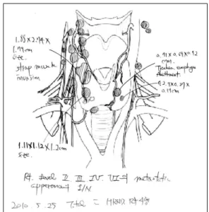
Fig. 1. Schematic picture of neck as a result of preoperative US.
disease; tumor invades prevertebral fascia or encases carotid artery or mediastinal vessel, N1a: metastasis to level VI (pretracheal, paratracheal, and prelar- yngeal/Delphian lymph nodes), N1b: metastasis to unilateral, bilateral, or contralateral cervical (level I, II, III, IV, or V) or retropharyngeal or superior mediastinal lymph nodes (level VII)). The longest tu- mor size, extrathyroidal extension and invasion of adjacent organs were each evaluated to determine T stage. The US criterion for extrathyroidal extension was contact with the adjacent thyroid capsule along more than 25% of the perimeter of the tumor.(10) In the case of multiple papillary thyroid carcinomas, the most extensive tumor had priority for determination of the T stage.
Both lateral compartments (level II-V) and the central compartment (level VI) were examined to de- termine N stage. The US criteria for lymph node me- tastasis was focal or diffuse hyperechogenicity, mi- cro-calcification, cystic change, round shape and abnormal vascular pattern (chaotic or periph- eral).(11,12)
The multifocality and bilaterality of the malignant thyroid nodules also were evaluated. Multifocality was defined as the “observation of multiple PTCs in one thyroid lobe”, and bilaterality was defined as
“multiple PTCs in both thyroid lobes”.
After finishing the full evaluation, all malignant lesions in the thyroid and metastatic lymph nodes were described on schematic picture of neck (Fig.1).
3. Surgical procedures
Total thyroidectomy was performed for these pa- tients who had, at least, one condition among them;
cancer size ≥ 1 cm, extrathyroidal extension, multi- focal papillary thyroid cancer, bilateral papillary thyroid cancer, suspicious lymph node metastasis on preoperative US, male patients; total 185 patients underwent total thyroidectomy. Near total thyroi- dectomy was performed for these patients who had all
these conditions; 1) only one micropapillary carcino- ma confined within ipsilateral thyroid lobe, 2) in- determinate nodules on preoperative US are in con- tralateral lobe, 3) no suspicious lymph node meta- stasis on preoperative US, 4) female patients; three patients underwent near total thyroidectomy. Ipsi- lateral lobectomy was performed for these patients who had all these condition; 1) only one micro- papillary carcinoma confined within ipsilateral lobe without any nodules in contralateral lobe, 2) no sus- picious lymph node metastasis on preoperative US, 3) female patients; ipsilateral lobectomy was performed on four patients. Central compartment dissection was routinely performed for all patients. Lymph node picking was performed on patients who had suspi- cious metastatic lymph node in lateral compartment as a result of preoperative US without cytologic confirmation. If the node picked up was metastatic node on frozen section, modified radical lymph node dissection was performed. Modified radical lymph node dissection was selectively performed on patients who had metastatic lymph node in lateral compart- ment confirmed by fine needle aspiration cytology preoperatively. Twenty-seven of the patients under- went lateral compartment dissection.
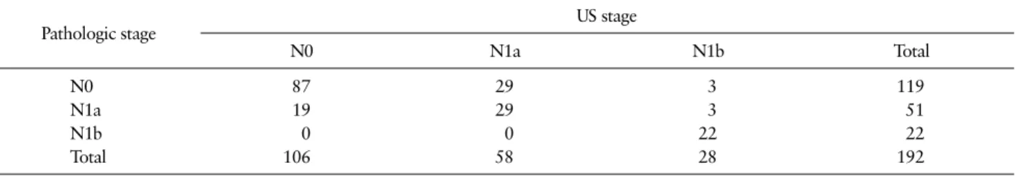
Table 2. Sonographic Versus Pathologic N Stage
Pathologic stage US stage
N0 N1a N1b Total
N0 87 29 3 119
N1a 19 29 3 51
N1b 0 0 22 22
Total 106 58 28 192
Table 1. Sonographic Versus Pathologic T Stage Pathologic stage
(n = patients)
US stage (n = patients)
T1 T2 T3 T4 Total
T1 74 1 14 0 89
T2 0 1 2 0 3
T3 9 0 80 0 89
T4 0 0 0 11 11
Total 83 2 96 11 192
4. Statistical analysis
The sonographic T stage, N stage, multifocaity, and bilaterality were compared with the histopatho- logic results, and sensitivity, specificity, positive pre- dictive value (PPV), negative predictive value (NPV), and accuracy were calculated. These values are based on number of patients.
RESULTS
Multifocality was identified in 82 patients (42.7%) pathologically. The sensitivity, specificity, PPV, NPV, and accuracy of preoperative US were 78.1% (64/82), 94.5% (104/110), 91.4% (64/70), 85.2% (104/122), and 87.5% (168/192) to predict multifocality. Bilaterality was identified in 59 patients (30.7%) pathologically.
The sensitivity, specificity, PPV, NPV and accuracy of preoperative US were 76.3% (45/59), 97.7% (130/133), 93.8% (45/48), 90.3% (130/144), and 91.1% (175/192) to predict bilaterality.
The overall accuracy of preoperative US for T stage was 86.5% (166/192). The individual accuracies of pre-
operative T stage for T1, T2, T3, and T4 were 89.2%
(74/83), 50% (1/2), 83.3% (80/96), and 100% (11/11) (Table 1). Extrathyroidal extension was detected in 101 patients (52.6%). The sensitivity, specificity, PPV, NPV, and accuracy of preoperative US were 91.1%
(92/101), 82.4% (75/91), 85.2% (92/108), 89.3% (75/84), and 87.0% (167/192).
The overall accuracy of preoperative US for N stage was 71.9% (138/192). The accuracies of preoperative US for N0, N1a, and N1b were 82.1% (87/106), 50.0%
(29/58), and 78.6% (22/28) (Table 2).
Lymph node metastasis in the central compartment was observed in 73 patients (38.0%). The sensitivity, specificity, PPV, NPV, and accuracy of preoperative US were 74.0% (54/73), 73.1% (87/119), 62.8% (54/86), 82.1% (87/106), and 73.4% (141/192). Lateral lymph node metastasis was detected in 22 patients (11.5%).
The sensitivity, specificity, PPV, NPV, and accuracy of preoperative US were 100% (22/22), 96.5%
(164/170), 78.6% (22/28), 100% (164/164), and 96.9%
(186/192).
DISCUSSION
The trend in thyroid surgery has been changed to a more conservative approach.(13) In this context, it has become important to determine the optimal sur- gical extent for patients with PTC preoperatively.
Risk factors (TNM stage, tumor size, extrathyroidal extension, lymph node metastasis, and multifocality), bilaterality and lymph node metastasis in central or lateral compartment should be incorporated into the decision-making process concerning the extent of the thyroidectomy and lymph node dissection to be performed.
Basically, no study has been conducted to check multifocality on the preoperative US. Our data showed that high specificity (94.5%), PPV (91.4%), and accu- racy (87.5%) of preoperative US for predicting multi- focality. Choi et al. (13) showed high accuracy (88.6%) and specificity (94.2%) of preoperative US for pre- dicting bilaterality. Our data showed higher specif- icity (97.7%), PPV (93.8%), NPV (90.3%), and 91.1%
(175/192) of preoperative US to predict bilaterality when compared to the other study.(13)
The sensitivity, specificity and accuracy of pre- operative US diagnosis of extrathyroidal extension were 91.1% (92/101), 82.4% (75/91), and 87.0%
(167/192). These results were better (or at least com- parable) to the findings yielded by other studies.(13,14) Some studies have shown relatively low sensitivity (9.5-61%) of preoperative staging US for lymph nodes in central compartment and high sensitivity (64-93.9%) for lymph nodes in lateral compartment.(14-16) Our data showed higher sensitivity to detect central and lateral lymph node metastasis compare to the other studies.(13,14) However, our data also showed sensi- tivity to detect central lymph node metastasis (N1a) is lower than sensitivity for lateral lymph node meta- stasis (N1a). The low sensitivity of US in the de- tection of central lymph node metastasis may be due to limitations in this area of the neck as a result of
surrounding structures such as the clavicle, ster- num, and tracheal air shadow.(17) Delicate scanning to detect lateral lymph node metastasis is more crit- ical to determine the extent of lymph node dissection in our institution, because the central compartment is routinely dissected for therapeutic cause and for prevention of recurrence of PTC. However, we at- tempted to identify and assess the central lymph node metastasis as much as possible, in an effort to figure out clinical risk of recurrence preoperatively and to demonstrate reasonable results of surgeon-per- formed US (SUS).
Since 2008, outpatient clinic-based SUS has been used routinely at outpatient institutions to evaluate patients who present with thyroid nodules. We were able to determine that the use of SUS is an indis- pensable part of the physical examination when eval- uating any patient who presents with thyroid pathology.
The SUS provides live images and immediate in- formation that is useful, even critical, for the specif- ic purpose of planning the best surgical approach for the surgeon.(18) However, US is a mixture of art and science, and the need for interpretation makes it a highly operator-dependent modality. Interpretation of images is subjective and individual, and that vari- ability in image interpretation between sonographers can be problematic.(19,20) Experienced thyroid sur- geons have advantages in terms of performing US.
Surgeons have access to all pertinent clinical in- formation at time of US, an excellent understanding of the relevant anatomy and the benefit of feedback from final pathologic results to continue to learn the finer nuances of US findings with the neck.(19,21,22) Radiology educational literature emphasizes the im- portance of repetition, in addition to familiarity with the key imaging characteristics, to promote greater accuracy of thyroid US interpretation.(23) Thyroid surgeons, by using US weekly in both the clinic and operating room, can quickly develop the skill set necessary to proficiently and accurately perform
Fig. 3. (A) The US finding showed the metastatic lymph node in lateral compartment. (B) The specimen taken during surgery was the same metastatic lymph node that we have detected when preoperative US was performed.
Fig. 2. (A) The US finding showed the metastatic lymph node in central compartment. (B) The specimen ex- cised during surgery was the same metastatic lymph node that we have detected when preoperative US was performed.
thyroid US.
The present study has several limitations. Firstly, most patients have T1, T3 tumor. Only one patient have T2 tumor and eleven patients have T4 tumor.
Also, only 22 patients had lateral lymph node meta- stasis. Thus, the accuracy of US for preoperative evaluation of PTC with T2, T4 tumor or lateral lymph node metastasis could not be determined accurately in this study. Secondly, basically, we tried to match metastatic lymph nodes (one by one) after finishing surgery (Fig. 2, 3). However, it was impossible in some cases. A compartment-oriented approach was therefore implemented to match in these cases. The findings of this study should be confirmed, lymph node by node, for all patients.
CONCLUSION
In this study, we evaluated the efficacy of sur- geons-performed preoperative US. Obviously, US is a good technique for preoperative staging of PTC and is helpful for accurate prediction of extrathyroidal ex- tension, multifocality and bilaterality. Surgeons-per- formed preoperative staging US might be accurate and is adequate for presurgical planning.
REFERENCES
1. Kim KM, Park JB, Bae KS, Kim CB, Kang DR, Kang SJ. Clinical prognostic index for recurrence of papil- lary thyroid carcinoma including intraoperative findings.
Endocr J 2013;60:291-7.
2. Sebastian SO, Gonzalez JM, Paricio PP, Perez JS, Flores DP, Madrona AP, et al. Papillary thyroid car- cinoma: prognostic index for survival including the histological variety. Arch Surg 2000;135:272-7.
3. Mazzaferri EL. Papillary thyroid carcinoma: factors influencing prognosis and current therapy. Semin Oncol 1987;14:315-32.
4. Mazzaferri EL, Jhiang SM. Long-term impact of ini- tial surgical and medical therapy on papillary and follicular thyroid cancer. Am J Med 1994;97:418-28.
5. Samaan NA, Maheshwari YK, Nader S, Hill CS Jr, Schultz PN, Haynie TP, et al. Impact of therapy for differentiated carcinoma of the thyroid: an analysis of 706 cases. J Clin Endocrinol Metab 1983;56:1131-8.
6. Kim KM, Park JB, Bae KS, Kang SJ. Analysis of prognostic factors in patients with multiple recur- rences of papillary thyroid carcinoma. Surg Oncol 2012;21:185-90.
7. Sherman SI. Thyroid carcinoma. Lancet 2003;361:
501-11.
8. Sosa JA, Udelsman R. Papillary thyroid cancer. Surg Oncol Clin N Am 2006;15:585-601.
9. Kim KM, Park JB, Bae KS, Kang SJ. Analysis of clin- icopathologic factors associated with bilateral thyroid micro papillary carcinoma. Korean J Endocr Surg 2011;11:18-21.
10. Kwak JY, Kim EK, Youk JH, Kim MJ, Son EJ, Choi SH, et al. Extrathyroid extension of well-differ- entiated papillary thyroid microcarcinoma on US.
Thyroid 2008;18:609-14.
11. Na DG, Lim HK, Byun HS, Kim HD, Ko YH, Baek JH.
Differential diagnosis of cervical lymphadenopathy:
usefulness of color Doppler sonography. AJR Am J Roentgenol 1997;168:1311-6.
12. Rosário PW, de Faria S, Bicalho L, Alves MF, Borges MA, Purisch S, et al. Ultrasonographic differentiation between metastatic and benign lymph nodes in pa- tients with papillary thyroid carcinoma. J Ultrasound Med 2005;24:1385-9.
13. Choi JS, Chung WY, Kwak JY, Moon HJ, Kim MJ, Kim EK. Staging of papillary thyroid carcinoma with ul- trasonography: performance in a large series. Ann Surg Oncol 2011;18:3572-8.
14. Park JS, Son KR, Na DG, Kim E, Kim S. Performance of preoperative sonographic staging of papillary thy- roid carcinoma based on the sixth edition of the
AJCC/UICC TNM classification system. AJR Am J Roentgenol 2009;192:66-72.
15. Choi JS, Kim J, Kwak JY, Kim MJ, Chang HS, Kim EK. Preoperative staging of papillary thyroid carci- noma: comparison of ultrasound imaging and CT. AJR Am J Roentgenol 2009;193:871-8.
16. Kim E, Park JS, Son KR, Kim JH, Jeon SJ, Na DG.
Preoperative diagnosis of cervical metastatic lymph nodes in papillary thyroid carcinoma: comparison of ultrasound, computed tomography, and combined ul- trasound with computed tomography. Thyroid 2008;
18:411-8.
17. Loevner LA, Kaplan SL, Cunnane ME, Moonis G.
Cross-sectional imaging of the thyroid gland. Neu- roimaging Clin N Am 2008;18:445-61, vii.
18. Méndez W, Rodgers SE, Lew JI, Montano R, Solórzano CC. Role of surgeon-performed ultrasound in pre- dicting malignancy in patients with indeterminate thyroid nodules. Ann Surg Oncol 2008;15:2487-92.
19. Hwang HS, Orloff LA. Efficacy of preoperative neck ultrasound in the detection of cervical lymph node metastasis from thyroid cancer. Laryngoscope 2011;
121:487-91.
20. Kabaker AS, Tublin ME, Nikiforov YE, Armstrong MJ, Hodak SP, Stang MT, et al. Suspicious ultrasound characteristics predict BRAF V600E-positive papil- lary thyroid carcinoma. Thyroid 2012;22:585-9.
21. Mazzaglia PJ. Surgeon-performed ultrasound in pa- tients referred for thyroid disease improves patient care by minimizing performance of unnecessary pro- cedures and optimizing surgical treatment. World J Surg 2010;34:1164-70.
22. Milas M, Stephen A, Berber E, Wagner K, Miskulin J, Siperstein A. Ultrasonography for the endocrine sur- geon: a valuable clinical tool that enhances diagnostic and therapeutic outcomes. Surgery 2005;138:1193- 200; discussion 1200-1.
23. Kim HG, Kwak JY, Kim EK, Choi SH, Moon HJ. Man to man training: can it help improve the diagnostic performances and interobserver variabilities of thy- roid ultrasonography in residents? Eur J Radiol 2012;
81:e352-6.