ARTICLE
J Korean Thyroid Assoc 2013 November 6(2): 91-95 http://dx.doi.org/10.11106/jkta.2013.6.2.91Received October 16, 2012 / Revised December 3, 2012 / Accepted December 4, 2012
Correspondence: Koon Soon Kim, MD, PhD, Department of Internal Medicine, Chungnam National University School of Medicine, 282 Munhwa-ro, Jung-gu, Daejeon 301-721, Korea
Tel: 82-42-280-7134, Fax: 82-42-280-7995, E-mail: kunsunkim@naver.com
Copyright ⓒ 2013, the Korean Thyroid Association. All rights reserved.
This is an open-access article distributed under the terms of the Creative Commons Attribution Non-Commercial License (http:// creative- commons.org/licenses/by-nc/3.0/), which permits unrestricted non-commercial use, distribution, and reproduction in any medium, provided the original work is properly cited.
L1 세포부착물질과 암의 증식, 이동, 침윤
충남대학교 의과대학 충남대학교병원 내분비대사질환 특성화 연구센터
김군순
Aberrant L1 Cell Adhesion Molecule Expression in Cancer:
In View of Proliferation, Migration and Invasion
Koon Soon Kim
Research Center for Endocrine and Metabolic Diseases, Division of Endocrinology, Department of Internal Medicine, Chungnam National University School of Medicine, Daejeon, Korea
L1 cell adhesion molecule (L1CAM) is a 220-kDa type I membrane glycoprotein and is normally expressed in neuronal cells, endothelial cells, and renal epithelial cells. Recent clinical studies demonstrated aberrant L1CAM expression in various cancers, especially at the invasive area of cancers. L1CAM has a key role in tumorigenesis, tumor invasion, and it is associated with a poor prognosis of cancer. Anaplastic thyroid carcinoma (ATC) has a highly poor outcome and it is resistant to conventional treatment. In this review, I discuss the biological role of L1CAM in proliferation, migration, and invasion in the ATC.
Key Words: L1CAM, Anaplastic thyroid carcinoma
서 론
우리나라의 갑상선암은 대부분 갑상선유두암(papillary thyroid carcinoma, PTC)이고 생존율이 매우 높은 반면 미분화갑상선암(anaplastic thyroid carcinoma, ATC)은 적은 유병률에도 가장 공격적이고 예후가 좋지 않은 암 중 하나로 현재까지도 평균 6개월의 생존율이 지속 되고 있다.1) 미분화갑상선암의 기원에 대해서는 논란 이 있지만 de novo 발생을 하거나 기존에 존재하던 분 화갑상선암(differentiated thyroid carcinoma, DTC)에서 발생하는 것으로 알려져 있으며,1-3) 임상과 연구 면에 서 두 가지의 unmet need가 있다. 첫째, 미분화갑상선 암 환자의 치료를 위해서 새로운 biomarker 발굴 및 표 적치료제 개발이 절실하고, 둘째, 미분화갑상선암의 기 원이 다른 점은 갑상선암의 발생 및 진행에 대한 좋은
연구 모델이 될 수 있다.
갑상선암의 발생 및 진행을 유도하는 RAS, BRAF 유 전자 변이, RET/PTC 유전자 재배열 등 다양한 유전자 변이에 대해서 활발하게 연구가 이루어지고 있으나 갑 상선암의 이동, 침윤 및 전이에 기여하는 세포부착물 질(cell adhesion molecules)의 연구는 적은 실정이다.
또한, 기존에 알려진 종양의 진행에 관련된 세포부착 물질들로는 cadherins, integrins, selectin과 immunoglo- bulin CAM superfamily 등이 있는데 대부분 종양의 진 행과 관련된 현상을 설명하는데 그치고 있으며,1) 임상 적인 예후 및 치료 타깃의 역할을 충족하기에는 한계 가 있다.
따라서 본 종설에서는 L1 세포부착물질(L1 cell adhesion molecule, L1CAM)과 암의 임상적인 예후와의 관련성 과 치료 타깃으로서의 역할, 종양의 증식(proliferation), 이동(migration), 침윤(invasion)을 유도하는 생물학적
Fig. 1. Structure of L1CAM.
인 역할에 대해서 지금까지 보고된 연구들을 기초로 고찰해 보고자 한다.
L1CAM의 임상적인 의의: 암의 불량한 예후인자 및 치료 타깃으로서의 역할
L1CAM은 정상적으로 세포막에 발현하여 신경의 분화, 발달 및 유지에 중요한 역할을 하는 것으로 알려 져 있고, L1CAM의 다양한 돌연변이로 인하여 신경계 발달에 장애가 와서 CRASH 증후군(정신지체, 뇌수 종, 경직성 마비, 내전엄지, 뇌량 형성부전)과 같은 심 각한 뇌기능 저하 및 운동장애가 발생하기도 한다.4,5) L1CAM은 일부 기관에서도 발견되지만 아직 그 역할 에 대해서 알려진 바가 미미하며,6) 비정상적으로 암세 포에 발현할 경우에 매우 불량한 예후와 연관되어 있 다.7,8) 즉 난소암, 자궁암,9) 대장암,10,11) 담도암,12) 담낭 암,13) 췌장암14) 등을 분석한 결과 암 조직의 10-40%에 서 L1CAM이 발현이 되었고, L1CAM이 발현되지 않는 각각의 대조군에 비해서 불량한 예후와 밀접한 관련이 있었다. 또한, 난소암을 가진 rodent를 대상으로 연구한 결과에서는 L1CAM을 억제하는 항체의 투여가 종양의 성장과 전이를 의미 있게 억제시켰다.15,16) 따라서, 기전 은 아직 불분명하지만 암세포의 세포막에 비정상적으 로 L1CAM이 발현하게 되면, 사망률이 증가하고, L1CAM을 억제하는 항체치료가 효과가 있는 점은 L1CAM이 진단 및 치료의 역할을 동시에 만족하는 생 물학적인 타깃이 됨을 알 수 있다.
L1CAM의 상호작용
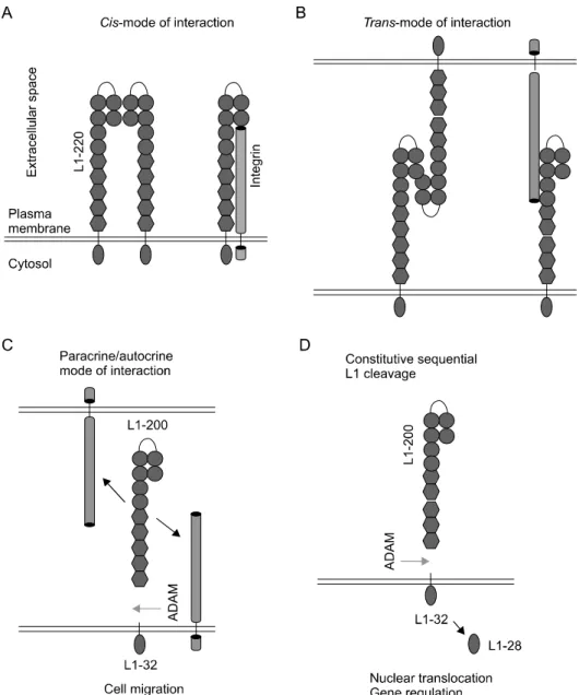
L1CAM은 세포막에 발현되는 220 kda 크기의 면역 글로불린 슈퍼가족의 하나인데, 세포막을 기준으로 세 포막외 영역(extracellular domain), 세포막을 통과하는 영역(transmembranous domain) 그리고 세포질내 영역 (cytoplasmic domain)의 3부분으로 구성되어 있다. 이 중 세포막외 영역은 6개의 면역글로불린(immuno- globulin)과 5개의 파이브로넥틴타입 III 반복 도메인 (fibronectin type III)으로 이루어져 있다(Fig. 1). 이러한 구조적인 특징은 동형친화성 상호작용(homophilic inte- raction)과 이종친화성 상호작용(heterophilic interaction) 을 유도해서 세포내, 세포상호 간 혹은 세포와 세포 외 기질 사이에 상호작용을 통하여 종양의 성장, 침윤과 전이에 중요한 역할을 할 수 있게 한다. 즉, 세포내 상 호작용은 L1CAM 간의 동형친화성 상호작용, L1CAM 과 다양한 티로신키나아제 수용체(receptor tyrosine kinase)나 인테그린(integrin)과의 이종친화성 상호작용 을 통하여 세포내 신호 전달을 증폭시킨다. 이러한 상 호 작용은 세포와 세포 사이에서 가능하며, 단백질 분 해 효소에 의해서 세포막외 영역이 분리된 후에도 같 은 상호작용을 통한 세포내 신호 전달 증폭을 유도할 수 있다(Fig. 2).17) L1CAM의 이러한 상호작용들은 불 량한 예후를 가지는 암들을 대변할 수 있는 구조적, 기 능적인 특징을 가지고 있음을 시사한다.
종양의 침윤 부위(invasive front)에서 L1CAM 발현
일반적으로 암과 관련된 사망의 90% 이상은 전이에 의하는 것으로 알려져 있다.18) 특히 상피세포암(epithelial cancer, carcinoma) 중에서 분화된 암의 침윤 및 전이가 일어날 때 원발 종양의 국소 침윤 부위에서 invasive front (역분화 혹은 탈분화 암세포군) 형태가 나타나는데, 이것은 epithelial-mesenchymal transition (EMT)의 특 징이며 국소 증식하던 암 덩어리에서 기원하여 암세포 의 이동성을 증가시켜서 전이를 유발하는 중요한 과정 으로 인식되고 있다.19) Invasive front에는 β-catenin이 핵으로 이동하여 발현이 증가되어 있는 것으로 알려져 있는데, L1CAM도 같은 부위의 invasive front에서 비정 상적으로 발현이 증가되어 있는 것이 관찰되었다.20) 이 러한 현상은 다양한 암 조직에서 동일한 패턴으로 나
타난다.8,12-14,21) 특히, 본 저자는 분석된 미분화갑상선암
Fig. 2. Interaction of L1CAM.
(anaplastic thyroid carcinoma) 환자의 9개 암 조직 모두 invasive front에 L1CAM의 발현이 의미 있게 증가한 것 을 증명하였다. 또한, 한 case는 처음에 분화갑상선유두 암으로 수술을 받고, 추적관찰 중에 재발한 갑상선암 이 미분화갑상선암으로 발현이 되었고, 처음 수술 당 시에 발현되지 않던 L1CAM이 재발한 미분화갑상선암 조직에서 강하게 발현이 되었으며, 일부 cases는 갑상 선유두암과 미분화갑상선암이 혼재하고 있는 패턴을 보이고 있었고, L1CAM이 강하게 발현되는 미분화갑 상선암 주변의 갑상선유두암 부위에서도 중등도의 L1CAM이 발현되고 있음을 확인하였다. 이러한 결과 들은 갑상선암의 역분화 과정에서 L1CAM이 중요한 역할을 하는 것을 시사한다.22)
L1CAM 과발현에 의한 정상 세포의 암화과정
마우스 섬유아세포(fibroblast)인 NIH3T3 세포에 L1CAM을 과발현시키면 대조군에 비해서 지속적인 Erk 활성화를 유도하여 세포의 증식과 생존율이 증가 하고, 이동성을 증가시키며, 제작된 세포들을 누드 마 우스에 접종하였을 때 L1CAM을 과발현시킨 그룹이 대조군에 비해서 종양생성이 의미 있게 증가하였다.20) 또한, 정상 췌관 세포인 H6c7세포에 TGF-β1을 처리 하면 대조군에 비해서 L1CAM 발현이 증가되어서 항 암제에 내성을 갖게 되고 이동성이 증가함을 증명하였
다.23) 이러한 현상은 전통적으로 알려진 다양한 종양유 전자(oncogene)에 의한 정상 세포의 암화과정 이론과 더불어서, 세포부착물질인 L1CAM의 비정상적인 발현 에 의해서도 상피세포의 특징을 잃어버리고 세포의 증 식 및 이동성이 증가하는 것을 증명한 실험으로 L1CAM이 정상 세포의 암화과정에 중요한 의의를 가 지고 있음을 알 수 있다.
L1CAM 발현에 의한 종양의 증식, 이동, 침윤에 미치는 생물학적인 역할
일반적으로 원발종양은 자가 증식을 통하여 암 덩어 리를 키운 후에 암의 이동, 침윤 및 전이 과정이 일어나 는 것으로 알려져 있는데, 암 덩어리가 작더라도 림프 선 전이나 장기전이가 나타나는 암들이 수술, 항암치 료 및 방사선치료에도 불구하고, 환자의 생존율을 급 격히 떨어뜨리는 현상이 많은 상피세포암에서 자주 관 찰된다. 또한, 전통적으로 수술 당시에 암의 분화도가 환자의 예후를 결정하는 것으로 알려져 있으나, 암의 분화도가 좋음에도 빠른 재발이나 예후가 좋지 않은 암들에 대해서도 다양한 접근에 의한 활발한 연구가 이루어지고 있으나 아직 정확한 기전이 알려져 있지 않다. 추정되는 기전들로는 암의 일차 치료 후에 항암 제에 내성을 가지는 세포들이 증식 및 전이되는 것과 일차 치료 당시부터 밝혀지지 않은 고도의 악성화 특 성을 가지는 암세포의 존재를 들 수 있다. 이러한 면에 서 세포부착물질인 L1CAM의 비정상적인 발현이 암세 포를 역분화시킴으로써 종양의 침윤과 전이에 기여하 는 연구 결과들이 중요함을 알 수 있다. 즉, 대장암세포 를 이용한 실험에서 L1CAM이 과발현되는 암세포가 발현이 없거나 적은 암세포에 비해서 증식성, 이동성 및 침윤성이 증가함을 증명하였다.20) 이러한 현상은 다 양한 암세포에서도 증명이 되고 있다.12-14,21,24-27)
갑상선암은 크게 분화갑상선암(DTC), 저분화갑상 선암(poorly differentiated thyroid carcinoma, PDTC), 갑 상선수질암(medullary thyroid carcinoma) 및 미분화갑상 선암(ATC)으로 분류되는데, 본 저자는 ATC에서 L1CAM 발현이 종양의 증식, 이동, 침윤 및 항암제 내성에 미치 는 영향을 알아보기 위해서 in vitro, in vivo 실험을 하 였다.22) DTC 세포주인 TPC1, BCPAP, primary cultured PTC에서는 L1CAM 발현이 없었고, 강력하게 L1CAM 을 발현하는 ATC 세포주인 FRO, 8505C에서 shRNA 기법을 이용하여 안정적으로 L1CAM 발현이 억제되는 세포주를 제작하였으며, L1CAM 발현이 억제된 ATC
세포주(L1 shRNA-FRO, L1 shRNA-8505C)가 대조군 (CTL shRNA-FRO, CTL shRNA-8505C)에 비해서 각 각 암세포의 증식, 이동, 침윤이 현저히 떨어짐을 확인 하였다. 또한, ATC 세포주인 FRO, 8505C에서 안정적 으로 L1CAM을 억제한 후에 gemcitabine, paclitaxel 항 암제를 처리하면 대조군에 비해서 의미 있게 항암제에 대한 반응이 증가하는 것을 증명하였다. 마지막으로 L1 shRNA-FRO, CTL shRNA-FRO 세포주(1.0×10⁷)를 각각 nude mice (n=6 per treatment group)에 피하 접종 을 한 후에 종양의 생성 및 생존에 대한 분석을 시행한 결과 대조군에 비해서 현저한 종양 생성의 감소 및 생 존의 증가가 있었다. 따라서 ATC에서 L1CAM 억제에 의한 종양 생성 감소, 암의 공격성 감소 및 항암제 반응 증가 결과를 바탕으로 향후에 L1CAM 표적 항체 치료 제를 개발한다면 기존의 치료 방법과 더불어서 ATC의 치료에 기여 할 수 있을 것으로 기대된다.
결 론
암세포가 비정상적인 L1CAM의 발현을 유도하는 기 전은 아직 명확하게 밝혀지지 않았으나 예후가 불량한 암들에서 L1CAM의 발현이 증명되고 있고, 암의 역분 화 과정에서 L1CAM의 생물학적인 중요성이 밝혀지고 있다. 따라서 향후 L1CAM 발현 조절 기작에 대한 연 구 및 치료 항체의 적용은 미분화갑상선암 환자의 생 존율을 증가시키는데 반드시 필요하다.
중심 단어: L1CAM, 미분화갑상선암.
References
1) Smallridge RC, Marlow LA, Copland JA. Anaplastic thyroid cancer: molecular pathogenesis and emerging therapies. Endocr Relat Cancer 2009;16(1):17-44.
2) Hadar T, Mor C, Shvero J, Levy R, Segal K. Anaplastic carcinoma of the thyroid. Eur J Surg Oncol 1993;19(6):511-6.
3) Spires JR, Schwartz MR, Miller RH. Anaplastic thyroid carcinoma. Association with differentiated thyroid cancer. Arch Otolaryngol Head Neck Surg 1988;114(1):40-4.
4) Dahme M, Bartsch U, Martini R, Anliker B, Schachner M, Mantei N. Disruption of the mouse L1 gene leads to malfor- mations of the nervous system. Nat Genet 1997;17(3):346-9.
5) Kenwrick S, Watkins A, De Angelis E. Neural cell recognition molecule L1: relating biological complexity to human disease mutations. Hum Mol Genet 2000;9(6):879-86.
6) Debiec H, Christensen EI, Ronco PM. The cell adhesion molecule L1 is developmentally regulated in the renal epithelium and is involved in kidney branching morphogenesis. J Cell Biol
1998;143(7):2067-79.
7) Huszar M, Moldenhauer G, Gschwend V, Ben-Arie A, Altevogt P, Fogel M. Expression profile analysis in multiple human tumors identifies L1 (CD171) as a molecular marker for differential diagnosis and targeted therapy. Hum Pathol 2006;37(8):1000-8.
8) Raveh S, Gavert N, Ben-Ze'ev A. L1 cell adhesion molecule (L1CAM) in invasive tumors. Cancer Lett 2009;282(2):137-45.
9) Fogel M, Gutwein P, Mechtersheimer S, Riedle S, Stoeck A, Smirnov A, et al. L1 expression as a predictor of progression and survival in patients with uterine and ovarian carcinomas.
Lancet 2003;362(9387):869-75.
10) Kaifi JT, Reichelt U, Quaas A, Schurr PG, Wachowiak R, Yekebas EF, et al. L1 is associated with micrometastatic spread and poor outcome in colorectal cancer. Mod Pathol 2007;20(11):
1183-90.
11) Boo YJ, Park JM, Kim J, Chae YS, Min BW, Um JW, et al. L1 expression as a marker for poor prognosis, tumor pro- gression, and short survival in patients with colorectal cancer.
Ann Surg Oncol 2007;14(5):1703-11.
12) Li S, Jo YS, Lee JH, Min JK, Lee ES, Park T, et al. L1 cell adhesion molecule is a novel independent poor prognostic factor of extrahepatic cholangiocarcinoma. Clin Cancer Res 2009;
15(23):7345-51.
13) Choi SY, Jo YS, Huang SM, Liang ZL, Min JK, Hong HJ, et al. L1 cell adhesion molecule as a novel independent poor prognostic factor in gallbladder carcinoma. Hum Pathol 2011;
42(10):1476-83.
14) Tsutsumi S, Morohashi S, Kudo Y, Akasaka H, Ogasawara H, Ono M, et al. L1 Cell adhesion molecule (L1CAM) expression at the cancer invasive front is a novel prognostic marker of pancreatic ductal adenocarcinoma. J Surg Oncol 2011;
103(7):669-73.
15) Arlt MJ, Novak-Hofer I, Gast D, Gschwend V, Moldenhauer G, Grunberg J, et al. Efficient inhibition of intra-peritoneal tumor growth and dissemination of human ovarian carcinoma cells in nude mice by anti-L1-cell adhesion molecule monoclonal antibody treatment. Cancer Res 2006;66(2):936-43.
16) Knogler K, Grunberg J, Zimmermann K, Cohrs S, Honer M, Ametamey S, et al. Copper-67 radioimmunotherapy and growth inhibition by anti-L1-cell adhesion molecule monoclonal anti-
bodies in a therapy model of ovarian cancer metastasis. Clin Cancer Res 2007;13(2 Pt 1):603-11.
17) Schafer MK, Altevogt P. L1CAM malfunction in the nervous system and human carcinomas. Cell Mol Life Sci 2010;67(14):
2425-37.
18) Gupta GP, Massague J. Cancer metastasis: building a framework.
Cell 2006;127(4):679-95.
19) Brabletz T. To differentiate or not--routes towards metastasis.
Nat Rev Cancer 2012;12(6):425-36.
20) Gavert N, Conacci-Sorrell M, Gast D, Schneider A, Altevogt P, Brabletz T, et al. L1, a novel target of beta-catenin signaling, transforms cells and is expressed at the invasive front of colon cancers. J Cell Biol 2005;168(4):633-42.
21) Min JK, Kim JM, Li S, Lee JW, Yoon H, Ryu CJ, et al. L1 cell adhesion molecule is a novel therapeutic target in intra- hepatic cholangiocarcinoma. Clin Cancer Res 2010;16(14):3571- 80.
22) Kim KS, Min JK, Liang ZL, Lee K, Lee JU, Bae KH, et al. Aberrant l1 cell adhesion molecule affects tumor behavior and chemosensitivity in anaplastic thyroid carcinoma. Clin Cancer Res 2012;18(11):3071-8.
23) Geismann C, Morscheck M, Koch D, Bergmann F, Ungefroren H, Arlt A, et al. Up-regulation of L1CAM in pancreatic duct cells is transforming growth factor beta1- and slug-dependent:
role in malignant transformation of pancreatic cancer. Cancer Res 2009;69(10):4517-26.
24) Meier F, Busch S, Gast D, Goppert A, Altevogt P, Maczey E, et al. The adhesion molecule L1 (CD171) promotes mela- noma progression. Int J Cancer 2006;119(3):549-55.
25) Zecchini S, Bianchi M, Colombo N, Fasani R, Goisis G, Casadio C, et al. The differential role of L1 in ovarian carcinoma and normal ovarian surface epithelium. Cancer Res 2008;68(4):1110-8.
26) Yang M, Li Y, Chilukuri K, Brady OA, Boulos MI, Kappes JC, et al. L1 stimulation of human glioma cell motility correlates with FAK activation. J Neurooncol 2011;105(1):27-44.
27) Hai J, Zhu CQ, Bandarchi B, Wang YH, Navab R, Shepherd FA, et al. L1 cell adhesion molecule promotes tumorigenicity and metastatic potential in non-small cell lung cancer. Clin Cancer Res 2012;18(7):1914-24.