ARTICLE
Clin Exp Thyroidol 2014 November 7(2): 129-135 http://dx.doi.org/10.11106/cet.2014.7.2.129Received March 25, 2014 / Revised June 16, 2014 / Accepted June 24, 2014
Correspondence: Seong Joon Kang, MD, Department of Surgery, Wonju Severance Christian Hospital, 20 Ilsan-ro, Wonju 220-701, Korea
Tel: 82-33-741-1303, Fax: 82-33-742-1815, E-mail: [email protected]
Copyright ⓒ 2014, the Korean Thyroid Association. All rights reserved.
This is an open-access article distributed under the terms of the Creative Commons Attribution Non-Commercial License (http:// creative- commons.org/licenses/by-nc/3.0/), which permits unrestricted non-commercial use, distribution, and reproduction in any medium, provided the original work is properly cited.
Regional Lymph Node Metastasis in Papillary Thyroid Cancer
Jae Hyun Park, Kang San Lee, Keum-Seok Bae and Seong Joon Kang
Department of Surgery, Yonsei University Wonju College of Medicine, Wonju, Korea
Papillary thyroid cancer is a common endocrine cancer and commonly presents with lymph node metastases.
It has been generally accepted that lymphatic drainage occurs from the thyroid primarily to the central lymphatic compartment and secondarily to the lateral compartment nodes. Recently, improvements in the resolution of imaging studies and the availability of highly sensitive thyroglobulin assays have highlighted the importance of identifying disease in the pre-operative assessment and dealing effectively with metastatic regional disease in order to prevent recurrence. However, there are limitations to diagnosing central lymph node metastases. With unreliable imaging modalities, prophylactic central lymph node dissection should be performed on all patients with papillary thyroid cancer. In comparison with the central compartment, prophylactic lateral node dissection has little or no effect on improving the prognosis of patients with papillary thyroid cancer. Therefore, lateral node dissection is recommended only as a part of the therapeutic procedure.
The extension of lateral neck dissection is recommended a comprehensive selective neck dissection of levels IIa, III, IV, and Vb. The rich lymphatic supply of the thyroid gland coupled with the propensity for nodal metastases in papillary thyroid cancer require the modern thyroid surgeon to be familiar with the indications for and techniques of regional lymph node dissection.
Key Words: Papillary thyroid cancer, Lymph node, Metastasis, Central compartment, Lateral neck
Introduction
Papillary thyroid carcinoma (PTC) is the most common endocrine malignancy, with an increasing incidence of, now, approximately 8.7 cases/100,000 per year.1) Overall, the 10-year disease-specific survival rate for PTC is 90%.2) Despite this excellent prognosis, cervical lymph node metastases are found in 40-90% of cases, andup to 20% of patients encounter recurrence of PTC after their primary operation.3-5) Almost four-fifths of all PTC recurrences occur in the neck alone, and, of these, three-fourths are associated with lymph node metas- tases.6,7) Because of its high incidence, regional lymph node metastasis has long been a critical issue with
PTC. However, the true impact of local lymph node metastasis on survival in PTC remains controversial.
Nonetheless, there are ample data in the literature as- sociating lymph node metastasis with decreased dis- ease-specific survival and increased risk of local recurrence. Outcome data from large institutional co- horts from the United States, Canada, and Germany have shown a significant and independent negative impact on survival with lymph node metastasis.8-10) Analysis of large cancer registry databases from the United States and Sweden has similarly shown poorer outcomes and increased mortality rate inpatients with locoregional lymph node metastases.11,12) Additionally, the presence of local lymph node metastasis has been associated with an increased risk of future local
recurrence.13)
Recently, improvements in the resolution of imaging studies and the availability of highly sensitive thyro- globulin assays have highlighted the importance of identifying disease in the pre-operative assessment and dealing effectively with metastatic regional disease in order to prevent recurrence. The aim of this review is to describe the contemporary approach to assess- ment and management of regional lymph node meta- stasis in PTC.
Lymphatic Drainage of the Thyroid
Accurate characterization and prediction of lym- phatic drainage of the thyroid gland is technically challenging because it is extensive and flows in many directions. Some authors have reported that each lobe of the thyroid gland has its own internal lymphatic system, and there is no communication with con- tralateral regional lymph nodes14,15) (although PTC can spread to regional lymph nodes bilaterally16-18)) It has been generally accepted that lymphatic drainage oc- curs from the thyroid primarily to the central lymphatic compartment and secondarily to the lateral compart- ment nodes.19,20) As a result, locoregional spread of thyroid cancer predominantly involves the ipsilateral, and sometimes also the contralateral central and lat- eral neck nodes.21-24)
Dralle and Machens25) suggested that lymphatic tu- mor cell dissemination is modulated by the anatomical location of the primary thyroid tumor. PTCs lodging in the upper thyroid pole often spread first to the upper parts of the ipsilateral lateral compartment, whereas primary tumors arising from the mid- and lower por- tions of the thyroid gland favor the central compartment.26,27) The different drainage of the supe- rior and inferior thyroid pole is a natural consequence of the peculiar anatomy of the thyroid gland’s lym- phatic system, which is organized in parallel to the gland’s venous drainage system: lymph vessels draining the upper thyroid pole accompany the supe- rior venous vessels to the ipsilateral lateral neck no- des; lymph vessels draining the middle portion of the thyroid gland follow the middle venous vessels to the
middle internal jugular nodes; and lymph vessels draining the inferior thyroid pole course together with the inferior pole veins to the central and lower ipsi- lateral lateral (supraclavicular) nodes.28,29)
Presentation of Nodal Metastases
In the past, the identification of cervical lymph node metastases was based primarily on palpation. However, enlarged cervical lymph nodes may not be easily palpable, especially when they are small, located behind the sternocleidomastoid muscles, or located behind a carotid artery or jugular vein and in level VI.
The introduction of diagnostic imaging modalities such as ultrasonography (US), computed tomography (CT), magnetic resonance imaging (MRI), and positron emis- sion tomography (PET) has increased detection of non-palpable cervical lymph node metastases.
US allows for accurate fine needle aspiration of suspicious nodes which will further aid the clinician in surgical planning. Fine needle aspiration samples may be evaluated with cytology and thyroglobulin assay on the aspirate sample. For these reasons it has be- come the gold standard for assessment of regional metastases. Also, Kim et al.30) attempted to create a new staging system by building a risk model asso- ciated with the disease-free survival rate using, with the factors that can be clinically ascertained with the operation, preoperative US as the basis and they veri- fied its usefulness by comparing this new staging sys- tem with 13 prior staging systems.
Despite the advantages of US, there are limitations to diagnosing central lymph node metastases. The sensitivity (27.3-55%) and specificity (69-90.0%) of preoperative US within the central compartment are lower than those (sensitivity 65-90.3%, specificity 82-94.8%) of the lateral compartment.31-34)
Recently several investigators have reported that preoperative US with CT had superior diagnostic perform ances to US alone, although the routine preoperative usage of CT is not recommended by the American Thyroid Association (ATA) guidelines under revision.33,35,36) However, US and CT together were still not sufficiently sensitive to detect small lymph node
metastases in the central neck.
Central Neck Dissection
Lymph nodes in the central compartment are from the thyroid notch to the innominate artery cepha- locaudal, and from one carotid artery to the other.
Central neck dissection refers to the systematic ex- cision of the prelaryngeal, pretracheal, and para- tracheal lymph nodes that reside in the level VI com- partment of the neck, and can be unilateral or bilateral.37)
In the published clinical guidelines of organizations such the Korean Thyroid Association (KTA), the ATA, the National Comprehensive Cancer Network (NCCN), and the British Thyroid Association (BTA), in cases of lymphadenopathy, the general recommendation is to perform lymph node dissection of the affected com- partments.38-41) However, there is virtually no consensus on the indications for prophylactic lymph node dissection. The NCCN guideline indicates prophylactic central neck dissection for patients aged <15 or
>45 years, with tumors >4 cm in diameter or with extrathyroidal extension.40) In the BTA guidelines, the above-mentioned indications for prophylactic central neck dissection are limited to male patients only.41) In the KTA and ATA guidelines, prophylactic central- compartment neck dissection (ipsilateral or bilateral) are recommended in patients with PTC with clinically uninvolved central neck lymph nodes, especially for advanced primary tumors (T3 or T4).38,39) Even among the major guidelines, there is no clear consensus re- garding the appropriate neck compartments to be dissected; therefore, many thyroid surgeons do not routinely perform systematic cervical lymph node dis- section due to elevated postoperative morbidities.42) This inconsistency between published guidelines for prophylactic lymph node dissection could be attribut- able to the lack of unbiased information on the pat- terns of lymph node metastases.
Proponents of routine prophylactic central neck dissection argue that lymph node metastases occur in 21-82% of cases43) and that removing involved nodes reduces disease persistence. An argument against
routine prophylactic central neck dissection is the ab- sence of evidence for improvement of long-term outcomes. Furthermore, clinically apparent metastatic lymphadenopathy (i.e., larger size, greater number, and extranodal extension) has a much worse prog- nosis than microscopic lymph node metastases,44-46) thus the long-term benefit of resection of occult mi- croscopic nodal metastases is unclear. Other important concerns include increased risks of temporary hypo- calcaemia after central neck dissection (compared with no neck dissection).47,48)
Some medical evidence suggests that prophylactic central neck dissection leads to decreased overall or disease-associated mortality in low-risk PTC. Two meta-analyses48,49) including mostly retrospective data have shown an absence of a clinically significant re- duction in disease recurrence from prophylactic cen- tral neck dissection. One single- center study has re- ported that routine central neck dissection might sig- nificantly reduce disease persistence or recurrence.50) In a multicenter observational study by Popadich and colleagues,51) fewer individuals treated with central neck dissection had central neck reoperations (1.5%), than did those not treated with central neck dissection (6.1%; p=0.004), although overall re-operation rates were no different. Permanent hypocalcaemia and vo- cal cord paralysis are not statistically significantly dif- ferent in some studies47,48) although temporary hypo- calcaemia is higher in patients with a history of a thy- roidectomy who are being treated with prophylactic central neck dissection compared with those without central neck dissection.47,48,52)
W hile it is clearly understood that mortality rates attributable to PTC are low, the problem of locore- gional recurrence can represent a major challenge to treating physicians and surgeons and is anissue that consumes considerable medical resources in the fol- low-up period. A serious problem caused by disease relapse in the central compartment might occlude the airway or esophagus, or paralyze the recurrent lar- yngeal nerve,53) and recurrent nerve injury frequently occurs during additional surgery for the dissection of recurrent nodes in the central compartment in com- parison to the initial surgery.54,55)
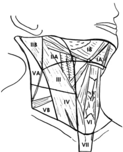
Fig. 1. Nodal levels with corresponding anatomic landmarks.
Accurate preoperative prediction of central lymph node metastases may be helpful in the management of patients with PTC, and may help to more carefully select patients for central neck dissection. But more sensitive and accurate methods are required to im- prove the detection of small lymph node metastasis in the central neck. With unreliable imaging modalities, prophylactic central lymph node dissection should be performed on all patients with PTC.
Lateral Neck Dissection
Metastatic PTC to the lateral compartment is clin- ically significant as it is thought to be a predisposing condition to distant metastasis. Due to the increasing incidence of thyroid cancer over the past four deca- des, there may also be a rise in the number of patients presenting with lateral neck disease and requiring neck dissection.1,56)
Historically, the management of lateral lymph node metastasis varies from “berry-picking” to modified radical neck dissection,57) and there is still no clear consensus regarding the appropriate neck level to which nodes must be removed.
The ATA in their 2009 thyroid nodule management guidelines advocated that a “therapeutic lateral neck compartmental lymph node dissection should be performed for patients with biopsy-proven metastatic lateral cervical lymphadenopathy.”35) The Triological Society followed in 2010, advocating a comprehensive selective neck dissection.58) Due to the variability in extent and definition of neck dissections, the ATA has recently published a consensus review on the anatomy, terminology, and rationale for lateral neck dissection in this population.59) The anatomic neck regions described include the upper jugular (II), the midjugular (III), the lower jugular (IV), and the posterior triangle (V). Level II can be divided into level IIa nodes, which surround the internal jugular vein, and level IIb nodes, which lie posterior to the spinal accessory.
Level V can also be divided into level Va nodes, which lie between the skull base and the level of the lower margin of the cricoid cartilage arch behind the posterior edge of the sternocleidomastoid muscle, and
level Vb nodes, which lie between the level of the lower margin of the cricoid cartilage and the level of the clavicle (Fig. 1).59)
In the absence of significant lymphadenopathy at levels IIa, III, IV and Vb, disease above the spinal accessory nerve (IIb or Va) or in level I is uncommon.
Sparing these lymph node basins will minimize risk of injury to the marginal mandibular and accessory nerves, whilst also preserving the submandibular salivary gland. Therefore, the ATA recommended a comprehensive selective neck dissection of levels IIa, III, IV, and Vb.59)
In comparison with the central compartment, the lateral neck harbors lower rates (23%)60) of occult metastasis and elective surgery results in higher postoperative complication rates. Despite this, some experts continue to advocate selective prophylactic lateral neck dissection.
Such groups report rates of occult disease greater than 20%, low rates of complication in their hands and more accurate staging of initial disease.60) However, even ex- pert groups report complication rates as high as 50%
following total thyroidectomy and lateral neck dissection, including hypocalcemia (30%), chronic neck pain (30%), chyle leak (3%) and recurrent laryngeal nerve palsy (10%).61)
There have been many studies suggesting that prophylactic lateral node dissection has little or no effect on improving the prognosis of patients with PTC.
A similarly good prognosis can be achieved by a sal- vage operation after lymph node recurrence in the lateral compartment, in comparison to prophylactic node dissection. Therefore, lateral node dissection is recommended only as a part of the therapeutic procedure.
Conclusion
The rich lymphatic supply of the thyroid gland cou- pled with the propensity for nodal metastases in PTC require the modern thyroid surgeon to be familiar with the indications for and techniques of regional lymph node dissection. Overt nodal metastasis is related with poor outcome in PTC, and they should be resected with the minimum of morbidity in order to reduce rates of regional recurrence and prolong survival. Accurate preoperative prediction of regional lymph node meta- stases by the variables obtained through the patient’s preoperative clinical information, imaging studies and intraoperative findings may be helpful in the manage- ment of patients with PTC, and may help to more carefully select patients for neck node dissection.
References
1) Davies L, Welch HG. Increasing incidence of thyroid cancer in the United States, 1973-2002. JAMA 2006;295(18):2164-7.
2) Malterling RR, Andersson RE, Falkmer S, Falkmer U, Nilehn E, Jarhult J. Differentiated thyroid cancer in a Swedish county--long-term results and quality of life. Acta Oncol 2010;49(4):454-9.
3) Grodski S, Cornford L, Sywak M, Sidhu S, Delbridge L.
Routine level VI lymph node dissection for papillary thyroid cancer: surgical technique. ANZ J Surg 2007;77(4):203-8.
4) Moo TA, McGill J, Allendorf J, Lee J, Fahey T 3rd, Zarnegar R. Impact of prophylactic central neck lymph node dissection on early recurrence in papillary thyroid carcinoma. World J Surg 2010;34(6):1187-91.
5) Enyioha C, Roman SA, Sosa JA. Central lymph node dissection in patients with papillary thyroid cancer: a population level analysis of 14,257 cases. Am J Surg 2013;205(6):655-61.
6) Caron NR, Clark OH. Well differentiated thyroid cancer. Scand J Surg 2004;93(4):261-71.
7) Ito Y, Tomoda C, Uruno T, Takamura Y, Miya A, Kobayashi K, et al. Ultrasonographically and anatomopathologically detectable node metastases in the lateral compartment as indicators of worse relapse-free survival in patients with papillary thyroid carcinoma. World J Surg 2005;29(7):917-20.
8) Mazzaferri EL, Jhiang SM. Long-term impact of initial surgical and medical therapy on papillary and follicular thyroid cancer. Am J Med 1994;97(5):418-28.
9) Simpson WJ, McKinney SE, Carruthers JS, Gospodarowicz MK, Sutcliffe SB, Panzarella T. Papillary and follicular thyroid cancer. Prognostic factors in 1,578 patients. Am J Med 1987;83(3):479-88.
10) Scheumann GF, Gimm O, Wegener G, Hundeshagen H, Dralle H. Prognostic significance and surgical management of locoregional lymph node metastases in papillary thyroid cancer.
World J Surg 1994;18(4):559-67; discussion 67-8.
11) Ries L YJ, Keel G, Eisner M, Lin Y, Horner M. SEER Survival Monograph Cancer Survival Among Adults: U.S. SEER Program, 1988-2001, Patient and Tumor Characteristics, 2007 NIH Pub. No. 07-6215 edition. National Cancer Institute, SEER Program, Bethesda, MD.
12) Lundgren CI, Hall P, Dickman PW, Zedenius J. Clinically significant prognostic factors for differentiated thyroid carcinoma:
a population-based, nested case-control study. Cancer 2006;
106(3):524-31.
13) Beasley NJ, Lee J, Eski S, Walfish P, Witterick I, Freeman JL. Impact of nodal metastases on prognosis in patients with well-differentiated thyroid cancer. Arch Otolaryngol Head Neck Surg 2002;128(7):825-8.
14) Qubain SW, Nakano S, Baba M, Takao S, Aikou T. Distribution of lymph node micrometastasis in pN0 well-differentiated thyroid carcinoma. Surgery 2002;131(3):249-56.
15) Fernandez-Cruz L, Astudillo E, Pera C. Lymphography of the thyroid gland: is intraglandular dissemination of thyroid carcinoma possible? World J Surg 1977;1(5):647-54.
16) Eichhorn W, Tabler H, Lippold R, Lochmann M, Schreckenberger M, Bartenstein P. Prognostic factors determining long-term survival in well-differentiated thyroid cancer: an analysis of four hundred eighty-four patients undergoing therapy and aftercare at the same institution. Thyroid 2003;13(10):949-58.
17) Noguchi M, Earashi M, Kitagawa H, Ohta N, Thomas M, Miyazaki I, et al. Papillary thyroid cancer and its surgical management. J Surg Oncol 1992;49(3):140-6.
18) Cranshaw IM, Carnaille B. Micrometastases in thyroid cancer.
An important finding? Surg Oncol 2008;17(3):253-8.
19) Noguchi S, Noguchi A, Murakami N. Papillary carcinoma of the thyroid. I. Developing pattern of metastasis. Cancer 1970;26(5):1053-60.
20) Ito Y, Miyauchi A. Lateral lymph node dissection guided by preoperative and intraoperative findings in differentiated thyroid carcinoma. World J Surg 2008;32(5):729-39.
21) Gimm O, Rath FW, Dralle H. Pattern of lymph node metastases in papillary thyroid carcinoma. Br J Surg 1998;85(2):252-4.
22) Machens A, Hinze R, Thomusch O, Dralle H. Pattern of nodal metastasis for primary and reoperative thyroid cancer.
World J Surg 2002;26(1):22-8.
23) Machens A, Holzhausen HJ, Dralle H. Contralateral cervical and mediastinal lymph node metastasis in medullary thyroid cancer: systemic disease? Surgery 2006;139(1):28-32.
24) Machens A, Hauptmann S, Dralle H. Prediction of lateral lymph node metastases in medullary thyroid cancer. Br J Surg 2008;95(5):586-91.
25) Dralle H, Machens A. Surgical management of the lateral neck compartment for metastatic thyroid cancer. Curr Opin Oncol 2013;25(1):20-6.
26) Park JH, Lee YS, Kim BW, Chang HS, Park CS. Skip lateral neck node metastases in papillary thyroid carcinoma. World J Surg 2012;36(4):743-7.
27) Zhang L, Wei WJ, Ji QH, Zhu YX, Wang ZY, Wang Y, et al. Risk factors for neck nodal metastasis in papillary thyroid microcarcinoma: a study of 1066 patients. J Clin Endocrinol Metab 2012;97(4):1250-7.
28) Von Lanz T, Wachsmuth W. Praktische anatomie, hals. Berlin:
Springer Verlag; 1955. p.243-4.
29) Foldi M, Kubik S. Lehrbuch der lymphologie. Stuttgart: Fischer Verlag; 1989. p.27-50.
30) Kim KM, Park JB, Bae KS, Kim CB, Kang DR, Kang SJ.
Clinical prognostic index for recurrence of papillary thyroid carcinoma including intraoperative findings. Endocr J 2013;
60(3):291-7.
31) Kouvaraki MA, Shapiro SE, Fornage BD, Edeiken-Monro BS, Sherman SI, Vassilopoulou-Sellin R, et al. Role of preoperative ultrasonography in the surgical management of patients with thyroid cancer. Surgery 2003;134(6):946-54; discussion 54-5.
32) Shimamoto K, Satake H, Sawaki A, Ishigaki T, Funahashi H, Imai T. Preoperative staging of thyroid papillary carcinoma with ultrasonography. Eur J Radiol 1998;29(1):4-10.
33) Ahn JE, Lee JH, Yi JS, Shong YK, Hong SJ, Lee DH, et al. Diagnostic accuracy of CT and ultrasonography for evaluating metastatic cervical lymph nodes in patients with thyroid cancer.
World J Surg 2008;32(7):1552-8.
34) Hwang HS, Orloff LA. Efficacy of preoperative neck ultrasound in the detection of cervical lymph node metastasis from thyroid cancer. Laryngoscope 2011;121(3):487-91.
35) American Thyroid Association (ATA) Guidelines Taskforce on Thyroid Nodules and Differentiated Thyroid Cancer, Cooper DS, Doherty GM, Haugen BR, Kloos RT, Lee SL, et al. Revised American Thyroid Association management guidelines for patients with thyroid nodules and differentiated thyroid cancer. Thyroid 2009;19(11):1167-214.
36) Kim E, Park JS, Son KR, Kim JH, Jeon SJ, Na DG.
Preoperative diagnosis of cervical metastatic lymph nodes in papillary thyroid carcinoma: comparison of ultrasound, computed tomography, and combined ultrasound with computed tomography.
Thyroid 2008;18(4):411-8.
37) Carty SE, Cooper DS, Doherty GM, Duh QY, Kloos RT, Mandel SJ, et al. Consensus statement on the terminology and classification of central neck dissection for thyroid cancer.
Thyroid 2009;19(11):1153-8.
38) Yi KH, Park YJ, Koong SS, Kim JH, Na DG, Ryu JS, et al. Revised Korean Thyroid Association management guidelines for patients with thyroid nodules and thyroid cancer. Endocrinol Metab 2010;25(4):270-97.
39) Hartl DM, Travagli JP. The updated American Thyroid Association Guidelines for management of thyroid nodules and differentiated thyroid cancer: a surgical perspective. Thyroid 2009;19(11):1149-51.
40) National Comprehensive Cancer Network. Clinical practice guidelines in oncology: thyroid carcinoma. [cited Nov 9, 2014]
http://www.nccn.org/professionals/physician_gls/f_guidelines.asp 41) British Thyroid Association and Royal College of Physicians.
Guidelines for the management of thyroid cancer, 2nd ed. [cited Nov 9, 2014] http://www.british-thyroid-association.org/news/Docs/
Thyroid_cancer_guidelines_2007.pdf, p.13-15.
42) Rotstein L. The role of lymphadenectomy in the management of papillary carcinoma of the thyroid. J Surg Oncol 2009;99(4):186-8.
43) Attie JN. Modified neck dissection in treatment of thyroid cancer:
a safe procedure. Eur J Cancer Clin Oncol 1988;24(2):315-24.
44) Bardet S, Malville E, Rame JP, Babin E, Samama G, De Raucourt D, et al. Macroscopic lymph-node involvement and neck dissection predict lymph-node recurrence in papillary thyroid carcinoma. Eur J Endocrinol 2008;158(4):551-60.
45) Ito Y, Tsushima Y, Masuoka H, Yabuta T, Fukushima M, Inoue H, et al. Significance of prophylactic modified radical neck dissection for patients with low-risk papillary thyroid carcinoma measuring 1.1-3.0 cm: first report of a trial at Kuma Hospital. Surg Today 2011;41(11):1486-91.
46) Moreno MA, Edeiken-Monroe BS, Siegel ER, Sherman SI, Clayman GL. In papillary thyroid cancer, preoperative central neck ultrasound detects only macroscopic surgical disease, but negative findings predict excellent long-term regional control and survival. Thyroid 2012;22(4):347-55.
47) Chisholm EJ, Kulinskaya E, Tolley NS. Systematic review and meta-analysis of the adverse effects of thyroidectomy combined with central neck dissection as compared with thyroidectomy alone. Laryngoscope 2009;119(6):1135-9.
48) Shan CX, Zhang W, Jiang DZ, Zheng XM, Liu S, Qiu M.
Routine central neck dissection in differentiated thyroid carcinoma: a systematic review and meta-analysis. Laryngoscope 2012;122(4):797-804.
49) Zetoune T, Keutgen X, Buitrago D, Aldailami H, Shao H, Mazumdar M, et al. Prophylactic central neck dissection and local recurrence in papillary thyroid cancer: a meta-analysis. Ann Surg Oncol 2010;17(12):3287-93.
50) Perrino M, Vannucchi G, Vicentini L, Cantoni G, Dazzi D, Colombo C, et al. Outcome predictors and impact of central node dissection and radiometabolic treatments in papillary thyroid cancers < or =2 cm. Endocr Relat Cancer 2009;16(1):201-10.
51) Popadich A, Levin O, Lee JC, Smooke-Praw S, Ro K, Fazel M, et al. A multicenter cohort study of total thyroidectomy and routine central lymph node dissection for cN0 papillary thyroid cancer. Surgery 2011;150(6):1048-57.
52) Giordano D, Valcavi R, Thompson GB, Pedroni C, Renna L, Gradoni P, et al. Complications of central neck dissection in patients with papillary thyroid carcinoma: results of a study on 1087 patients and review of the literature. Thyroid 2012;22(9):911-7.
53) Machens A, Hinze R, Lautenschlager C, Thomusch O, Dralle H. Thyroid carcinoma invading the cervicovisceral axis: routes of invasion and clinical implications. Surgery 2001;129(1):23-8.
54) Shaha AR. Revision thyroid surgery-technical considerations.
Otolaryngol Clin North Am 2008;41(6):1169-83, x.
55) Lefevre JH, Tresallet C, Leenhardt L, Jublanc C, Chigot JP, Menegaux F. Reoperative surgery for thyroid disease. Langenbecks Arch Surg 2007;392(6):685-91.
56) Davies L, Welch HG. Thyroid cancer survival in the United
States: observational data from 1973 to 2005. Arch Otolaryngol Head Neck Surg 2010;136(5):440-4.
57) Shaha AR, Shah JP, Loree TR. Patterns of nodal and distant metastasis based on histologic varieties in differentiated carcinoma of the thyroid. Am J Surg 1996;172(6):692-4.
58) Hasney CP, Amedee RG. What is the appropriate extent of lateral neck dissection in the treatment of metastatic well-differentiated thyroid carcinoma? Laryngoscope 2010;120(9):1716-7.
59) Stack BC Jr, Ferris RL, Goldenberg D, Haymart M, Shaha A, Sheth S, et al. American Thyroid Association consensus review and statement regarding the anatomy, terminology, and
rationale for lateral neck dissection in differentiated thyroid cancer. Thyroid 2012;22(5):501-8.
60) Hartl DM, Leboulleux S, Al Ghuzlan A, Baudin E, Chami L, Schlumberger M, et al. Optimization of staging of the neck with prophylactic central and lateral neck dissection for papillary thyroid carcinoma. Ann Surg 2012;255(4):777-83.
61) Roh JL, Park JY, Park CI. Total thyroidectomy plus neck dissection in differentiated papillary thyroid carcinoma patients:
pattern of nodal metastasis, morbidity, recurrence, and postoperative levels of serum parathyroid hormone. Ann Surg 2007;245(4):
604-10.