J. Exp. Biomed. Sci. 2012, 18(3): 227~236 pISSN : 1738-3226
Regulation of PPAR and SREBP-1C Through Exercise in White Adipose Tissue of Female C57BL/6J Mice
Sunhyo Jeong
†Division of Biohealth, Mokwon University, Daejeon 302-729, Korea
Previous study showed that swimming improved obesity but was not through PPARα activation in liver and skeletal muscle in high fat diet-fed female mice with functioning ovaries as an animal model of obese premenopausal women.
Thus, this study was aimed at investigation of the effects of swimming on the promotion of health and its molecular mechanism in adipose tissue of high fat diet-fed female mice. Eight-week-old female C57BL/6J mice were randomly divided into two groups (a non-swim control group and a swim group, n=8/group). Mice in the swim group swam for 2 h daily for 6 weeks in water bath with temperature of 35 ± 1℃. All the animals received high fat diet (45% kcal fat) for 6 weeks. Reverse transcription-polymerase chain reaction was used to elucidate the molecular mechanism. Female mice subjected to swimming had significantly decreased body weight gain and white adipose tissue mass compared with the female control mice. Histological studies illustrated that swimming decreases the hepatic lipid accumulation. As expected, swimming did not affect the expression of mRNA levels of peroxisome proliferator-activated receptor (PPAR) α and PPARα target genes responsible for mitochondrial fatty acid β-oxidation, such as carnitine palmitoyltransgerase-1 and medium chain acyl-CoA dehydrogenase in the white adipose tissue. However, mice that underwent 6-weeks of swimming exercise had decreased the mRNA expression of lipogenic genes, such as sterol regulatory element-binding proteins-1C and fatty acid synthase in comparison to sedentary control mice, with decreased PPARγ target genes involved in adipocyte-specific marker genes, such as adipocyte fatty acid binding protein and leptin in the white adipose tissue.
These results suggest that swimming can effectively prevent obesity induced by high fat diet-fed, in part through down-regulation of adipogenesis and lipogenesis in white adipose tissue of female obese mice. Moreover, these results suggest that swimming maybe contributing the promotion of health through regulation of adipogenesis and lipogenesis in overweight premenopausal women.
Key Words: Swimming, Female, PPAR, SREBP-1C, White adipose tissue
INTRODUCTION
Obesity has become one of the leading health care pro- blems in most industrialized countries. Apart from cosmetic reasons, obesity is undesirable because it increases the risk for numerous chronic diseases. Indeed, obesity hardly occurs
in isolation but is most often a part of an array of metabolic abnormalities including Type 2 diabetes, hypertension and hypertriglyceridemia (Bray, 2003). Obesity is the result of an energy imbalance caused by an increased ratio of caloric intake to energy expenditure, leading to a positive energy balance. Obesity is characterized by increased adipose tissue mass that results from both increased fat cell number and increased fat cell size (Couillard et al., 2000). In particular, accumulation of visceral fat is thought to play a major role in the pathogenesis of metabolic syndrome because the occurrence of the syndrome correlates with the amount of intra-abdominal fat, and intra-abdominal adipose tissue is lipolytically active (kissebah, 1997; Jensen, 2006).
Original Article
*Received: July 17, 2012 / Revised: August 16, 2012 Accepted: August 19, 2012
†Corresponding author: Sunhyo Jeong. Division of Biohealth, Mokwon University, Daejeon 302-729, Korea.
Tel: +82-42-829-7595, Fax: +82-42-829-7590 e-mail: [email protected]
○CThe Korean Society for Biomedical Laboratory Sciences. All rights reserved.
Many programs and strategies to prevent and treat obesity such as diet cure, exercise and pharmacological therapy have been developed (Müller et al., 2001; Swinburn and Egger, 2002; Halpern and Mancini, 2003). Exercise is a powerful modifier of the manifestations of metabolic syndrome in the direction of health enhancement (Pedersen and Saltin, 2006). Physical exercise rapidly increases energy expenditure and has been associated with improved weight control (King et al., 2001; Wier et al., 2001). Low-intensity and long-duration exercise also causes a preferential utilization of fat, resulting in less visceral fat in active individuals than sedentary controls (Thompson et al., 1998;
Morifuji et al., 2006; Redinger, 2009). Physical activity elicits physiological responses. Therefore, improved under- standing of the molecular mechanisms will help guide the proper use of regular exercise and physical activity in daily life, resulting in reduced incidence of lifestyle-related disease in modern society (Rogge, 2009; Saltin and Pilegaard, 2002).
The family of peroxisome proliferator-activated receptors (PPARs) plays a central role in energy balance. PPAR heterodimerizes with retinoid X receptor (RXR) and binds to PPAR response elements (PPREs) in the promoter region of target genes (Sander et al., 2000). Among the three PPAR isoforms, PPARαseems to be important in obesity and fat catabolism (Staels et al., 1998; Schoonjans et al., 2000). PPARα target genes include those involved in the hydrolysis of plasma triglycerides, fatty acid uptake and binding, and fatty acid β-oxidation (Zhang et al., 1992;
Hertz et al., 1995; Auwerx et al., 1996; Osumi et al., 1996;
Schoonjans et al., 1996; Martin et al., 1997; Nicolas-Frances et al., 2000). Therefore, the activation of PPARα target genes promotes increased fatty acids oxidation and the increased breakdown, reduced synthesis, and secretion of triglycerides. The PPARγ is a major regulator of glucose and lipid metabolism (adipogenesis and lipid storage) by modulating energy homeostasis in adipose tissue (Rosen et al., 2000; Semple et al., 2006). The activation of PPARγ is both necessary and sufficient to induce an adipose phenotype, which is defined by lipid accumulation and the expression of adipocyte-specific marker genes such as adipocyte fatty acid binding protein (aP2), lipoprotein lipase and adipsin (Rosen et al., 2000). Sterol regulatory element-binding
protein-1C (SREBP-1C) is also a transcription factor that stimulates the expression of numerous genes connected with lipogenesis, such as ATP-citrate lyase, acetyl-CoA carboxylase and fatty acid synthase (FAS) (Shimano et al., 1996; Shimano et al., 1997).
Previous studies did not clarify the molecular mechanisms through which swimming regulates lipid metabolism and protects development of obesity in high fat diet-fed female mice with functioning ovaries (Jun et al., 2010; Jeong and Yoon, 2011). Thus, this study determined that swimming regulates body weight gain and adipose tissue mass through the molecular mechanism involved in lipid metabolism in white adipose tissue of female C57BL/6J mice.
MATERIALS AND METHOD
Treatment of and exercise program (swimming) for animal
For all experiments, eight-week-old female mice (C57BL/
6J) were housed and bred at the Korea Research Institute of Bioscience and Biotechnology under pathogen-free conditions with a standard 12-h light/dark cycle. Prior to administration of special diets, mice were fed standard rodent chow and water ad libitum. Female mice are randomly divided into two groups (a non-swim control group and the swim group, n=8/group). Mice in the swim group swam for 2 h daily for 6 weeks in a water bath with temperature of 35 ± 1℃ (1 × 1 m, Jeiotech, Seoul, Korea);
during the first two weeks, the duration of daily training was increased from 10 min to 2 h. All the animals received high fat diet (45% kcal fat, Research Diets, New Brunswick, NJ) for 6 weeks and were sacrificed by cervical dislocation.
Tissues were harvested, weighed, snap frozen in liquid nitrogen and stored at -80℃ until use. An additional section of liver tissue was fixed in phosphate-buffered formalin for histological analysis.
Histological analysis
Liver tissues were fixed in 10% phosphate-buffered
formalin for l day and processed in a routine manner for
paraffin sectioning. Sections (5 μm) were stained with
hematoxylin and eosin for microscopic examination.
Analysis of target gene expression
Total RNA was isolated from parametrial adipose tissue using Trizol reagent (Gibco-BRL, Grand Island, NY), and relative levels of specific mRNA were assessed by reverse transcription-polymerase chain reaction (RT-PCR). Com- plementary DNA was synthesized from RNA samples by mixing 2 μg of total RNA and 0.5 μg of the reverse primer in water with total volume of 14 μl, heating the mixture at 75℃ for 15 min, cooling the mixture immediately in ice for 5 min, and adding 5X M-MLV reaction buffer, 10 mM dNTP mixture (Promega, Madison, WI, USA) and 200 units M-MLV RT (Promega, Madison, WI, USA) to total volume of 25 μl. Samples were incubated at 42℃ for 60 min. A 5 μl aliquot of the RT reaction was then used for subsequent PCR amplification with specific primers.
Twenty five microliters PCR sample contained 5 μl of the RT reaction, 10X buffer with MgCl
2, 10 mM dNTP, 5 units of Tag polymerase (Solgent, Taejon, Korea) and 10 μM of each primer. Primer sequences and PCR conditions are shown in Table 1. PCR was performed in a PTC-100
TMProgrammable Thermal Controller (MJ Research, Watertown, MA, USA). PCR products were electophoresed on 1%
agarose gel.
Table 1. Sequences of oligonucleotide primers and PCR conditions
Genes Primer sequences AT (℃) Cycle
F : 5'-attctggcccaccaacttcgg-3' PPARγ
R : 5'-tggaagcctgatgctttatcccca-3' 58 28
F : 5'-caaaatgtgtgatgcctttgtg-3'
aP2 R : 5'-ctcttcctttggctcatgcc-3' 58 24
F : 5'-ccaagaagagggatccctgctccagcagc-3' Leptin
R : 5'-agaatggggtgaagcccagga-3' 58 26
F : 5'-gcagctcgtacaggtcatca-3' PPARα
R : 5'-ctcttcatccccaagcgtag-3' 58 45
F : 5'-gacatttggaaagctgctagtg-3' MCAD
R : 5'-tcacgagctatgatcagcctctg-3' 58 43
F : 5'-tatgtgaggatgctgcttcc-3' CPT-1
R : 5'-ctcggagagctaagcttgtc-3' 52 40
F : 5'-cggctgtcgtctaccataagct-3' SREBP-1C
R : 5'-ccagtgttgccatggatatag-3' 58 28
F : 5'-tccaaggaagcctttgagaa-3'
FAS R : 5'-ccatcctcagtcccagaaaa-3' 55 30
F : 5'-tggaatcctgtggcatccatgaaac-3' β-actin
R : 5'-taaaacgcagctcagtaacagtccg-3' 58 28
AT: Annealing Temperature
Fig. 1. Effects of swimming on body weight gain in female mice at 6 weeks. Female C57BL/6J mice (n=8/group) with similar initial body weight were either subjected to swimming for 2 h daily or kept sedentary for 6 weeks. All values are expressed as mean
± SD. *P<0.05, as compared with their respective control groups.
Statistics
Unless otherwise noted, all values are expressed as mean
± standard deviation (SD). All data were analyzed by unpaired student's t-test for statistically significant differences between each of the groups.
RESULTS
Effects of swimming on body weight gain, white adipose tissue mass and hepatic lipid accumulation
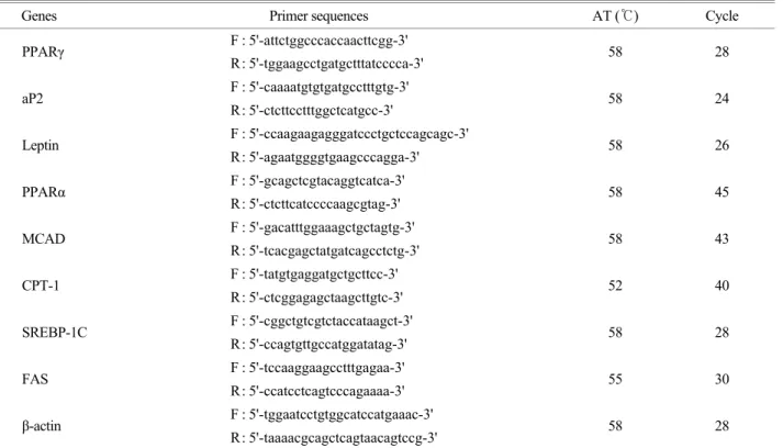
Swimming for 6 weeks had significantly decreased body weight gain by 36.7%, compared with sedentary controls (P<0.05) (Fig. 1). Also, compared with sedentary controls, swimming significantly decreased total white adipose tissue weight by 27.5%, and significantly decreased inguinal, parametrial and mesenteric adipose tissue weights by 26.0%, 35.9%, and 33.3%, respectively (Fig. 2). Moreover, it was found that hepatic lipid accumulation was lower in mice subjected to swimming than in sedentary controls (Fig. 3).
Effects of swimming on the expression of genes involved in lipid metabolism in the white adipose tissue
To evaluate whether the effects of swimming on body weight gain, adipose tissue mass and hepatic lipid accumu- lation are associated with alteration of genes expression in
Fig. 2. Effects of swimming on white adipose tissue mass in female mice at 6 weeks. Female C57BL/6J mice (n=8/group) with similar initial body weight were either subjected to swimming for 2 h daily or kept sedentary for 6 weeks. All values are expressed as mean ± SD. * P<0.05, as compared with their respective control groups.Fig. 3. Effects of swimming on hepatic lipid accumulation in female mice at 6 weeks. Representative hematoxylin and eosin- stained liver sections are shown (original magnification ×200).
Female C57BL/6J mice (n=8/group) with similar initial body weight were either subjected to swimming for 2 h daily or kept sedentary for 6 weeks. Arrows indicate the lipid droplets in hepatocytes.
lipid metabolism, measurement was taken on mRNA levels of the PPARα and PPARα target genes for mitochondrial fatty acid β-oxidation in parametrial adipose tissue (Fig. 4).
Compared with the control group mice, the swam group did not show elevations in mRNA levels of PPARα and PPARα target genes involved in mitochondrial fatty acid β-oxidation, such as CPT-1 and MCAD.
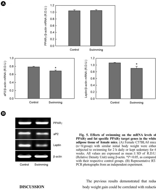
However, swimming decreased the expression of genes involved in adipogenesis and lipogenesis in parametrial adipose tissue of female mice (Fig. 5 and 6). Compared with sedentary controls, the swam group displayed significant
decreases in mRNA levels of adipocyte-specific PPARγ target genes, such as aP2 and leptin, by 12.8% and 16.0%, respectively (P<0.05), but there was no decrease in the expression of PPARγ mRNA. In addition, the effects of swimming on the expression of lipogenic genes in white adipose tissue of high fat diet-fed female mice were tested.
Swimming significantly decreased the levels of SREBP-1C and FAS, by 15.1% and 19.3%, respectively, in comparison to the control mice (P<0.05).
A
B
Fig. 4. Effects of swimming on the mRNA levels of PPARα and PPARα target genes involved in fatty acid β-oxidation in the white adipose tissue of female mice.
(A) Female C57BL/6J mice (n=8/group) with similar initial body weight were either subjected to swim training for 2 h daily or kept sedentary for 6 weeks. All values are expressed as mean ± SD of R.D.U. (Relative Density Unit) using β-actin. *P<0.05, as compared with their respective control groups. (B) Representative RT-PCR photographs from an independent experiment.
PPARα/β-actin mRNA (R.D.U.) MCAD/β-actin mRNA (R.D.U.)
CPT-1/β-actin mRNA (R.D.U.)
PPARα
β-actin
DISCUSSION
Obesity arises from the imbalance between energy intake and energy expenditure, leading to a pathological accumu- lation of lipids in adipocytes. Physical exercise rapidly increases energy expenditure and has been associated with improved weight control (Brook et al., 1995; King et al., 2001; Wier et al., 2001). In this study, it was demonstrated that swimming reduces body weight gain, white adipose tissues mass and hepatic lipid accumulation in high fat diet-fed female C57BL/6J mice.
The previous results demonstrated that reductions in body weight gain could be correlated with reductions in fat mass, indicating that reduced fat may lead to reduced body weight, and that the capacity for fat utilization is related to the development of obesity (Yoon et al., 2002; Yoon et al., 2003; Jeong et al., 2004a). Therefore, understanding of effect of exercise on lipid metabolism may assist in preventing obesity and in prescribing effective body or fat weight-loss strategies.
Adipocytes play a role in maintaining lipid homeostasis and energy balance in vertebrates by storing triglycerides or releasing free fatty acids in response to changing energy
AB
Fig. 5. Effects of swimming on the mRNA levels of PPARγ and fat specific PPARγ target genes in the white adipose tissue of female mice. (A) Female C57BL/6J mice (n=8/group) with similar initial body weight were either subjected to swimming for 2 h daily or kept sedentary for 6 weeks. All values are expressed as mean ± SD of R.D.U.
(Relative Density Unit) using β-actin. *P<0.05, as compared with their respective control groups. (B) Representative RT- PCR photographs from an independent experiment.
PPARγ/β-actin mRNA (R.D.U.)
aP2/β-actin mRNA (R.D.U.) Leptin/β-actin mRNA (R.D.U.)
PPARγ
β-actin
needs. So the study of adipose tissue is central to the under- standing of the metabolic abnormalities with development of obesity (Vázquez-Vela et al., 2008). For the purpose of examination of regulation of body weight gain and adipose tissue mass through what is molecular mechanism involved in lipid metabolism by swimming, this study determined
the expression of genes involved in lipid metabolism in white adipose tissue of female C57BL/6J mice.
This study found no differences in the expression of PPARα target genes involved in fat catabolism, or of PPARα between swam and sedentary control groups in white adipose tissue. PPARα is known to be activated by fatty acids (Kliewer et al., 1997), whose concentrations in plasma increase immediately with exercise (Mougios et al., 2003). The binding of fatty acids to PPARα increases the DNA binding activity of PPARα (Mochizuki et al., 2006).
However, the action of PPARα on lipid metabolism and obesity becomes negative due to estrogens, major ovarian factors in female mice (Yoon et al., 2003; Jeong et al., 2004b), because of a bidirectional signal cross-talk exists between PPARα and ERs (Wang and Kilgore, 2002; Jeong and Yoon, 2007). Accordingly, the unchanged expression levels of PPARα target genes by swimming in female mice with functioning ovaries may be due to interference of estrogens. Several previous studies also assist this suggestion.
Exercise increased mRNA expression of PPARα in male and female ovariectomized rats (Morifuji et al., 2006;
Pighon et al., 2011), but did not lead to increase in female with functional ovaries (Kannisto et al., 2006; Jun et al., 2010).
As weight is gained, adipose tissue mass has considerable capacity to expand through a complex interplay between proliferation and differentiation of preadipocytes into functional adipocytes (adipogenesis), and an increase in individual adipocyte size (hypertrophy) by accumulated increase in the levels of triglycerides in adipocytes (Bertrand et al., 1978). SREBP-1C is a transcription factor that stimulates the expression of numerous genes involved in triglyceride synthesis and lipogenesis (Shimano et al., 1996; Shimano et al., 1997). In this study, the swam group displayed decreased expression of SREBP-1C and FAS mRNA in white adipose tissue compared with sedentary controls. These results suggest that swimming decreased adipose tissue mass through inhibition of lipogenesis by down-regulation of SREBP-1C.
PPARγ has also attracted a lot of interest from researchers studying obesity. PPARγ is not only highly expressed in adipose tissue, but also plays a very important role in
Fig. 6. Effects of swimming on the mRNA levels of lipogenicgenes in the white adipose tissue of female mice. (A) Female C57BL/6J mice (n=8/group) with similar initial body weight were either subjected to swimming for 2 h daily or kept sedentary for 6 weeks. All values are expressed as mean ± SD of R.D.U. (Relative Density Unit) using β-actin. *P<0.05, as compared with their respective control groups. (B) Representative RT-PCR photographs from an independent experiment.
A
B
SREBP-1C/β-actin mRNA (R.D.U.)FAS/β-actin mRNA (R.D.U.)
β-actin
adipogenesis and lipogenesis (Kubota et al., 1999; Rosen et al., 1999; Rosen et al., 2000). However, this study illustrated that the swam group did not decrease PPARγ mRNA levels in white adipose tissue in comparison to the sedentary control group. This concept is supported by data from Kump et al. (2005) and Petridou et al. (2007), who showed that the PPARγ activity in exercised mice did not differ significantly from unexercised mice in terms of epididymal fat. However, the swam group displayed decrease in the expression of adipocyte-specific marker genes including aP2 and leptin. These genes are under transcriptional control of CCAAT/enhanced-binding protein α (C/EBPα), as well as PPARγ. C/EBPα is a key regulator of adipose cell development and binds to promoters and activates adipocyte-specific genes (Hwang et al., 1996; Hollenberg et al., 1997; Tang et al., 2004; Alonso-Vale et al., 2009).
Interestingly, it was reported that exercise decreased C/EBPα protein levels in epididymal adipose tissue, but not the PPARγ (Kump and Booth, 2005). Thus, these researchers support the present results on the presence of changes in the expression of adipocyte-specific PPARγ target genes with exercise despite the lack of changes in the PPARγ mRNA levels of adipose tissue. Moreover, the possibility that purpose of down-regulation of adipocyte-specific marker genes by swimming may be to reduce adipose C/EBPα activities in female C57BL/6J mice was suggested.
The previous report illustrated that swimming signifi- cantly decreased serum triglycerides and the average size of parametrial adipocytes in female mice with functional ovaries (Jun et al., 2010). With respect to morphological changes in which the increase in adipose tissue mass was due to the enlargement of the preexisting adipocytes with increased lipid accumulation (Ogawa et al., 2004; Villena et al., 2004; Yagi et al., 2004), the present study suggests that down-regulation of genes involved in adipogenesis and lipogenesis in white adipose tissue by swimming may be decrease adipocyte size, resulting in decrease of adipose mass and body weight gain. Moreover, these effects of swimming on lipogenic genes may contribute to decrease in serum lipid levels and hepatic lipid accumulation.
In conclusion, these results suggest that swimming can effectively prevent body weight gain induced by high fat
diet, and adipose mass and hepatic lipid accumulation, in part, through down-regulation of adipogenesis and lipo- genesis in the white adipose tissue of female mice with functional ovaries. Thus, this study may make contribution towards the understanding that exercise physiology is important for the maintenance of premenopausal women health.
Acknowledgements
This work was supported by a grant No. KRF-2009- 351-G00135 from the Korea Research Foundation.
REFERENCES
Alonso-Vale MI, Peres SB, Vernochet C, Farmer SR, Lima FB.
Adipocyte differentiation is inhibited by melatonin through the regulation of C/EBPbeta transcriptional activity. J Pineal Res. 2009. 47: 221-227.
Auwerx J, Schoonjans K, Fruchart JC, Staels B. Transcriptional control of triglyceride metabolism: fibrates and fatty acids change the expression of the LPL and apo C-III genes by activating the nuclear receptor PPAR. Atherosclerosis 1996.
124: S29-S37.
Bertrand HA, Masoro EJ, Yu BP. Increasing adipocyte number as the basis for perirenal depot growth in adult rats. Science 1978. 201: 1234-1235.
Bray GA. Risks of obesity. Endocrinol Metab Clin North Am.
2003. 32: 787-804.
Brook GA, Fahey TD, White TP. Human bioenergetics and its applications. Exerc Physiol. 1995. 2: 151-158.
Couillard C, Mauriège P, Imbeault P, Prud'homme D, Nadeau A, Tremblay A, Bouchard C, Després JP. Hyperleptinemia is more closely associated with adipose cell hypertrophy than with adipose tissue hyperplasia. Int J Obes Relat Metab Disord.
2000. 24: 782-788.
Halpern A, Mancini MC. Treatment of obesity: an update on anti- obesity medications. Obes Rev. 2003. 4: 25-42.
Hertz R, Bishara-Shieban J, Bar-Tana J. Mode of action of peroxisome proliferators as hypolipidemic drugs: suppression of apolipoprotein C-III. J Biol Chem. 1995. 270: 13470-13475.
Hollenberg AN, Susulic VS, Madura JP, Zhang B, Moller DE, Tontonoz P, Sarraf P, Spiegelman BM, Lowell BB. Functional antagonism between CCAAT/Enhancer binding protein-alpha
and peroxisome proliferator-activated receptor-gamma on the leptin promoter. J Biol Chem. 1997. 272: 5283-5290.
Hwang CS, Mandrup S, MacDougald OA, Geiman DE, Lane MD.
Transcriptional activation of the mouse obese (ob) gene by CCAAT/enhancer binding protein alpha. Proc Natl Acad Sci U S A. 1996. 93: 873-877.
Jensen MD. Is visceral fat involved in the pathogenesis of the metabolic syndrome? Human model. Obesity (Silver Spring).
2006. 14: 20S-24S.
Jeong S, Kim M, Han M, Lee H, Ahn J, Kim M, Song YH, Shin C, Nam KH, Kim TW, Oh GT, Yoon M. Fenofibrate prevents obesity and hypertriglyceridemia in low-density lipoprotein receptor-null mice. Metabolism 2004a. 53: 607-613.
Jeong S, Han M, Lee H, Kim M, Kim J, Nicol CJ, Kim BH, Choi JH, Nam KH, Oh GT, Yoon M. Effects of fenofibrate on high-fat diet-induced body weight gain and adiposity in female C57BL/6J mice. Metabolism 2004b. 53: 1284-1289.
Jeong S, Yoon M. Inhibition of the actions of peroxisome proliferator-activated receptor alpha on obesity by estrogen.
Obesity (Silver Spring). 2007. 15: 1430-1440.
Jeong S, Yoon M. Differential regulation of Obesity by swim training in female Sham-operated and ovariectomized mice.
J Exp Biomed Sci. 2011. 17: 13-20.
Jun J, Lee WL, Lee YR, Jeong S. Swim training fitness in high fat diet-fed female mice. J Exp Biomed Sci. 2010. 16: 151-159.
Kannisto K, Chibalin A, Glinghammar B, Zierath JR, Hamsten A, Ehrenborg E. Differential expression of peroxisomal pro- liferator activated receptors alpha and delta in skeletal muscle in response to changes in diet and exercise. Int J Mol Med.
2006. 17: 45-52.
King GA, Fitzhugh EC, Bassett Jr DR, McLaughlin JE, Strath SJ, Swartz AM, Thompson DL. Relationship of leisure-time physical activity and occupational activity to the prevalence of obesity. Int J Obes Relat Metab Disord. 2001. 25: 606-612.
Kissebah AH. Central obesity: measurement and metabolic effects.
Diabetes Reviews. 1997. 5: 8-20.
Kliewer SA, Sundseth SS, Jones SA, Brown PJ, Wisely GB, Koble CS, Devchand P, Wahli W, Willson TM, Lenhard JM, Lehmann JM. Fatty acids and eicosanoids regulate gene expression through direct interactions with peroxisome proliferator-activated receptors alpha and gamma. Proc Natl Acad Sci U S A. 1997. 94: 4318-4323.
Kubota N, Terauchi Y, Miki H, Tamemoto H, Yamauchi T, Komeda K, Satoh S, Nakano R, Ishii C, Sugiyama T, Eto K, Tsubamoto Y, Okuno A, Murakami K, Sekihara H, Hasegawa
G, Naito M, Toyoshima Y, Tanaka S, Shiota K, Kitamura T, Fujita T, Ezaki O, Aizawa S, Kadowaki T. PPAR gamma mediates high-fat diet-induced adipocyte hypertrophy and insulin resistance. Mol Cell. 1999. 4: 597-609.
Kump DS, Booth FW. Sustained rise in triacylglycerol synthesis and increased epididymal fat mass when rats cease voluntary wheel running. J Physiol. 2005. 565: 911-925.
Martin G, Schoonjans K, Lefebvre AM, Staels B, Auwerx J.
Coordinate regulation of the expression of the fatty acid transport protein and acyl-CoA synthetase genes by PPAR- alpha and PPARgamma activators. J Biol Chem. 1997. 272:
28210-28217.
Mochizuki K, Suruga K, Fukami H, Kiso Y, Takase S, Goda T.
Selectivity of fatty acid ligands for PPARalpha which correlates both with binding to cis-element and DNA binding- independent transactivity in Caco-2 cells. Life Sci. 2006. 80:
140-145.
Morifuji M, Sanbongi C, Sugiura K. Dietary soya protein intake and exercise training have an additive effect on skeletal muscle fatty acid oxidation enzyme activities and mRNA levels in rats. Br J Nutr. 2006. 96: 469-475.
Mougios V, Ring S, Petridou A, Nikolaidis MG. Duration of coffee- and exercise-induced changes in the fatty acid profile of human serum. J Appl Physiol. 2003. 94: 476-484.
Müller MJ, Mast M, Asbeck I, Langnäse K, Grund A. Prevention of obesity--is it possible? Obes Rev. 2001. 2: 15-28.
Nicolas-Frances V, Dasari VK, Abruzzi E, Osumi T, Latrufle N.
The peroxisome proliferator response element (PPRE) present at positions -681/-669 in the rat liver 3-ketoacyl-CoA thiolase B gene functionally interacts differently with PPARalpha and HNF-4. Biochem Biophys Res Commun. 2000. 269: 347-351.
Ogawa R, Mizuno H, Watanabe, Migita M, Hyakusoku H, Shimada T. Adipogenic differentiation by adipose-derived stem cell harvested from GFP transgenic mice-including relationship of sex differences. Biochem Biophys Res Commun. 2004.
319: 511-517.
Osumi T, Osada S, Tsukamoto T. Analysis of peroxisome proliferator-responsive enhancer of the rat acyl-CoA oxidase gene. Ann N Y Acad Sci. 1996. 804: 202-213.
Pedersen BK, Saltin B. Evidence for prescribing exercise as therapy in chronic disease. Scand J Med Sci Sports. 2006. 16:
3-63.
Petridou A, Tsalouhidou S, Tsalis G, Schulz T, Michna H, Mougios V. Long-term exercise increases the DNA binding activity of peroxisome proliferator-activated receptor gamma in rat
adipose tissue. Metabolism. 2007. 56: 1029-1036.
Pighon A, Gutkowska J, Jankowski M, Rabasa-Lhoret R, Lavoie JM. Exercise training in ovariectomized rats stimulates estrogenic-like effects on expression of genes involved in lipid accumulation and subclinical inflammation in liver.
Metabolism. 2011. 60: 629-639.
Redinger RN. Is enhanced energy utilization the answer to prevention of excessive adiposity? J Ky Med Assoc. 2009.
107: 211-217.
Rogge MM. The role of impaired mitochondrial lipid oxidation in obesity. Biol Res Nurs. 2009. 10: 356-373.
Rosen ED, Sarraf P, Troy AE, Bradwin G, Moore K, Milstone DS, Spiegelman BM, Mortensen RM. PPAR gamma is required for the differentiation of adipose tissue in vivo and in vitro.
Mol Cell. 1999. 4: 611-617.
Rosen ED, Waikey CJ, Puigserver P, Spiegelman BM. Trans- criptional regulation of adipogenesis. Genes Dev. 2000. 14:
1293-1307.
Saltin B, Pilegaard H. Metabolic fitness: physical activity and health. Ugeskr Laeger. 2002. 164: 2156-2162.
Sander K, Beatrice D, Walter W. Roles of PPARs in health and disease. Nature 2000. 405: 421-424.
Schoonjans K, Peinado-Onsurbe J, Lefebvre AM, Heyman RA, Briggs M, Deeb S, Staels B, Auwerx J. PPARalpha and PPARgamma activators direct a distinct tissue-specific trans- criptional response via a PPRE in the lipoprotein lipase gene.
EMBO J. 1996. 15: 5336-5348.
Schoonjans K, Staels B, Auwerx J. Role of peroxisome proliferator- activated receptor (PPAR) in mediating the effects of fibrates and fatty acids on gene expression. J Lipid Res. 2000. 37:
907-925.
Semple RK, Chatterjee VK, O'Rahilly S. PPARgamma and human metabolic disease. J Clin Invest. 2006. 116: 581-589.
Shimano H, Horton JD, Hammer RE, Shimomura I, Brown MS, Goldstein JL. Overproduction of cholesterol and fatty acids causes massive liver enlargement in transgenic mice expressing truncated SREBP-1a. J Clin Invest. 1996. 98:
1575-1584.
Shimano H, Horton JD, Shimomura I, Hammer RE, Brown MS, Goldstein JL. Isoform 1c of sterol regulatory element binding protein is less active than isoform 1a in livers of transgenic mice and in cultured cells. J Clin Invest. 1997. 99: 846-854.
Staels B, Dallongeville J, Auwerx J, Schoonjans K, Leitersdorf E,
Fruchart JC. Mechanism of action of fibrate on lipid and lipoprotein metabolism. Circulation 1998. 98: 2088-2093.
Swinburn B, Egger G. Preventive strategies against weight gain and obesity. Obes Rev. 2002. 3: 289-301.
Tang QQ, Zhang JW, Daniel Lane M. Sequential gene promoter interactions by C/EBPbeta, C/EBPalpha, and PPARgamma during adipogenesis. Biochem Biophys Res Commun. 2004.
318: 213-218.
Thompson DL, Townsend KM, Boughey R, Patterson K, Bassett DR Jr. Substrate use during and following moderate- and low-intensity exercise: Implications for weight control. Eur J Appl Physiol Occup Physiol. 1998. 78: 43-49.
Vázquez-Vela ME, Torres N, Tovar AR. White adipose tissue as endocrine organ and its role in obesity. Arch Med Res. 2008.
39: 715-728.
Villena JA, Viollet B, Andreelli F, Kahn A, Vaulont S, Sul HS.
Induced adiposity and adipocyte hypertrophy in mice lacking the AMP-activated protein kinase-alpha2 subunit. Diabetes.
2004. 53: 2242-2249.
Yagi K, Kondo D, Okazaki Y, Kano K. A novel preadipocyte cell line established from mouse adult mature adipocytes. Biochem Biophys Res Commun. 2004. 321: 967-974.
Wang X, Kilgore MW. Signal cross-talk between estrogen receptor alpha and beta and the peroxisome proliferator-activated receptor gamma1 in MDA-MB-231 and MCF-7 breast cancer cells. Mol Cell Endocrinol 2002. 194: 123-133.
Wier LT, Ayers GW, Jackson AS, Rossum AC, Carlos Poston WS, Foreyt JP. Determining the amount of physical activity needed for long-term weight control. Int J Obes Relat Metab Disord.
2001. 25: 613-621.
Yoon M, Jeong S, Nicol CJ, Lee H, Han M, Kim JJ, Seo YJ, Ryu C, Oh GT. Fenofibrate regulates obesity and lipid metabolism with sexual dimorphism. Exp Mol Med. 2002. 34: 481-488.
Yoon M, Jeong S, Lee H, Han M, Kang JH, Kim EY, Kim M, Oh GT. Fenofibrate improves lipid metabolism and obesity in ovariectomized LDL receptor-null mice. Biochem Biophys Res Commun. 2003. 302: 29-34.
Zhang B, Marcus SL, Sajjadi FG, Alvares K, Reddy JK, Subramani S, Rachubinski RA, Capone JP. Identification of a peroxisome proliferator-responsive element upstream of the gene encoding rat peroxisomal enoyl-CoA hydratase/3-hydroxyacyl-CoA dehydrogenase. Proc Natl Acad Sci USA. 1992. 89: 7541 -7545.