ABSTRACT
A 57-year-old male had cardiac arrest during an operation of traumatic acute subdural haematoma (ASDH) and intraparenchymal haemorrhage in the infratentorium due to a great amount of bleeding from the pre-injured venous sinus. After effective bleeding control using a gauze, the patient recovered without additional neurological sequelae. The operation of traumatic ASDH in the infratentorium always poses a risk of excessive bleeding from the injured venous sinus that could be life-threatening to the patient. This risk could be avoided with the effective first method that can immediately control the bleeding.
Keywords: Acute subdural haematoma; Infratentorium; Venous sinus; Bleeding control; Gauze
INTRODUCTION
In about 71% cases, acute subdural haematoma (ASDH) occurs mainly due to a trauma.4) Traumatic ASDH in the infratentorium is very rare, and its incidence rate is 0.5–1.6% of all traumatic ASDH.2) The mainstay of management for a significant amount of ASDH is a surgical operation.3) However, the outcome of ASDH in the infratentorium is poor.4) There can be a risk of excessive blood loss from the injured venous sinus and even a disastrous result during an operation for ASDH. In the present report, we operated a patient with traumatic ASDH in the infratentorium accompanied by intraparenchymal cerebellar haemorrhage.
There was profuse bleeding from the subdural aspect of the pre-injured venous sinus leading to cardiac arrest during the operation. The patient was resuscitated after bleeding control with a gauze packing and recovered alert mentality without additional neurological sequelae.
In the present study, we report this case and discuss the implications.
CASE REPORT
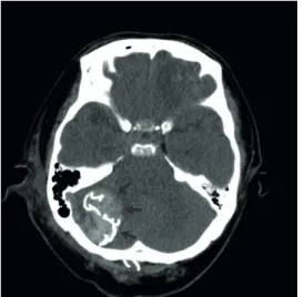
A 59-year-old male was transferred to our hospital presenting with a headache after falling backward. No neurological deficit was observed. The brain computerized tomography (CT) at the first hospital showed a small amount of ASDH in the right side of posterior fossa (FIGURE 1). The patient was admitted to our intensive care unit for careful observation of Received: Mar 10, 2019
Revised: Jul 4, 2019 Accepted: Sep 2, 2019 Address for correspondence:
Tae Ki Yang
Department of Neurosurgery, Jeju National University, 102 Jejudaehang-ro, Jeju 63243, Korea.
E-mail: [email protected]
Copyright © 2019 Korean Neurotraumatology Society
This is an Open Access article distributed under the terms of the Creative Commons Attribution Non-Commercial License (https://
creativecommons.org/licenses/by-nc/4.0/) which permits unrestricted non-commercial use, distribution, and reproduction in any medium, provided the original work is properly cited.
ORCID iDs Tae Ki Yang
https://orcid.org/0000-0003-2473-5880 Conflict of Interest
The author has no financial conflicts of interest.
Department of Neurosurgery, Jeju National University, Jeju, Korea
his neurological status without operation. Several hours after the admission, the patient's condition got worse to drowsy from alert mentality. The follow-up brain CT revealed a newly developed intraparenchymal haemorrhage in the right cerebellar hemisphere and an increased amount of the right subdural haematoma (FIGURE 2). The right intracerebellar parenchymal haemorrhage was considered to be the main cause of the deterioration of mentality.
After informed consent, the patient was transferred to an operation room. A midline incision and right suboccipital craniotomy were performed in a prone position under general anaesthesia.
Most of the intraparenchymal hemorrhage was smoothly removed. However, as soon as we removed the haematoma located around the right lower lateral corner covered with unresected bone, a sudden excessive blood poured into the operation field. Due to the huge amount of bleeding and the bone, which had not been resected at the beginning of the operation, concealing the bleeding point, the location of the bleeding point could not be determined. The bleeding seemed to be from a venous sinus at the far lower lateral corner of right posterior fossa. To stop FIGURE 1. A brain computerized tomography scan from the first hospital shows a small amount of acute subdural haematoma in the right infratentorium (arrows).
FIGURE 2. The brain computerized tomography scan checked in the intensive care unit after the deterioration of mentality reveals a newly developed intraparenchymal haemorrhage and an increased amount of acute subdural haematoma.
and showed a prompt light reflex. On day 2 after the operation, the brain CT showed no significant intraparenchymal hemorrhage around the gauze, ischemic lesion, and brain stem compression except the hyperdense shadow of the gauze (FIGURE 3). In the next several days, the patient gradually recovered to drowsy mentality without significant motor weakness. The patient became febrile over 38°C from the second post-operation day.
On day 10 after the operation, the patient got the second operation to remove the previously packed gauze. The enhanced preoperative brain CT showed occlusion of the right transverse sinus at the distal end, and we confirmed that again with transfemoral cerebral angiography for a safe surgery (FIGURE 4). While reviewing the images before the second surgery, we found that there was a diastatic fracture of right occipitomastoid suture around the subdural hematoma (FIGURE 5). The fracture might have caused the sigmoid sinus injury and the development of the subdural haematoma. During the removal of the gauze, no recurrent haemorrhage from the sinus, which had been expected through the enhanced preoperative CT and the cerebral angiography, was observed. There was no intraparenchymal hemorrhage inside and around the gauze. The patient improved to the light drowsy status on day 2 after the operation and became afebrile immediately after the second operation. He was getting better over time and became alert. On day 26 after the second operation, he was transferred to the rehabilitation department for the management of gait difficulty.
FIGURE 3. The postoperative brain computerized tomography exhibits the hyperdense gauze shadow with radiopaque markers (arrows), but no significant intraparenchymal haemorrhage, ischemic lesion, or mass effect against brain stem.
DISCUSSION
During an operation for ASDH in the posterior fossa, bleeding from an injured venous sinus can be profuse, thereby even resulting in fatal consequences. In some cases, it is not easy to control the abundant amount of subdural sinus bleeding from the unexposed area covered with bone in the infratentorium. Spending too much time with ineffective methods trying stop the pouring subdural blood from the venous sinus could result in instant cardiac arrest.
Therefore, in the event of an excessive amount of subdural sinus bleeding in spite of using all measures to stop the bleeding, and if there is no enough time to prevent hypovolemic shock, we have to use the most reliable and easily applicable method first to prevent cardiac arrest.
In our first operation, the bleeding was very rapidly and effectively controlled using the gauze packing into the bleeding site. Only some blood oozing through the packed gauze was observed during the suturing of the skin. This method was not only prompt, but also safe, as there was no significant intraparenchymal haemorrhage around the gauze, ischemic lesion or mass effect
B A
FIGURE 4. (A) The enhanced brain computerized tomography before the second operation shows occlusion of transverse sinus at the distal end (arrow). (B) The cerebral angiography before the second operation shows no contrast filling from the distal end of the right transverse sinus (arrow).
FIGURE 5. The review of the brain computerized tomography images with bone setting windows checked at the first hospital reveals a diastatic fracture of the right occipitomastoid suture (arrow).
If the sinus is not occluded after the gauze packing, it can be occluded using the coil embolization technique before the second operation to prevent the dangerous rebleeding after the consideration of the benefit to risk ratio for the sinus embolization.1) The acute venous sinus occlusion can cause several complications, such as venous infarction, intracranial hemorrhage, edema, subarachnoid hemorrhage, as well as rapidly progressive illness and coma.1) According to a recent report, the venous sinus remained patent three days after the operation of acute epidural hematoma removal and gauze packing.1) To prevent a dangerous rebleeding, the operators successfully occluded the injured sinus using the coil embolization technique. There was no sign of contrast leakage and venous hypertension in the post-embolization angiograms. Considering the high-risk of rebreeding, this technique can be a good option to ensure safety of the second operation.
In a trauma patient with an operable amount of intraparenchymal haemorrhage accompanying a small amount of ASDH near the dural sinus in the posterior fossa, it is necessary to try not to overlook the small amount of subdural haematoma and not to focus only on intraparenchymal haemorrhage. An effort should be made to find a fracture line, including diastatic fracture crossing the dural sinus over the subdural haematoma. This will help the operator to prepare and avoid an unexpected catastrophic bleeding from the injured venous sinus.
CONCLUSION
During the operation of traumatic ASDH in the posterior fossa, the operator should keep in mind that there can be a sudden unexpected profuse subdural bleeding from the pre-injured venous sinus which can cause cardiac arrest and a gauze packing can be the most simple and efficient method to choose first for bleeding control. And the fact that venous sinus can be occluded spontaneously several days after gauze packing gives us another option on future planning after the sinus bleeding control with gauze packing.
REFERENCES
1. Kim JH, Yu SH, Kim BC, Lee JH, Lee JI, Choi HJ. Endovascular treatment following gauze packing for the control of massive bleeding from traumatic transverse sinus lesion. Korean J Neurotrauma 14:150-154, 2018 PUBMED | CROSSREF
2. Motohashi O, Kameyama M, Shimosegawa Y, Fujimori K, Sugai K, Onuma T. Single burr hole evacuation for traumatic acute subdural hematoma of the posterior fossa in the emergency room. J Neurotrauma 19:993-998, 2002
PUBMED | CROSSREF
3. Phan K, Moore JM, Griessenauer C, Dmytriw AA, Scherman DB, Sheik-Ali S, et al. Craniotomy versus decompressive craniectomy for acute subdural hematoma: systematic review and meta-analysis. World Neurosurg 101:677-685.e2, 2017
PUBMED | CROSSREF
4. Vega RA, Valadka AB. Natural history of acute subdural hematoma. Neurosurg Clin N Am 28:247-255, 2017 PUBMED | CROSSREF