Acute Rupture of Flexor Tendons as a Complication of Distal Radius Fracture
Youn Moo Heo, M.D., Sang Bum Kim, M.D. , Kwang Kyoun Kim, M.D., Doo Hyun Kim, M.D., and Won Keun Park, M.D.
Department of Orthopedic Surgery, Konyang University College of Medicine, Daejeon, Korea
Acute rupture of flexor tendons following distal radius fracture is very rare. We experienced four cases of acute rupture of flexor tendons that were treated surgically. Injured tendons included flexor pollicis longus, flexor carpi radialis, palmaris longus and third flexor digitorum profundus. A severely displaced fracture with a volar spike of the distal radius was detected on plain radiographs in all cases. Ruptures of flexor pollicis longus and third flexor digitorum profundus were diagnosed on preoperative examination but ruptures of other tendons were identified during the operation. Repairs of fractures and ruptured tendons were performed simultaneously and good functional outcomes were achieved.
Key words: distal radius, flexor tendon, fracture, acute rupture
Tendon rupture is a rare complication of a distal radius fracture. Al- though the previously reported incidence of rupture of the extensor pollicis longus tendon after distal radius fractures varies from 0.07%
to 0.88%, the incidence of rupture of flexor tendons following distal radius fractures is lesser than that of rupture of extensor tendons.1) Recently, a number of cases and mechanisms for chronic rupture of flexor tendons caused by attrition from volar plating have been reported.2) However, acute rupture of flexor tendons occurs less fre- quently than chronic rupture of flexor tendons. We experienced four cases of acute rupture of flexor tendons in association with distal radius fractures and have reported these cases along with literature review.
CASE REPORTS
1. Case 1
A 29-year-old right-handed male presented with painful swelling of his left wrist after rolling down the stairs. Physical examination
revealed a loss of active flexion of the interphalangeal (IP) joint of the left thumb but other fingers did not have any limitation of active motion. The plain radiographs showed a dorsally displaced, com- minuted intra-articular fracture with volar spike of distal radius and a fracture of the styloid process of the ulna (Fig. 1A, 1B).
The operation was performed through a volar radial approach.
The rupture of flexor pollicis longus (FPL) tendon (Fig. 1C) and the laceration of pronator quadratus (PQ) muscle were identified at the level of the fracture of distal radius. The fracture was fixed with a volar locking plate and the ruptured FPL tendon was repaired with four-strand cruciate suture (Nylon 3-0) and epitendinous suture (Prolene 5-0) (Fig. 1D). After surgery, posterior thumb spica splint with extension block was applied for four weeks and gentle active motion of the IP joint of the left thumb was permitted immediately.
Four weeks after the operation, a protective splint was applied for an additional two weeks. At the 13 months follow-up, the wrist was pain-free and a range of motion (ROM) was 80o palmar flexion, 80o dorsal extension, 80o supination and 80o pronation. And active mo- tion of the IP joint of left thumb was not limited (Fig. 1E, 1F).
2. Case 2
A 22-year-old right-handed male presented to the emergency de- partment after a car accident. He had severe swelling and tenderness
Copyright © 2015 by The Korean Orthopaedic Association
“This is an Open Access article distributed under the terms of the Creative Commons Attribution Non-Commercial License (http://creativecommons.org/licenses/by-nc/3.0/) which permits unrestricted non-commercial use, distribution, and reproduction in any medium, provided the original work is properly cited.”
The Journal of the Korean Orthopaedic Association Volume 50 Number 1 2015 Received September 15, 2014 Revised October 22, 2014
Accepted October 25, 2014
Correspondence to: Sang Bum Kim, M.D.
Department of Orthopedic Surgery, Konyang University Hospital, 158 Gwanjeodong- ro, Seo-gu, Daejeon 302-812, Korea
TEL: +82-42-600-9120 FAX: +82-42-545-2373 E-mail: hurym1973@hanmail.net
over his right wrist and an intact active motion of all fingers. The plain radiographs showed a dorsally displaced fracture with volar spike of distal radius and a fracture of the styloid process of ulna (Fig.
2A, 2B). Surgical treatment was planned because his fracture was unstable. Simultaneously, he also sustained a contralateral unstable fracture of distal radius.
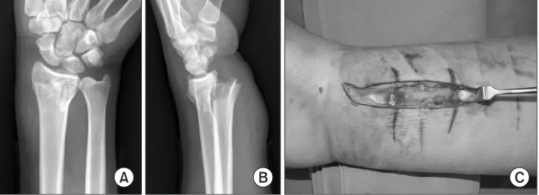
Seven days after the trauma, open reduction and internal fixation through the volar radial approach was performed. The incision was made just radial to the flexor carpi radialis (FCR) tendon and the ruptured FCR tendon was identified immediately (Fig. 2C). Both ends of the ruptured FCR tendon were retracted in the opposite direction until about 3.5 cm. Also, the forearm fascia under the FCR
tendon and PQ muscle was injured. Volar spike of the distal radius was visible through the torn PQ muscle. The fracture was reduced and fixed with a volar locking plate and the FCR tendon was re- paired with cruciate suture and epitendinous suture. And the ac- companying scapholunate dissociation was fixed with two Kirschner wires. After surgery, the sugar tong splint was applied for five days and it was replaced with a short arm thumb spica cast for another six weeks. Six weeks after surgery, the cast was taken off and the two Kirschner wires were removed. Then the patient underwent gradual mobilization with passive ROM exercise. At the 12 months follow-up, there was no wrist pain and the ROM was 80o palmar flexion, 80o dorsal extension, 80o supination and 80o pronation.
Figure 1. (A, B) Plain radiographs of the left wrist showing an intra-articular distal radius fracture with an ulnar styloid fracture. (C, D) Intraoperative photographs show the rupture of the flexor pollicis longus tendon and the repair of the injured tendon using cruciate suture and epitendinous suture.
(E, F) The photograph shows a normal range of motion of the interphalangeal joint of the left thumb at six months after surgery.
Figure 2. (A, B) Plain radiographs showing a distal radius fracture with severe dorsal displacement and a sharp anterior bony spike. (C) Rupture of the flexor carpi radialis tendon was confirmed intraoperatively.
3. Case 3
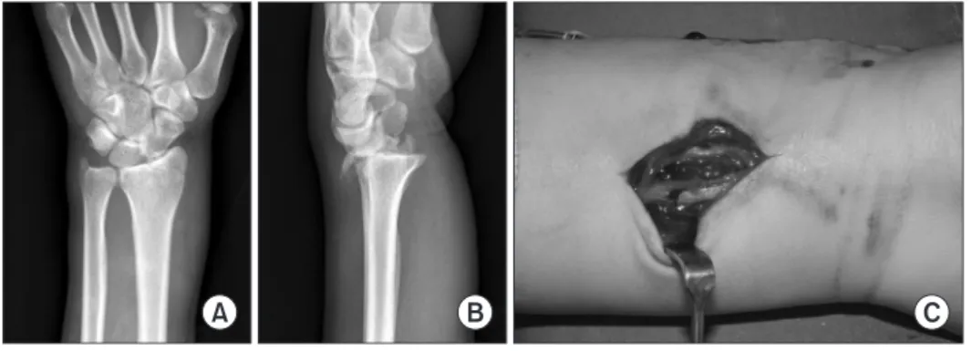
A 25-year-old right-handed male presented to the emergency de- partment after a fall during mountain climbing. He had a limitation of motion of the left wrist due to pain but active motion of all fin- gers was intact. A laceration of about 1 cm in size was identified on the volar aspect of the wrist. And, tendon injury was not identified through the wound. Tingling or sensory changes in the fingers were not noted. Radiographs showed a fracture of the styloid process of the left distal radius and a fracture of the styloid process of the left ulna. The radiocarpal joint was dorsally dislocated and volar spike of the proximal margin of fractured radius was detected on radiographs (Fig. 3A, 3B). Simultaneously, he also sustained an ipsilateral cla- vicular fracture and a contralateral humeral shaft fracture.
The fracture of the radial styloid were exposed through a longi- tudinal incision over the radial styloid. It was fixed with a precon- toured plate on the radial aspect of the radial styloid. An additional incision was made for the exploration of wound on the volar wrist.
Intraoperatively, total rupture of the palmaris longus (PL) tendon was identified and repaired with cruciate suture (Fig. 3C). But, injury of other structures was not detected. Postoperatively, a short arm cast was applied for four weeks and then the patient underwent gradual mobilization with active assisted and gentle passive exer- cises. At the 16 months follow-up, the ROM of the left wrist was almost full and there were no other complications.
4. Case 4
A 34-year-old right-handed male presented to the emergency department after a motorcycle accident. He sustained fractures of bilateral distal radius, fracture of the shaft of left radius and ulna and fracture of the shaft of left femur. The active flexion of right third finger was partially limited and the pain was aggravated by passive extension and flexion. Moreover, he complained of tingling sensa- tion in his right palm in the dermatome of median nerve. Radio- graphs of the right wrist showed a dorsally displaced, comminuted intra-articular fracture with volar spike of distal radius and a fracture
of the styloid process of ulna (Fig. 4).
The next day after the trauma, an operation was performed on right wrist. We planned an open reduction and internal fixation and exploration of the median nerve and flexor tendons. During carpal tunnel release, a partial rupture (>50%) of third flexor digitorum profundus (FDP) tendon was identified but an injury of median nerve was not found. The fracture was fixed with a volar locking plate and Kirschner wires and the ruptured FDP tendon was also repaired with four-strand cruciate suture (Nylon 3-0). Postopera- tively, the sugar tong splint was applied for four weeks. After the splint was taken off, the wrist underwent gradual mobilization with active ROM exercise. At 18 months after the operation, the mo- tion of the right third finger was intact and the wrist was 55o palmar flexion, 70o dorsal extension, 90o supination and 60o pronation.
DISCUSSION
Flexor tendon injuries following distal radius fractures are less com- mon than extensor tendon injuries. Flexor tendons may be protected
Figure 3. (A, B) Plain radiographs showing a styloid fracture of the distal radius with dorsal displacement and a sharp anterior bony spike. (C) Intraoperative photograph shows the injury of the palmaris longus tendon through the volar laceration.
Figure 4. (A) Antero-posterior radiograph showing a comminuted intra- articular distal radius fracture with a distal ulnar fracture. (B) Lateral radiograph showing a dorsal displacement of the distal fragment and a sharp anterior bony spike of the proximal part.
by the PQ and less tight enclosure over the distal radial aspect com- pared with extensor tendon.3) However, the incidence of rupture of the flexor tendons is unknown and has not been reported so far.
Between March 2007 and December 2011, we experienced four cases with acute injury of flexor tendons including FCR, FPL, PL, and 3rd FDP in 474 patients with distal radius fractures that were treated with open reduction and internal fixation. And the incidence of acute rupture of the flexor tendons at our institute was 0.84%.
A literature search revealed one case of FCR rupture in associa- tion with distal radius fracture. DiMatteo and Wolf4) described a case of an acute FCR tendon rupture identified intraoperatively and found that it was caused by a volar spike of bone through the torn PQ. Because it is hard to detect the rupture of FCR tendon on physical examination of distal radius fractures, closed acute injury to the FCR tendon may be missed except the exploration using volar approach. In our case, torn PQ, bone spike of distal radius and two frayed ends of the FCR tendon with a 3.5 cm gap were intraopera- tively identified with volar approach and this bone spike might have caused direct laceration of the FCR tendon.
The injury of FPL tendon in distal radius fracture may occur as acute or chronic rupture. Most of cases were chronic rupture after 4 weeks of distal radius fractures.5,6) McMaster,5) who first described a rupture of FPL tendon after distal radius fracture, suggested that two different pathological processes were responsible for this occur- rence. First process involved a direct partial laceration of FPL ten- don by the bone spur at the time of fracture and the laceration was not completely healed during the period of immobilization, thereby resulting in a weak spot and later a strong exertion on the tendon caused the tendon rupture. Second process was a local necrosis due to a decrease of blood supply caused by rupture or obstruction of the blood supply with additional pressure due to the hematoma or callus. Only two cases were reported for acute rupture of the FPL tendon that occurred within four weeks after distal radius fracture.
However, these two cases had no immediate evidence of total rup- ture of the FPL tendon after fracture. Ruptures of the FPL tendon were indentified after 4 weeks in Wong and Pho’s case7) and 3 days in Kim et al.’s case8), respectively. In our case, the patient could not flex the IP joint of his thumb on initial examination and the rupture of FPL tendon was intraoperatively confirmed on the next day.
Although the rupture of PL tendon into the palmaris fascia as a cause of an acute carpal tunnel syndrome has been reported, there is no report of a case of rupture of the PL tendon following distal ra- dius fracture.9) In this case, there was initially no evidence of rupture of PL tendon. However, the rupture was found by the exploration
of laceration during operation for internal fixation.
The rupture of FDP or superficialis tendons has been reported more frequently than that of FPL tendon. However, most of these reports were chronic rupture of these tendons. Only Southmayd et al.10) reported a case of rupture of second FDP tendon that oc- curred soon after a distal radius fracture. In our case, the rupture of third FDP tendon was preoperatively not identified. The injury of third FDP tendon rupture was intraoperatively identified and it was repaired. Most of distal radius fractures in our clinics have been im- mobilized for 4 weeks. The postoperative immobilized period for distal radius fractures was not changed because of the rupture of flexor tendon. The repair of FPL or FDP injury was protected by extension block splint and active motion of injured finger was per- mitted.
In our cases, all acute ruptures occurred in young adults by high- energy injuries. Although this ruptures can caused by low-energy injuries, most of reported cases were related with high-energy inju- ries.4,7,8,10) However, we think that it is hard to confirm the effect of the mechanism of injury because acute ruptures of flexor tendons following distal radius fracture is very rare. Rather, all distal radius fractures of this study showed a severe displaced and volar-spiked pattern that might be likely to cause acute ruptures of flexor tendon.
And acute pain by the fracture may interrupt active motion of ipsi- lateral fingers. Therefore, the authors think that the careful exami- nation have to be performed in order to identify acute ruptures of flexor tendon that may complicates in distal radius fractures.
CONFLICTS OF INTEREST
The authors have nothing to disclose.
REFERENCES
1. Hove LM. Delayed rupture of the thumb extensor tendon.
A 5-year study of 18 consecutive cases. Acta Orthop Scand.
1994;65:199-203.
2. Arora R, Lutz M, Hennerbichler A, Krappinger D, Espen D, Gabl M. Complications following internal fixation of unstable distal radius fracture with a palmar locking-plate. J Orthop Trauma. 2007;21:316-22.
3. Davis DI, Baratz M. Soft tissue complications of distal radius fractures. Hand Clin. 2010;26:229-35.
4. DiMatteo L, Wolf JM. Flexor carpi radialis tendon rupture as a complication of a closed distal radius fracture: a case report.
J Hand Surg Am. 2007;32:818-20.
5. McMaster PE. Late ruptures of extensor and flexor pollicis longus tendons following colles' fracture. J Bone Joint Surg Am. 1932;14:93-101.
6. Cooney WP 3rd, Dobyns JH, Linscheid RL. Complications of Colles' fractures. J Bone Joint Surg Am. 1980;62:613-9.
7. Wong FY, Pho RW. Median nerve compression, with tendon ruptures, after Colles' fracture. J Hand Surg Br. 1984;9:139-41.
8. Kim DY, Seo EM, Nam WD, Park SJ, Lee SS. Flexor pollicis
longus tendon rupture as a complication of a closed distal ra- dius fracture: a case report. J Korean Fract Soc. 2011;24:191-4.
9. Lourie GM, Levin LS, Toby B, Urbaniak J. Distal rupture of the palmaris longus tendon and fascia as a cause of acute car- pal tunnel syndrome. J Hand Surg Am. 1990;15:367-9.
10. Southmayd WW, Millender LH, Nalebuff EA. Rupture of the flexor tendons of the index finger after Colles' fracture. Case report. J Bone Joint Surg Am. 1975;57:562-3.
원위 요골 골절에 동반된 급성 굴곡건 파열
허윤무 • 김상범 • 김광균 • 김두현 • 박원근
건양대학교 의과대학 정형외과학교실
원위 요골 골절에서 급성 굴곡건 파열은 드물게 동반된다. 저자들은 원위 요골 골절에서 수술적 치료를 시행한 급성 굴곡건 파열을 4 예 경험하였다. 손상된 건은 장무지 굴곡건, 요수근 굴건, 장 장건 및 제3 심수지 굴곡건이었다. 모든 증례는 방사선 검사에서 전위가 심한 원위 요골 골절과 장측의 날카로운 골절단이 관찰되었다. 장무지 굴곡건과 제3 심수지 굴곡건 손상은 수술 전 신체 검사에서 진 단되었으나 요수근 굴건과 장 장건 손상은 수술 과정에서 발견되었다. 골절 고정과 건 봉합을 동시에 시행하였고 만족스러운 결과를 얻었다.
색인단어: 원위 요골, 굴곡건, 골절, 급성 파열
접수일 2014년 9월 15일 수정일 2014년 10월 22일 게재확정일 2014년 10월 25일 책임저자 김상범
대전시 서구 관저동로 158, 건양대학교병원 정형외과
TEL 042-600-9120, FAX 042-545-2373, E-mail hurym1973@hanmail.net
Copyright © 2015 by The Korean Orthopaedic Association
“This is an Open Access article distributed under the terms of the Creative Commons Attribution Non-Commercial License (http://creativecommons.org/licenses/by-nc/3.0/) which permits unrestricted non-commercial use, distribution, and reproduction in any medium, provided the original work is properly cited.”
대한정형외과학회지:제 50권 제 1호 2015

