두개내 동맥류(intracranial aneurysm)의 진단에는 고식적 뇌혈관조영술(conventional cerebral angiography, 이하 C A로 줄 임)이 기본적인 검사법으로 사용되고 있으나 이는 검사에 많 은 시간이 소요되고 침습적으로 여러 가지 합병증이 발생할 수 있다(1). 근래 이를 대체할 수 있는 기법으로 자기공명영 상 및 전산화단층촬영을 이용한 비침습적인 혈관조영술이 이 용되고 있다. 최근에는 나선식 전산화 단층촬영(helical CT)을 이용한 3차원 혈관 조영술(3-dimensional CT angiography, 이 하 C T A로 줄임)이 체내의 여러 혈관질환 진단에 많이 이용 되고 있으며(2-5), 또한 C A와 비교하여 두개내 동맥류의 진 단에 있어 양호한 결과들이 보고되고 있다(6-11). 그러나 C A 의 대체 가능성이 너무 강조되어 C A를 시행하지 않고 C T A 만 시행한 후 치료에 임한 경우를 경험할 수가 있었다. 이 연
구는 C T A를 CA 및 수술소견과 비교 분석하여 수술을 전제 로한 동맥류 환자에 있어서 C T A의 유용성을 평가하고, CTA 가 C A를 대체 할 수 있는지 알아보고자 하였다.
대상 및 방법
1 9 9 4년 1 0월부터 1 9 9 8년 8월까지 본원에서 두개내 동맥류가 의심되어 C T A를 시행한 2 4 3명의 환자중 수술과 추적 C A로 확진된 1 1 6명 1 4 1예의 동맥류를 대상으로 하였다. 이중 남자 는 4 6명 여자는 70 명이었으며 연령은 2 8세에서 7 8세로 평균 5 4세였다.
수술을 시행했던 환자를 2개 군으로 나누었고, 1군은 C T A 와 C A를 시행한 후 수술을 한 7 7명 9 4예를 대상으로 하였다.
2군은 C T A만 시행한 후 수술 및 추적 C A ( 2 8명에서 시행)를 한 3 9명의 환자 4 7예를 대상으로 하였다.
C A는 디지털 감산 혈관 촬영기(Sire graph-D2 and Digi- trone 3VA, Siements, Erlangen, Germany) 또는 필름 스크린 혈 관 촬영기(Biplane Angiography with TV system, GE Medical 목적: 두개내 동맥류의 발견과 평가에 있어서 3차원 전산화 단층촬영(3-dimensional CT
angiography: CTA)의 유용성을 고식적 혈관조영술(conventional angiography: CA)및 수술소 견과 비교하고 C T A가 C A를 대체 할 수 있는지를 알아보고자 하였다.
대상 및 방법: 뇌동맥류가 의심되어 C T A를 시행한 2 4 3명의 환자 중 수술로써 확진된 1 1 6명
을 대상으로 하였다. 대상환자를 두개의 군으로 나누어 1군은 C T A와 C A를 함께 시행한 후 수술을 한 7 7명 9 4예를 대상으로 하였고, 2군은 C T A만 시행한 후 수술 및 추적 C A를 한 3 9 명 4 7예로 하였다. 아무런 정보를 갖지 못한 2명의 방사선과 의사가 CTA 소견을 CA 및 수 술 소견과 비교하여 동맥류의 발견률과 양상(크기, 경부 및 주위 혈관의 형태변화등) 파악 의 우열성을 일정등급을 정하여 후향적으로 평가하였다.
결과: 1군의 경우 동맥류의 발견률은 전체 9 4예중 C T A에서 8 4예( 89%), CA에서 8 9예( 95% ) 였다. CTA에서 발견하지 못한 1 0예의 동맥류 중 4예는 3mm 이하였고, 6예는 윌리스환 이 외의 부위에 위치하였다. 2군의 경우 C T A에서 동맥류의 발견률은 4 7예중 4 4예( 93 % )였고, 발견하지 못하였던 경우는 3m m미만인 경우가 1예였고, 3m m이상의 크기였던 2예는 C A와 수술소견에서 윌리스환 이외 부위에 위치하였다. 동맥류의 전체적인 양상파악에서는 8 2예중 7 5예( 92 % )가 C T A가 C A보다 동등 혹은 우수한 소견을 보였다.
결론: C T A는 동맥류의 평가에 있어서 윌리스환 주변 병변에서는 유용하지만 두개강내 전체 혈관을 조영하지 못함으로 복수동맥류가 의심되거나 수술을 전제로 한 경우 C A와 함께 상 호보완적으로 시행하는 것이 유용할 것으로 생각된다.
1전남대학교 의과대학 진단방사선과학교실
2전남대학교 의과대학 신경외과학교실
이 논문은 전남대학교병원 진단방사선과 영상의학연구소 1 9 9 9년도 임상 연구비 일부 보조로 이루어졌음
이 논문은 1 9 9 9년 4월 1 6일 접수하여 1 9 9 9년 7월 1 5일에 채택되었음.
뇌동맥류의 진단에 있어서 전산화단층뇌혈관조영술과 고식적 뇌혈관조영술의 비교
1서정진・강형근・이영철・정광우・김재규・정태웅・김태선2・강삼석2・이제혁2
systems, Milwaukee, U.S.A.)를 사용하였으며 모든 환자에서 대 퇴동맥을 통하여 양측 내경동맥 및 일측 혹은 양측 척추동맥 을 각각 선택하여 혈관촬영을 시행하였다. 각각의 내경동맥 및 척추동맥 촬영시 전후면과 측면 영상을 기본으로 얻었고, 필요 에 따라 사위면, 경안면(transorbital view) 또는 악하경정위 (submentovertical view)와 같은 추가 영상을 얻었다.
C T A는 나선식 C T기기(Hispeed AdvantageⓇ, GE M e d i c a l systems, Milwaukee, U.S.A.)를 이용하였으며 두경부의 기본영 상을 얻은 후 안와 외이도선을 기준으로 하여 터키안 부분으 로부터 상방으로 조영전 스캔하였다. 조영전 영상을 얻은 후 1 5 0 - 1 80mL 비이온성 조영제(Ultravist 370Ⓡ, 75.9% o f Lopromide, Schering, Berlin, Germany)를 주전정맥( a n t e c u b i t a l v e i n )을 통하여 초당 2 . 3 - 3 .5m l의 속도로 급속 투입하고 조영제 주입 시작후 2 4 - 3 4초 후에 윌리스환 부위를 절편두께 1 m m , 피치 1 : 1로 35mm 길이를 스캔한 후 이를 다시 0 .5mm 간격으
로 재구성하였다. 이러한 기본 영상을 독립영상 구성장치 (Independent Console, GE Medical systems, Milwaukee, U.S.A.) 상에서 1 8 0 - 4 00 H U의 역치(threshold level)를 기준으로, SSD(shaded-surface display)기법을 이용, 재구성하여 3차원 영 상을 얻었다.
아무런 임상 정보를 모르는 2명의 방사선과 의사가 C T A 소견을 판독하고 고식적 혈관 조영술 및 수술소견과 비교하 였다. 동맥류의 위치에 따라서 발견율을 비교하였고 동맥류의 크기를 3 m m이내, 3mm 부터 9 m m이내, 9mm 이상으로 나누 어 크기에 따른 각각의 발견율을 비교하였다. 또한 동맥류의 전체적인 양상(방향, 동맥류의 경부 소견)의 파악에 있어 C- T A가 C A보다 좋다, 같다, 나쁘다 등 3등급으로 나누어 분석 하였다.
─ 6 4 4 ─
Table 1. Comparison of Aneurysm Numbers Detected by CT Angiography, Conventional Angiography and Operation
L o c a t i o n CT Angiography C o n v e n t i o n a l Number of
A n g i o g r a p h y A n e u r y s m s
Antrior communcating a. 3 8 3 6 4 0
Posterior communicating a. 1 8 1 8 1 8
Middle cerebral a. 1 3 1 3 1 3
Internal carotid a.* 05 08 08
Anterior cerebral a. 04 05 05
Anterior choroidal a. 02 02 03
Ophthalmic a. 01 02 02
Superior cerebellar a. 02 02 02
Post. inferior cerebellar a. 01 02 02
Hypophyseal a. 00 01 01
T o t a l 0 0 00 8 4 ( 89% ) 0 0 00 8 9 ( 95 % ) 0 0 00 9 4 ( 1 00 % )
Post.=posterior
a.=artery, * cavernous portion and proximal portion of internal carotid artery
A B
Fig. 1. A 47 years-old-male patient with multiple aneurysms.
A . Anteriorposterior view of CT angiogram demonstrates an aneurysmal sac(arrows) at bifurcation site of left middle cerebral a r t e r y .
B . Anteriorposterior view of conventional angiogram shows an aneurysmal sac(arrow) at bifurcation of left middle cerebral artery and the other (arrow head) from internal carotid artery that was not depicted on CT angiogram.
결 과
1군의 경우 7 7명의 환자에서 총 9 4예의 동맥류를 발견할 수 있었고 이중 1 5명의 환자에서 2개씩, 1명의 환자에서 3개의 다 발성 동맥류가 발견되었다. 수술전 C T A에서 전교통동맥을
위시하여 hypophyseal artery 까지 다양한 위치에서 8 4예의 동 맥류를 발견하여 89%의 발견율을 보였다(Table 1). 후교통동 맥과 중대뇌동맥에서 1 00%, 전교통동맥에서는 오히려 C A보 다 2예를 더 발견할 수 있었다. 그러나 CT 스캔영역 밖에 위 치한 동맥류가 많았던 내경동맥의 3예와 후하소뇌동맥 1예 등은 발견하지 못하였다(Fig. 1).
2군의 경우 수술 및 추적 C A상 3 9명의 환자에서 4 7예의 동 맥류를 발견하였으며 이중 4명의 환자에서 2개씩, 2명의 환자 에서 각각 3개씩의 다발성 동맥류가 발견되었다(Table 2). C- T A에서 모두 4 4예( 93 % )를 발견하였는데 전교통동맥과 중대 뇌동맥에서 1 00% 발견율을 보였다.
크기에 따른 동맥류의 발견은 1군에서 C T A상 3mm 미만일 때 4예를, 3mm 이상인 경우 6예에서 발견하지 못하였는데 이 경우는 C A나 수술 소견에서 주로 윌리스환 이외 부위에서 발 견되었던 동맥류였다(Table 3). 2군에서도 C T A에서 3예의 동 맥류를 발견하지 못하였는데 3m m미만인 1예가 있었고, 3m m 이상의 크기였던 2예는 C A와 수술소견에서 윌리스환 이외의 부위에 위치하였다(Fig. 2 & Table 4).
Table 3. Numbers of Detected Aneurysms according to the Size in Group 1
S i z e ( m m ) C T Conventional Operation
A n g i o g r a p h y ( % ) A n g i o g r a p h y ( % ) f i n d i n g s ( % )
< 3 1 5 ( 7 9 ) 1 7 ( 8 9 ) 1 9 ( 1 0 0 ) 3-9 6 0 ( 9 2 ) 6 2 ( 9 5 ) 6 5 ( 1 0 0 )
> 9 09 ( 9 0 ) 1 0 ( 1 0 0 ) 1 0 ( 1 0 0 ) T o t a l 8 4 ( 8 9 ) 8 9 ( 9 5 ) 9 4 ( 1 0 0 ) Table 2. Comparison of Aneurysm Number Detected by CT Angiography and Operation findings with Follow up Conven- tional Angiography
L o c a t i o n CT Angiography Number of A n e u r y s m s
Antrior communcating a. 2 1 2 1
Posterior communicating a. 01 01
Middle cerebral a. 1 9 1 9
Anterior cerebral a. 01 02
Internal carotid a. 01 01
Anerior choroidal a. 01 01
Posterior cerebral a. 00 01
Vertebral a. 00 01
T o t a l 0 0 00 4 4 ( 9 3 % ) 0 0 0 004 7 ( 1 0 0 % ) a . = a r t e r y
A B
Fig. 2. A patient with recurred subarachnoidal hemorrhage following operation. A new aneurysm was diagnosed by conventional a n g i o g r a p h y .
A . Pre-operative oblique anteroposterior view of CT angiogram demonstrates an aneurysmal sac(arrow) from anterior communi- cating artery.
B . Post-operative oblique conventional angiogram shows an aneurysmal sac(large arrows) from distal anterior cerebral artery(A2 portion) that was not depicted on CT angiogram previously. Aneurysmal clip(small arrows) is faintly seen at anterior communicat- ing artery.
Table 4. Numbers of Detected Aneurysms according to the Size in Group 2
S i z e ( m m ) CT Angiography(%) Operation findings +Follow up CA(28)*
< 3 0 ( 0 ) 01 ( 1 0 0 )
3-9 2 9 ( 9 3 ) 3 1 ( 1 0 0 )
> 9 1 5 ( 1 0 0 ) 1 5 ( 1 0 0 )
T o t a l 4 4 ( 9 3 ) 4 7 ( 1 0 0 )
CA=conventional angiography,
* = 28 patients were performed with postoperative conventional a n g i o g r a p h y
전체적으로 C T A에서 수술 및 추적 C A와 비교하여 91%의 동맥류를 발견할 수 있었다. 또한 C T A에서 발견할 수 없었던 동맥류는 1군과 2군에서 1 3예로 이중 5예는 3 m m미만의 작은 크기의 동맥류였으며 8예는 CTA 스캔 영역 밖에 위치한 경 우였다.
C T A와 CA 모두에서 발견되었던 8 2예의 동맥류를 대상으로 동맥류의 전체적 양상파악에서 C T A가 C A에 비해 우수하였던 경우가 2 1예( 26%), 동일 하였던 경우가 5 4예( 66%), 그리고 C A 가 더 우수하였던 경우가 7예(8% )였다(Fig. 3, 4 & Table 5).
고 찰
현재까지는 동맥류를 포함한 두개내 혈관질환의 진단에 있 어 카테터를 이용한 C A가 표준검사로 이용되고 있으나 이들 검사자체가 침습적이고, 검사 시간이 비교적 오래 걸리며 검
사에 따른 합병증유발을 할 수 있다(1). 특히 동맥류 환자의 경우 동맥류 파열의 위험이 있으며 지주막하 출혈을 동반한 환자에서 전신상태가 좋지 않은 때는 합병증의 빈도는 더 높 기때문에 이를 대체할 수 있는 진단방법이 필요하게 되었다.
최근 개발된 자기공명혈관조영술은 이에 적합한 유용한 진단 방법으로(12-17) 비침습적이기는 하지만 가격이 비싸고, 검사 시간이 오래 걸리며, 지주막하출혈과 두개내 혈종 등이 있는 환자에서 진단이 어려운 단점이 있을 뿐만 아니라 폐쇄공포 증이나 심실세동기등을 부착한 환자에서 검사가 불가능한 점 등의 제한이 있다( 1 8 ) .
이에 비해 C T A는 주위 상자성물질(paramagnetic sub- s t a n c e )에 영향을 받지 않고 환자의 상태가 양호하지 못한 경 우에도 빠른 시간 내에 시행이 가능하기 때문에 선별검사로 서 많이 이용되었다. 그러나 이에 대한 과신으로 C A를 시행 하지 않고 수술을 바로 시행하는 경우가 있어 C T A의 이용에 Table 5. Comparison between CT Angiography and Conventional Angiography in the Delineation of Aneurysms
L o c a t i o n No. of
C T A > C A C T A = C A C T A < C A a n e u r y s m
Anterior communcating a. 3 6 1 1 2 4 1
Posterior communicating a. 1 8 05 1 2 1
Middle cerebral a. 1 3 04 08 1
Internal carotid a. 05 01 02 2
Anterior cerebral a. 04 00 03 1
Anterior choroidal a. 02 00 02 0
Superior cerebellar a. 02 00 01 1
Opthalmic a. 01 00 01 0
Post. inferior cerebellar a. 01 00 01 0
T o t a l 0 0 0 0 0 08 2 ( 1 0 0 % ) 0 0 0 0 02 1 ( 2 6 % ) 0 0 0 05 4 ( 6 6 % ) 0 007 ( 8 % ) CTA=CT angiography, CA=conventional angiography, Post.=posterior, a.=artery
A B
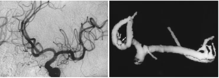
Fig. 3. Advantage of CT angiogram in delineation of giant aneurysm of left internal carotid artery.
A . Anteriorposterior view of conventional angiogram demonstrates a giant aneurysm(arrows), but not accurately depicts the a- neurysmal neck and parent artery.
B . Oblique superior anteriorposterior view of CT angiogram delineates more clearly aneurysmal neck(arrow heads) and parent artery(left internal carotid artery, large arrows) and relation to adjacent left anterior cerebral artery(medium sized arrows) and an- terior commnicating artery(small arrows) than CA.
─ 6 4 6 ─
대한 정확한 평가가 필요하다고 하겠다.
V i e c o등( 1 2 )은 C A와 비교하여 C T A의 민감도를 각각 7 7 - 97%, 87-100%로 보고 하였고, 다른 대부분의 연구에서도 높 은 민감도와 특이도를 보고하였다(7,8, 10-12). 이 연구에서도 전체적으로 91%의 높은 발견율을 보였다.
L i a n g등( 1 9 )은 조영제의 양과 촬영기법에 따라서 윌리스환 에 위치한 동맥류중 2 m m까지도 발견할 수 있었다고 하였다.
즉 직경 2 m m에서 부터 2 .4 m m까지 세개의 동맥류를 발견할 수 있었는데 이는 조영제를 3 00mg I/ml(40.5g )인 1 35 m l를 주 입하여 혈관들의 농도가 진하게 (densely) 조영증강 되었기 때 문으로 생각하고 있다. 본 연구에서도 1 50ml 이상 조영제를 주 입하였기 때문에 작은 동맥류의 발견율이 높았으리라 생각된 다. 또한 고관전류(higher amperage, 250-280m A )와 1m m의 c o l- l i m a t i o n등의 촬영기법이 작은 동맥류의 발견에 도움을 주는 요소로 생각하고 있는데 본 연구에서도 2 40 m A의 고관전류, 그리고 1 m m의 collimation 촬영기법을 사용하였다. Schwartz등 ( 1 1 )도 C T A를 이용하여 직경 3 m m이상의 동맥류는 대부분 발견할 수 있다고 하였다. 그러나 본 연구에서는 C T A에서 C A 및 수술소견과 비교하여 총 1 4 1예의 동맥류중 1 3예를 발견하 지 못하였는데 이 중 5예는 3 m m미만의 작은 동맥류로서 크기 가 작은 경우 진단에 여전히 어려운 문제가 있다.
C A에 비해 C T A는 복잡한 혈관구조를 다양한 각도에서 관 찰이 가능하며, 특히 상하 방향의 투사영상은 C A에서 얻을 수 없는 영상으로 윌리스환 부위를 평가하는데 큰 잇점이 있 다. 크기가 큰 동맥류의 경우 C A에서 동맥류내의 조영제에 가려서 동맥류의 경부 및 주위혈관과 동맥류와의 관계를 정 확히 알 수 없어 다른 각도로 여러번 C A를 시행할 필요가 있 다. 그러나 C T A는 한번 영상을 얻은 후 검사자가 원하는 각 도로 돌려볼 수 있기 때문에 동맥류의 전체적인 양상을 파악
하는데 유리하다. 이 외에도 다면적 영상을 이용하여 실제 수 술하는 것과 같은 방향으로 투사한 영상은 수술계획을 세우 는데 도움을 줄 수 있다(10). 이 연구에서도 C T A와 C A로 동 시에 진단하였던 8 2예에서 동맥류의 전체적 양상파악은 C T A 가 C A에 비해 우수하였거나 같은 경우가 7 5예( 92 % )로 나타 나 C A에 비해 이용가치가 높을 수 있다 하겠다.
그러나, CTA는 C A에 비해 동맥류의 크기가 작은 경우에 있어 그 유용성이 떨어지며, 역동적 스캔을 시행할 수 있는 범위의 제한으로 인하여 스캔영역 밖의 일부 동맥류를 발견 하지 못한다는 것이 단점으로 지적되어 왔다(8, 10-12).
V e l t h u i s등( 2 0 )은 본 연구에서처럼 안와 외이도선을 기준으로 하여 35 m m길이로 스캔하지 않고 1번 경추부위부터 1 .5 m m c o l l i m a t i o n으로 60m m길이로 최대한 스캔범위를 늘려 C T A의 대체 가능성을 보고하고 있지만 숙련도와 장비문제, 그리고 해상도등의 문제점이 있다. 이 연구에서도 C T A에서 CA 및 수술소견과 비교하여 총 1 4 1예의 동맥류중 1 3예를 발견하지 못하였는데 이중 8예는 C T A의 스캔 범위밖에 위치하였던 경 우였다. 이 중 3 예는 C T A만 시행하고 수술을 시행하여 수술 후 1 - 3일 이내에 C T A로 발견하지 못하였던 다른 복수동맥류 에서 재출혈이 있어 C A로 다른 동맥류를 발견하고 재수술을 했던 경우였다. 본 연구에서도 C T A만을 시행한 후 수술을 하 였던 경우에 있어 C T A에서 발견율이 4 7예중 4 4예( 93 % )로, C T A와 C A를 함께 시행한 후 수술하였던 9 4예중 8 9예( 95 % ) 를 발견할 수 있었던 것과 비교하여 후자에 있어 동맥류의 발 견율이 높았던 이유는 동맥류의 크기가 적거나 윌리스환 이 외의 부위에 있어 C T A상 동맥류가 발견되지 않아 C A를 병 행하여 정확한 진단이 가능하였기 때문으로 생각된다. 따라서 C T A는 C A를 시행한 후 수술계획을 수립하기 위하여 혹은 중재적방사선학적 치료를 하려고 할 때 동맥류의 경부등 양
A B
Fig. 4. Advantage of CT angiogram in detection of aneurysm less than 3 mm in diameter.
A . Oblique anteriorposterior view of conventional angiogram shows an aneurysmal sac from anterior communicating artery. It is overlapped by parent artery.
B . Oblique superior anteriorposterior view of CT angiogram demonstrates aneurysmal sac from anterior communicating artery less than 3 mm in diameter.
상파악이 어렵거나 환자가 도저히 C A를 시행하기 어려울 정 도로 상태가 불량하거나 조영제에 의한 부작용등이 있을 가 능성이 높은 경우 등으로 그 이용이 제한되어야 할 것이다.
또한 C T A는 영상을 재구성하는 단계에서 시간이 많이 걸리 고, 오류가 있을 수 있으므로 두개내 혈관 해부학에 지식이 있는 숙련된 방사선과 의사가 시행하는 것이 바람직하다고 생각한다.
결론적으로 C T A는 두개내 동맥류가 의심되는 환자에서 윌 리스환 주변 병변에서는 C A를 대체할 가능성이 있지만 두개 강내 전체 혈관을 조영하지 못하여 윌리스환 이외의 복수동 맥류를 간과할 수 있으므로, 특히 복수동맥류가 의심되거나 수술을 전제로 한 경우 C A와 함께 상호보완적으로 시행하는 것이 유용할 것으로 생각된다.
참 고 문 헌
1 . Earnest F, Forbes G, Sandok BA, et al. Complications of cerebral angiography: prospective assessment of risk. AJR 1 9 8 4 ; 1 4 2 : 2 4 7 - 253
2 . Schwartz RB, Jones KM, Chernoff DM, et al. Common carotid artery bifurcation: evaluation with spiral CT. Radiology 1 9 9 2 ; 1 8 5 : 513-519
3 . Marks MP, Nepel S, Jardan LE, Enzmann DR. Diagnosis of carotid artery disease: preliminary experience with maximum-intensity- projection spiral CT angiography. AJR 1 9 9 3 ; 1 6 0 : 1 2 6 7 - 1 2 7 1 4 . Castillo M. Diagnosis of disease of the common carotid artery bi-
furcation: CT angiography vs catheter angiography. AJR 1 9 9 3 ; 1 8 9 : 1 8 5 - 1 9 2
5 . Galanski M, Prokop M, Chavan A, et al. Renal artery stenosis: spi- ral CT angiography. Radiology 1 9 9 3 ; 1 8 9 : 1 8 5 - 1 9 2
6 . Rubin GD, Dake MD, Napel S, McDonnell CH, Jeffrey RB. Three- dimenal spiral CT angiography of the abdomen: initial clinical ex- perience. Radiology 1993;186:147-152
7 . 김규선, 윤대영, 김호철, 윤구섭등. 나선식 C T를이용한두개내 C T 혈관조영술: 예비보고. 대한방사선의학회지1 9 9 5 ; 3 3 ( 2 ) : 1 8 3 - 1 8 8 8 . 박수민, 서정진, 김윤현, 강형근등. 뇌동맥류진단에있어서 3차원
전산화단층촬영혈관조영술의유용성 : 고식적혈관조영술과비교 . 대한방사선의학회지1 9 9 6 ; 3 4 : 3 1 3 - 3 1 9
9 . 김종민, 김소선, 허진도등. 뇌동맥류의발견을위한얇은절편의고 속주입 CT 기법의유용성. 대한방사선의학회지1 9 9 3 ; 2 9 : 1 1 1 6 - 1 1 2 0 1 0 . Aoki S, Sasaki Y, Machida T, Ohkubo T, Minami M, Sasaki Y.
Cerebral aneurysm: detection and delineation using 3-D CT an- giography. AJNR 1 9 9 2 ; 1 3 : 1 1 1 5 - 1 1 2 0
1 1 . Schwartz, RB. Tice HM, Hooten SM, Hsu L, Stieg PE. Evaluation of cerebral aneurysms with helical CT: correlation with conven- tional angiography and MR angiography. Radiology 1 9 9 4 ; 1 9 2 : 7 1 7 - 722
1 2 . Vieco PT, Shuman WP, Alsofrom GF, Gross CE. Detection of cir- cle of Willis aneurysms in patients with acute subarachnoid hem- orrhage: a comparison of CT angiography and digital subtraction angiography. AJR 1 9 9 5 ; 1 6 5 : 4 2 5 - 4 3 0
1 3 . 최대섭, 장기현, 정혜원, 한문희등. 경부및두개내혈관질환에서 자기공명혈관조영술과고식적혈관조영술과의비교 . 대한방사선 의학회지 1 9 9 5 ; 3 2 : 2 0 9 - 2 1 4
1 4 . 윤대영, 김호철, 이정근, 배상훈등. 두개내동맥류의평가에서자기 공명혈관조영술과 C T혈관조영술의비교 . 대한방사선의학회지 1 9 9 6 ; 3 5 : 2 8 5 - 2 9 1
1 5 . Edleman RR, Mattle HP, Atkinson DJ, Hoogenwoud HM. MR an- giography. AJR 1 9 9 0 ; 1 5 4 : 9 3 7 - 9 4 6
1 6 . Masaryk TJ, Modic MT, Ross JS, et al. Intracranial circulation: pre- liminary clinical results with three-dimensional (volume) MR Angiography. Radiology 1 9 8 9 ; 1 7 1 : 7 9 3 - 7 9 9
1 7 . Katz DA, Marks MP, Nepel S, Bracci PM, Roberts SL. Circle of Willis: evaluation with spiral CT angiography, MR angiography, and conventional angiography. Radiology 1 9 9 5 ; 1 9 5 : 4 4 5 - 4 4 9 1 8 . Marchal G, Bosmans H, Van Fraeyenhoven L, et al. Intracranial
vascular lesions: optimization and clinical evaluation of three-di- mensional time-of-flight MR angiography. Radiology 1 9 9 0 ; 1 7 5 : 4 4 3 - 4 4 8
1 9 . Liang EY, Chan M, Hsiang JHK, Walkden SB, et al. Detection and assessment of intracranial aneurysms: value of CT angiography with shaded-surface display. AJR 1995;165:1497-1502
2 0 . Velthuis BK, Rinkel GJE, Ramos LMP, WitkampTD, et al.
Subarachnoid hemorrhage: aneurysm detection and preoperative evaluation with CT angiography. Radiology 1 9 9 8 ; 2 0 8 : 4 2 3 - 4 3 0
─ 6 4 8 ─
J Korean Radiol Soc 1999;4 1:6 43- 6 4 9
Comparison of CT Angiogra p hy and Conventional Angiogra p hy in Detection of Intra c ranial Aneurs y m s
1Jeong Jin Seo, M.D., Heoung Keun Kang, M.D., Young Chul Lee, M.D., Tae Woong Chung, M.D., Gwang Woo Jeong, Ph.D., Jae Kyu Kim, M.D.,
Tae Sun Kim, M.D.2, Sam Suck Kang, M.D.2, Jae Hyuck Lee, M.D.2
1Department of Diagnostic Radiology, Chonnam University Medical School
2Department of Neurosurgery Chonnam University Medical School
P u r p o se:To evaluate the usefulness of CT angiography (CTA) for the detection and assessment of intracranial aneurysms, compared with the findings of conventional angiography (CA) and surgery.
Materials and Methods:Among 243 patients who underwent CTA because of suspected intracranial a- neurysm, 116 who underwent surgery were studied. The patients were divided into two groups. Group 1 con- sisted of 77 patients (94 aneurysms) who underwent both preoperative CTA and CA, while group 2 comprised 39 patients (47 aneurysms) who underwent preoperative CTA only. The detection rate, size, shape and direc- tion of the neck and its relationship to the adjacent vessel of the intracranial aneurysm seen during CTA were retrospectively compared with those seen during CA and surgery. Two radiologists worked in a blinded fash- ion without access to clinical information.
R e s u l ts: In group 1, the detection rate of aneurysms seen during CTA was 89 % (84 of 94 cases), and for CA was 95 % (89 of 94 cases). Among ten undetected aneurysms, four cases were less than 3mm in size and six were out of the scanning area. In group 2, the detection rate of aneurysms seen during CTA was 94 %(44 of 47cases). Among three undetected aneurysms, one case was less than 3mm in size and two were out of the s- canning area. For delineation of an aneurysm (the direction and shape of the neck, for example), CTA was e- qual or superior to CA in 75 of 82cases(91 % ) .
C o n c l u s i on: CTA is a useful technique for the evaluation of intracranial aneurysms in the circle of Willis, but cannot depict all vessels in the brain. CTA performed in conjunction with CA is useful for the detection of in- tracranial aneurysms.
Index words :Cerebral blood vessel, aneurysm Cerebral blood vessel, CT Cerebral angiography
Address reprint requests to : Jeong Jin Seo, M.D., Department of Diagnostic Radiology, Chonnam University Medical School
#8 Hakdong, Dongku, Kwangju 501-757, Korea.
Tel. 82-62-220-5751 Fax. 82-62-226-4380