ABSTRACT
Purpose: This study was performed to investigate the effect of the interval between the start of gonadotropin-releasing hormone agonist (GnRHa) and the start of chemotherapy on ovarian protection in patients with breast cancer.
Methods: This was a prospective observational cohort study that included 136 patients with breast cancer below 40 years who received GnRHa during chemotherapy for fertility preservation. Plasma anti-Müllerian hormone (AMH) levels were measured before chemotherapy (baseline) and after chemotherapy. Subjects were divided into 3 groups according to the interval between the start of GnRHa and the start of chemotherapy for analysis: 1–6 days, 7–13 days, and ≥ 14 days. The ratio of the post-chemotherapy AMH value to the baseline AMH (pcAMH) at each time point were compared among the 3 groups.
Ranked analysis of covariance was used for statistical analysis, adjusted for age, body mass index (BMI), and the existence of polycystic ovaries (PCOs). In addition, recovery of ovarian function (AMH ≥ 1 ng/mL) at 12 months was evaluated.
Results: The median age of the patients was 32 years. There was no difference in the baseline AMH levels among the 3 groups (mean ± standard error: 5.0 ± 0.4 ng/mL [1–6 days], 5.3 ± 0.7 ng/mL [7–13 days], and 8.1 ± 1.3 ng/mL [≥ 14 days]; p = 0.250). The pcAMH at 3, 6, 12, 24, and 36 months were not significantly different among the 3 groups (p-values were 0.332, 0.732, 0.830, 0.148, and 0.393, respectively). In multivariate analysis, young age (p = 0.024), low BMI (p = 0.013), and the existence of PCO (p = 0.015) were predictors for AMH ≥ 1 ng/mL at 12 months.
Conclusion: There was no difference in the ovarian protective effect according to the difference in the timing of administration of GnRHa.
Keywords: Breast neoplasms; Drug therapy; Fertility preservation;
Gonadotropin-releasing hormone
Original Article
Received: Dec 1, 2019 Accepted: Apr 21, 2020 Correspondence to
Young Min Choi
Department of Obstetrics and Gynecology, Seoul National University College of Medicine, 101 Daehak-ro, Jongno-gu, Seoul 03080, Korea.
E-mail: [email protected]
© 2020 Korean Breast Cancer Society This is an Open Access article distributed under the terms of the Creative Commons Attribution Non-Commercial License (https://
creativecommons.org/licenses/by-nc/4.0/) which permits unrestricted non-commercial use, distribution, and reproduction in any medium, provided the original work is properly cited.
ORCID iDs
Jae Jun Shin
https://orcid.org/0000-0002-6110-7552 Young Min Choi
https://orcid.org/0000-0003-1245-0378 Jong Kwan Jun
https://orcid.org/0000-0002-0242-1736 Kyung-Hun Lee
https://orcid.org/0000-0002-2390-3240 Tae-Yong Kim
https://orcid.org/0000-0002-3930-6766 Wonshik Han
https://orcid.org/0000-0001-7310-0764 Seock-Ah Im
https://orcid.org/0000-0002-5396-6533
Conflict of InterestThe authors declare that they have no competing interests.
Jae Jun Shin
1,2, Young Min Choi
1,3, Jong Kwan Jun
1, Kyung-Hun Lee
4,5, Tae-Yong Kim
4,5, Wonshik Han
5,6, Seock-Ah Im
4,51
Department of Obstetrics and Gynecology, Seoul National University College of Medicine, Seoul, Korea
2
Fertility Center, Heryoojae Women's Hospital, Goyang, Korea
3
Medical Research Center, the Institute of Reproductive Medicine and Population, Seoul National University College of Medicine, Seoul, Korea
4
Department of Internal Medicine, Seoul National University College of Medicine, Seoul, Korea
5
Cancer Research Institute, Seoul National University College of Medicine, Seoul, Korea
6
Department of Surgery, Seoul National University College of Medicine, Seoul, Korea
Effect of Timing of Gonadotropin- Releasing Hormone Agonist
Administration for Ovarian Protection
in Patients with Breast Cancer
Author Contributions
Conceptualization: Choi YM; Data curation:
Shin JJ, Lee KH, Kim TY, Han W, Im SA; Formal analysis: Shin JJ; Investigation: Shin JJ;
Methodology: Shin JJ, Im SA; Resources: Lee KH, Kim TY, Han W, Im SA; Software: Shin JJ;
Validation: Shin JJ; Writing - original draft:
Shin JJ; Writing - review & editing: Jun JK, Im SA.
INTRODUCTION
Preserving fertility has become an important issue in young women who are breast cancer survivors. Breast cancer is the most commonly diagnosed cancer in young women aged <
40 years. The 5-year survival rate in this age group has been reported to be 85% [1,2]. The depletion of the germ cell pool or the ovarian reserve is accelerated by chemotherapeutic agents, resulting in transient amenorrhea, infertility, or permanent loss of ovarian function (menopause). The probability of ovarian damage is greater in young patients diagnosed with breast cancer since they are more likely to receive cytotoxic chemotherapy due to the aggressiveness of the disease at that age than older patients. The risk for ovarian damage is higher with old age, poor ovarian reserve before chemotherapy, and is dependent on the type, duration, and total dosage of chemotherapeutic agents [3].
Various parameters have been used in previous studies to assess the ovarian reserve [4].
Menstrual status is the most widely used method, although it is not the most ideal marker of ovarian reserve. The duration of amenorrhea used in the definition of ovarian failure markedly varies from 3 to 12 months among studies [5]. Furthermore, amenorrhea does not always represent permanent ovarian failure because patients may experience transient amenorrhea for a long time after chemotherapy. On the other hand, restoration of regular menstruation does not always represent sufficient ovarian reserve. Patients with regular, albeit short menstrual periods may have diminished ovarian reserve [6,7]. The use of follicle-stimulating hormone is limited due to the fact that it has to be tested only in the early follicular phase of the menstrual cycle and can be easily affected by hormonal medications administered to the patient.
Measurement of serum anti-Müllerian hormone (AMH), which is produced by the granulosa cells of small growing follicles, has proven to be the most accurate indicator of the antral follicle pool, indirectly reflecting the remaining ovarian reserve [8,9]. It is known to be independent of the menstrual cycle. In addition to reflecting post-chemotherapy damage, AMH is capable of predicting ongoing ovarian activity after chemotherapy [10-13]. Moreover, it is less affected by the administration of gonadotropin-releasing hormone agonist (GnRHa) than other indicators, which makes it an ideal marker of ovarian reserve in cases where GnRHa is used.
Cryopreservation of oocytes and embryos following oocyte retrieval is the only established method for fertility preservation in breast cancer; however, it cannot be applied to all patients [14]. Although the efficacy of GnRHa for fertility preservation is controversial, recent research has supported its efficacy and safety [15].
The use of GnRHa induces an initial release of gonadotropins that induces ovulation and increase the ovarian estradiol production (i.e., “flare-up phase”) followed by desensitization of gonadotropin-releasing hormone receptors in the pituitary gland, which then blocks the secretion of gonadotropins [12]. The flare-up phase usually lasts for several days; however, it is unknown whether GnRHa still has an ovarian protective effect if chemotherapy is initiated during this flare-up phase.
Therefore, in this study, we investigated the efficacy of the GnRHa when first administered at
different time points before the start of chemotherapy.
METHODS
Study population and participants
Women with cancer who were scheduled to receive chemotherapy and desired counseling for future fertility were referred to the fertility clinic at Seoul National University Hospital, Seoul, Korea. As a routine procedure, before counseling at the clinic, all subjects underwent pelvic ultrasound examination and plasma AMH levels were evaluated to identify women with decreased ovarian reserve. Initially, oocyte/embryo/ovary tissue cryopreservation was recommended for eligible subjects. Subsequently, ovarian suppression with monthly administration of long-acting GnRHa (Goserelin 3.6 mg subcutaneous injection, Zoladex
®; AstraZeneca, London, UK) during chemotherapy was offered to the appropriate candidates.
Plasma AMH levels were evaluated at regular intervals after the completion of chemotherapy, if needed, along with a description of patients' menstrual status. Since October 2009, the fertility preservation cohort included various patients with cancer who decided to receive long-acting GnRHa for fertility preservation. The cohort did not receive any intervention, and the treatments, laboratory exams, and follow-ups were performed as per the physician's judgement.
From this large cohort, operable premenopausal breast cancer patients who were ≤ 40 years old and diagnosed between October 2009 and February 2016 were eligible for this study.
Premenopausal status before chemotherapy was confirmed by spontaneous menstruation, visible follicles on pelvic ultrasound examination, and premenopausal hormone status.
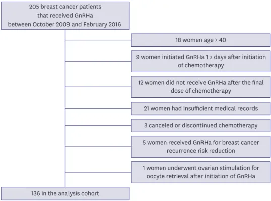
To ensure full coverage of the monthly administration of GnRHa treatment over chemotherapy, only patients who received GnRHa at least 1 day before the initiation of chemotherapy and at least once after the last dose of chemotherapy were included. Patients who did not have relevant medical records (n = 21), who did not receive chemotherapy as planned (n = 3), who received sequential GnRHa to prevent breast cancer recurrence (n = 5), and who underwent ovarian stimulation after initiation of GnRHa (n = 1) were excluded. Finally, 136 patients were included in the present analysis (Figure 1).
Various chemotherapeutic regimens were used: the 2 most common regimens were 5-fluorouracil (5-FU, 500 mg/m
2) plus doxorubicin (50 mg/m
2) and cyclophosphamide (500–600 mg/m
2) every 3 weeks for 6 cycles and doxorubicin (60 mg/m
2) plus
cyclophosphamide (600 mg/m
2) every 3 weeks for 4 cycles, followed by 4 cycles of paclitaxel (175 mg/m
2) or docetaxel (75 mg/m
2). The regimens used were classified into anthracycline- based, anthracycline-taxane-based, taxane-based, or others including CMF (6 cycles of cyclophosphamide [50 mg per oral thrice a day for 14 days] plus methotrexate [40 mg/m
2on days 1 and 8] plus 5-FU [600 mg/m
2on days 1 and 8] every 4 weeks).
Plasma AMH was measured using a commercially available kit (Gen II ELISA; Beckman Coulter, Brea, USA). The assay has a measurement range of 0.08–22.50 ng/mL. The intra- and inter- assay coefficient of variation were 5.4% and 5.6%, respectively. AMH values ≤ 0.08 ng/mL were imputed to one-half the threshold value (0.04 ng/mL) in the analyses.
The number of follicles in each ovary and the ovarian volume recorded in the initial
pelvic ultrasound examination were used to identify polycystic ovaries (PCOs). The PCO
morphology was defined as the presence of 12 or more follicles in each ovary measuring 2–9
mm in diameter and/or increased ovarian volume (> 10 cm
3) on transvaginal or transrectal
ultrasound examination [16].
The primary endpoint was to compare ovarian function among different timings of GnRHa administration. The main outcome measure was the percentage change in post- chemotherapy AMH (pcAMH) level, which was calculated using the following equation:
Patients were divided into 3 groups according to the interval between the initiation of GnRHa and chemotherapy (T
interval, 1–6, 7–13, or ≥ 14 days). In addition, recovery of ovarian function, defined as AMH ≥ 1.0 ng/mL, was evaluated based on previous reports [17,18].
Statistical analysis
Continuous variables were expressed as mean ± standard error, and the median values were shown when necessary. Baseline continuous variables were compared using the Kruskal- Wallis test. Qualitative variables were expressed as frequencies and percentages and
compared using the χ
2test. Potential confounders of pcAMH were evaluated using correlation analysis. Ranked analysis of covariance (ANCOVA) was used to compare the pcAMH values at different time points in each of the 3 groups while adjusting for the covariates identified in the correlation analyses. A multivariate analysis was performed to identify factors that were predictors of the recovery of ovarian function (AMH ≥ 1.0 ng/mL). Statistical significance was defined as differences with p < 0.05. All analyses were performed using SPSS version 23 (IBM Corp., Armonk, USA).
pcAMH value (ng/mL)
Baseline AMH value (ng/mL) × 100 (%) 136 in the analysis cohort
18 women age > 40 205 breast cancer patients
that received GnRHa
between October 2009 and February 2016
9 women initiated GnRHa 1 ≥ days after initiation of chemotherapy
12 women did not receive GnRHa after the final dose of chemotherapy
21 women had insufficient medical records 3 canceled or discontinued chemotherapy 5 women received GnRHa for breast cancer
recurrence risk reduction 1 women underwent ovarian stimulation for
oocyte retrieval after initiation of GnRHa
Figure 1. Flow diagram of subject selection.
GnRHa = gonadotropin-releasing hormone agonist.
Ethics statement
The Institutional Review Board of Seoul National University Hospital (Seoul, Korea) approved the prospective cohort, and informed consent was obtained from each patient (study
registered as H-0802-043-234).
RESULTS
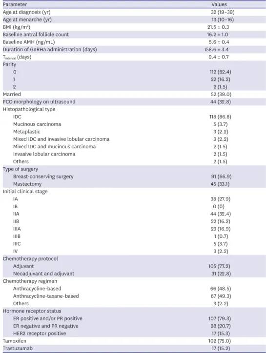
The baseline characteristics of the 136 subjects are summarized in Table 1. The median age was 32 years, and most subjects were nullipara (82.4%). The median follow-up duration of plasma AMH was 24 months. GnRHa was administered for an average of 158.6 days. Patients with hormone receptors positive tumors were very common in the present cohort (79.3%), and most of the hormone receptor positive patients received adjuvant tamoxifen (95.3%).
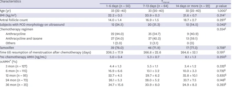
Baseline characteristics were similar among the 3 groups, except for the polycystic ovary (PCO) morphology on ultrasound (p = 0.042), where the PCO morphology was significantly more frequent in the 14-days-or-more group than in the 1-6-days group in the post-hoc analysis (p = 0.013) (Table 2). In the correlation analysis, age (Pearson coefficient, −0.414;
p < 0.001), body mass index (BMI, Pearson coefficient, −0.251; p = 0.015), and PCO on ultrasound (Spearman coefficient, 0.358; p = 0.001) were independently correlated with the AMH level (Table 3). Therefore, the pcAMH values shown in Table 2 were compared after adjustment for age, BMI, and PCO. Of note, the chemotherapy regimens and the use of tamoxifen did not demonstrate a significant correlation with AMH level (Table 3).
The AMH and pcAMH were nearly undetectable at 3 months and increased slowly thereafter, as shown in Table 2 and Figure 2. The pcAMH levels at each time point following chemotherapy
Time (mo) 0
Me an AMH (ng /mL )
13
12
A
3 10
8
5
36 24
Baseline 3 6
1–6 days 7–13 days 14 days or more Time interrval
Time (mo) 0
Me an pcAMH (%)
100
12
B
75
50
25
36 24
Baseline 3 6
1–6 days 7–13 days 14 days or more Time interrval
Figure 2. Comparison of changes in variables before and after chemotherapy among the 3 Tinterval
groups. (A) AMH, (B) pcAMH. The pcAMH was calculated by the equation:
pcAMH value (ng/mL)Baseline AMH value (ng/mL) × 100 (%)
. Error bars represent 95% confidence interval.
AMH = anti-Müllerian hormone; pcAMH = post-chemotherapy anti-Müllerian hormone; Tinterval = the interval between the initiation of gonadotropin-releasing hormone agonist and chemotherapy.
was not significantly different among the 3 T
intervalgroups (Table 2 and Figure 2). Comparison women with and without PCO, the difference in AMH gradually increased with time and was significantly different at baseline (p < 0.001), 12 months (p = 0.041), and 24 months (p = 0.009;
ranked ANCOVA, adjusted for age and BMI) (Figure 3A). Moreover, the difference in pcAMH between the 2 groups showed a gradual increase over time, and eventually became significant at 36 months (p = 0.046; ranked ANCOVA, adjusted for age and BMI) (Figure 3B).
Table 1. Patient characteristics
Parameter Values
Age at diagnosis (yr) 32 (19–39)
Age at menarche (yr) 13 (10–16)
BMI (kg/m
2) 21.5 ± 0.3
Baseline antral follicle count 16.2 ± 1.0
Baseline AMH (ng/mL) 5.6 ± 0.4
Duration of GnRHa administration (days) 158.6 ± 3.4
T
interval(days) 9.4 ± 0.7
Parity
0 112 (82.4)
1 22 (16.2)
2 2 (1.5)
Married 52 (39.0)
PCO morphology on ultrasound 44 (32.8)
Histopathological type
IDC 118 (86.8)
Mucinous carcinoma 5 (3.7)
Metaplastic 3 (2.2)
Mixed IDC and invasive lobular carcinoma 3 (2.2)
Mixed IDC and mucinous carcinoma 2 (1.5)
Invasive lobular carcinoma 2 (1.5)
Others 2 (1.5)
Type of surgery
Breast-conserving surgery 91 (66.9)
Mastectomy 45 (33.1)
Initial clinical stage
IA 38 (27.9)
IB 0 (0)
IIA 44 (32.4)
IIB 22 (16.2)
IIIA 23 (16.9)
IIIB 1 (0.7)
IIIC 5 (3.7)
IV 3 (2.2)
Chemotherapy protocol
Adjuvant 105 (77.2)
Neoadjuvant and adjuvant 31 (22.8)
Chemotherapy regimen
Anthracycline-based 66 (48.5)
Anthracycline-taxane-based 67 (49.3)
Others 3 (2.2)
Hormone receptor status
ER positive and/or PR positive 107 (79.3)
ER negative and PR negative 28 (20.7)
HER2 receptor positive 17 (15.3)
Tamoxifen 102 (75.0)
Trastuzumab 17 (15.2)
Data are shown as mean ± standard error, median (range), or number (%).
PCO = polycystic ovary; BMI = body mass index; IDC = invasive ductal carcinoma; GnRHa = gonadotropin- releasing hormone agonist; AMH = anti-Müllerian hormone; ER = estrogen receptor; PR = progesterone receptor;
HER2 = human epidermal growth factor receptor 2; T
interval= the interval between the initiation of gonadotropin-
releasing hormone agonist and chemotherapy.
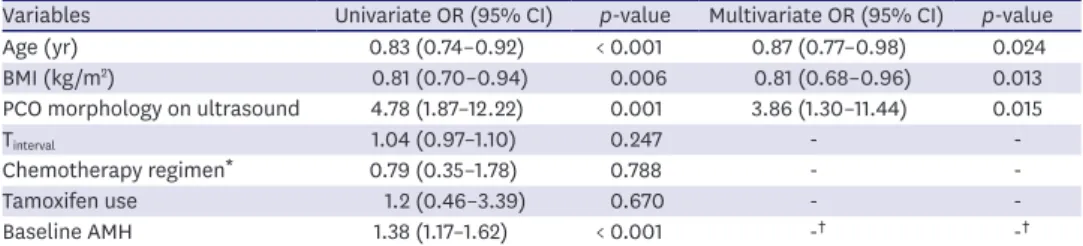
The factors associated with AMH ≥ 1 ng/mL at 12 months are summarized in Table 4. The proportion of patients with AMH ≥ 1 ng/mL at 12 months was 44.2%. Young age (adjusted odds ratio [OR], 0.87; 95% confidence interval [CI], 0.77–0.98]), low BMI (adjusted OR, 0.81; 95% CI, 0.68–0.96), and presence of PCO (adjusted OR, 3.86; 95% CI, 1.30–11.44) were associated with AMH ≥ 1 ng/mL. However, tamoxifen use and chemotherapy regimen did not have a significant association with AMH ≥ 1 ng/mL.
Table 2. Comparison of baseline characteristics, AMH and pcAMH* according to Tinterval
Characteristics T
interval1–6 days (n = 50) 7–13 days (n = 64) 14 days or more (n = 22)
p-valueAge (yr) 33 (22–40) 33 (20–40) 32 (22–40) 1.000
†BMI (kg/m
2) 22.2 ± 0.5 20.9 ± 0.3 21.6 ± 0.7 0.314
†Antral follicle count 14.0 ± 1.4 16.9 ± 1.5 18.7 ± 2.7 0.297
†Subjects with PCO morphology on ultrasound 12 (24.5) 20 (31.3) 12 (54.5) 0.042
‡Chemotherapy regimen 0.554
‡Anthracycline 22 (44.0) 35 (54.7) 9 (40.9)
Anthracycline and taxane 27 (54.0) 27 (42.2) 13 (59.1)
Others 1 (2.0) 2 (3.1) 0 (0)
Tamoxifen 39 (78.0) 46 (71.9) 17 (77.3) 0.728
‡Time till resumption of menstruation after chemotherapy (days) 206.5 ± 17.9 266.8 ± 22.6 264.6 ± 52.1 0.191
‡Pre-chemotherapy AMH (ng/mL) 5.0 ± 0.4 5.3 ± 0.7 8.1 ± 1.3 0.250
§pcAMH
*(%)
3 mon (n = 127) 4.4 ± 1.3 5.5 ± 1.1 3.4 ± 1.2 0.332
§6 mon (n = 115) 16.9 ± 6.6 13.1 ± 3.2 10.0 ± 3.3 0.732
§12 mon (n = 95) 22.7 ± 4.5 29.7 ± 6.2 32.8 ± 10.1 0.830
§24 mon (n = 73) 26.1 ± 5.3 28.0 ± 5.2 33.7 ± 7.5 0.148
§36 mon (n = 35) 34.7 ± 15.6 30.9 ± 8.0 24.9 ± 8.2 0.393
§Data are shown as mean ± standard error, median (range), or number (%).
BMI = body mass index; PCO = polycystic ovary; AMH = anti-Müllerian hormone; pcAMH = post-chemotherapy anti-Müllerian hormone; T
interval= the interval between the initiation of gonadotropin-releasing hormone agonist and chemotherapy.
*
pcAMH was calculated by the equation:
pcAMH value (ng/mL)Baseline AMH value (ng/mL) × 100 (%)
;
†Kruskal-Wallis test;
‡χ
2test;
§Ranked analysis of covariance test, adjusted for age, BMI, and PCO.
Time (mo) 0
Me an AMH (ng /mL )
12
12
A
2 10
6 8
4
36 24
Baseline 3 6
Without PCO With PCO
Time (mo) 0
Me an pcAMH (%)
100
12
B
20 60 80
40
36 24
Baseline 3 6
Without PCO With PCO
Figure 3. Comparison of changes in variables before and after chemotherapy between women with PCO and without PCO (A) AMH, (B) pcAMH. The pcAMH was
calculated by the equation:
pcAMH value (ng/mL)Baseline AMH value (ng/mL) × 100 (%)
. Error bars represent 95% confidence interval.
PCO = polycystic ovary; AMH = anti-Müllerian hormone; pcAMH = post-chemotherapy anti-Müllerian hormone.
DISCUSSION
In this prospective cohort study, we report that changes in AMH level after chemotherapy were not significantly different, regardless of the timing of GnRHa administration. In other words, there was no effect on the ovarian protective effect even if the chemotherapeutic agent was administered during the “flare-up phase” of GnRHa. This suggests that there may be little to no advantage in waiting 1–2 weeks before chemotherapy until pituitary suppression. A possible explanation for this is that although theoretically, the ovarian protective effect relies on pituitary suppression, there may be other direct actions of GnRHa on the ovaries that need to be elucidated [19].
One particular strength of our study is that we used AMH as a surrogate marker to evaluate ovarian function in patients with breast cancer. AMH is considered to be the most valuable marker for ovarian reserve and function, particularly in breast cancer survivors. A large proportion of breast cancer survivors receive either tamoxifen or GnRHa, both of which do not affect plasma AMH levels but may affect other surrogate markers of ovarian function, such as resumption of menses or other female hormones. Therefore, previous studies that evaluated ovarian function using these markers in breast cancer carry the risk of misclassifying women with declined ovarian function [20].
Although AMH appears to be an ideal marker of ovarian reserve, it can be misleading if PCO is not considered. Women with PCO generally exhibit AMH levels 2 or 3 times higher than the average value at their age, which can be misinterpreted as 2 or 3 times greater ovarian reserve than their actual value [21]. In this study, we demonstrated that there is not only a difference in the absolute value, but also differences in the pattern of change over time compared with baseline AMH values according to the presence of PCO.
Table 4. Predictors for AMH ≥ 1 ng/mL at 12 months
Variables Univariate OR (95% CI)
p-valueMultivariate OR (95% CI)
p-valueAge (yr) 0.83 (0.74–0.92) < 0.001 0.87 (0.77–0.98) 0.024
BMI (kg/m
2) 0.81 (0.70–0.94) 0.006 0.81 (0.68–0.96) 0.013
PCO morphology on ultrasound 4.78 (1.87–12.22) 0.001 3.86 (1.30–11.44) 0.015
T
interval1.04 (0.97–1.10) 0.247 - -
Chemotherapy regimen
*0.79 (0.35–1.78) 0.788 - -
Tamoxifen use 1.2 (0.46–3.39) 0.670 - -
Baseline AMH 1.38 (1.17–1.62) < 0.001 -
†-
†AMH = anti-Müllerian hormone; BMI = body mass index; OR = odds ratio; CI = confidence interval; PCO = polycystic ovary; T
interval= the interval between the initiation of gonadotropin-releasing hormone agonist and chemotherapy.
*
Two group analysis: anthracycline-based chemotherapy versus anthracycline and taxane-based chemotherapy;
†
Baseline AMH was discarded from multivariate analysis due to multicollinearity with age, BMI, and PCO as shown in Table 3.
Table 3. Identification of covariates for anti-Müllerian hormone at 12 months
Characteristics Correlation coefficient
p-valueAge (yr) −0.414
*< 0.001
BMI (kg/m
2) −0.251
*0.015
PCO morphology on ultrasound 0.358
†0.001
Tamoxifen 0.029
†0.781
Chemotherapy regimen
‡−0.044
†0.675
BMI = body mass index; PCO = polycystic ovary.
*
Pearson correlation coefficient;
†Spearman correlation coefficient;
‡Two group analysis: anthracycline-based
chemotherapy versus anthracycline and taxane-based chemotherapy.
Another strength of our study was that we used ranked ANCOVA to compare variables while adjusting for PCO and other factors. The pcAMH data were positively skewed where the mean was greater than the median; thus, a non-parametric analysis was required. With ranked ANCOVA, the confounders were successfully controlled while enabling a non-parametric comparison of variables. In addition, we used pcAMH as the dependent variable to eliminate the need for adjustment for baseline AMH, which may cause multicollinearity issues with age (i.e., adjusted twice).
In the study by Zhang et al., [22] the authors compared sequential versus simultaneous use of chemotherapy and GnRHa in estrogen receptor-positive patients with breast cancer. In that study, the main purpose of GnRHa was therapeutic ovarian suppression to prevent cancer recurrence, with the treatment lasting > 2 years. There was no difference in the median time to resumption of menstruation between the 2 groups. However, resumption of menstruation was not a suitable marker for that study because all patients received tamoxifen for 5 years which can cause menstrual irregularities, vaginal bleeding, or cessation of menstrual periods.
On the other hand, we have the advantage of assessing ovarian function using AMH which is a significantly more reliable marker than resumption of menstruation and in addition, was adjusted for age, BMI, and PCO.
To our knowledge, this was the third study to use AMH as a surrogate marker for ovarian reserve in patients with breast cancer who received GnRHa for fertility preservation.
Moreover, this was the first study to use AMH to compare the effect of different timings of administration GnRHa on ovarian reserve in patients with breast cancer. In a recent study, amenorrhea and AMH was used as a surrogate marker of ovarian reserve. They described the chronological change of AMH, but they did not compare AMH between groups [23].
Another previously published study that used AMH as a surrogate maker of ovarian function in patients with breast cancer following chemotherapy and concomitant administration GnRHa for ovarian protection focused on determining factors that influenced or predicted good ovarian reserve [18] and reported that tamoxifen was associated with low serum AMH level at 12 months in the multivariate analysis (adjusted OR, 0.156; 95% CI, 0.032–0.766).
In previous studies, tamoxifen was not associated with decreased AMH levels, and some studies reported associations with high AMH levels [24-26]. The authors addressed the issue in the discussion but did not describe the reason. A possible explanation is that they did not take PCO into account. As shown in our study, PCO is significantly associated with not only increase in the absolute AMH value but also an increase in the rate of AMH recovery relative to baseline AMH. In our multivariate analysis, young age, low BMI, and the presence of PCO were associated with high AMH levels at 12 months. High baseline AMH level was undoubtedly associated with high AMH level at 12 months; however, it was discarded from multivariate analysis due to multicollinearity (Table 3). Low BMI was associated with high AMH after chemotherapy in our study. Although the exact mechanism underlying this is unclear, it is consistent with a previous study that obesity adversely affects serum AMH when adjusted for PCO [27].
One limitation of our study was that the efficacy of the GnRHa was not assessed by
comparing with patients who did not receive GnRHa. However, because our cohort included
only patients who received GnRHa, acquiring data from patients with breast cancer who did
not receive GnRHa would require another study protocol, which was beyond the scope of the
present investigation.
An important consideration to keep in mind when interpreting our results is that the composition of our cohort may be different from the general young breast cancer population.
These were women diagnosed with breast cancer but were selectively referred to the fertility clinic for fertility preservation. Patients with severe disease who required prompt initiation of chemotherapy may not have been included in the cohort. Furthermore, the reason that there was only one patient who received CMF is that it is known for having a higher risk for ovarian failure than other regimens, and medical oncologists generally avoid this regimen in women having a significant interest in preserving fertility.
In conclusion, regardless of the timing of administration before chemotherapy, there was no difference in the efficacy of the GnRHa in preserving the ovarian reserve assessed using AMH as a surrogate marker in patients with breast cancer.
REFERENCES
1. Fidler MM, Gupta S, Soerjomataram I, Ferlay J, Steliarova-Foucher E, Bray F. Cancer incidence and mortality among young adults aged 20–39 years worldwide in 2012: a population-based study. Lancet Oncol 2017;18:1579-89.
PUBMED | CROSSREF
2. Zolton JR, Parikh TP, Hickstein DD, Holland SM, Hill MJ, DeCherney AH, et al. Oocyte cryopreservation for women with GATA2 deficiency. J Assist Reprod Genet 2018;35:1201-7.
PUBMED | CROSSREF
3. Brydøy M, Fosså SD, Dahl O, Bjøro T. Gonadal dysfunction and fertility problems in cancer survivors.
Acta Oncol 2007;46:480-9.
PUBMED | CROSSREF
4. Clemons M, Simmons C. Identifying menopause in breast cancer patients: considerations and implications. Breast Cancer Res Treat 2007;104:115-20.
PUBMED | CROSSREF
5. Zavos A, Valachis A. Risk of chemotherapy-induced amenorrhea in patients with breast cancer: a systematic review and meta-analysis. Acta Oncol 2016;55:664-70.
PUBMED | CROSSREF
6. Klein NA, Battaglia DE, Fujimoto VY, Davis GS, Bremner WJ, Soules MR. Reproductive aging: accelerated ovarian follicular development associated with a monotropic follicle-stimulating hormone rise in normal older women. J Clin Endocrinol Metab 1996;81:1038-45.
PUBMED
7. Xue C, Wei W, Sun P, Zheng W, Diao X, Xu F, et al. Pretreatment anti-Mullerian hormone-based nomogram predicts menstruation status after chemotherapy for premenopausal women with hormone receptor-positive early breast cancer. Breast Cancer Res Treat 2019;173:619-28.
PUBMED | CROSSREF
8. Fréour T, Barrière P, Masson D. Anti-Müllerian hormone levels and evolution in women of reproductive age with breast cancer treated with chemotherapy. Eur J Cancer 2017;74:1-8.
PUBMED | CROSSREF
9. Perdrix A, Saint-Ghislain M, Degremont M, David M, Khaznadar Z, Loeb A, et al. Influence of adjuvant chemotherapy on anti-Müllerian hormone in women below 35 years treated for early breast cancer.
Reprod Biomed Online 2017;35:468-74.
PUBMED | CROSSREF
10. van Rooij IA, Broekmans FJ, Scheffer GJ, Looman CW, Habbema JD, de Jong FH, et al. Serum
antimullerian hormone levels best reflect the reproductive decline with age in normal women with proven fertility: a longitudinal study. Fertil Steril 2005;83:979-87.
PUBMED | CROSSREF
11. Anderson RA, Nelson SM, Wallace WH. Measuring anti-Müllerian hormone for the assessment of ovarian reserve: when and for whom is it indicated? Maturitas 2012;71:28-33.
PUBMED | CROSSREF
12. Sandow J, Von Rechenberg W, Jerzabek G, Stoll W. Pituitary gonadotropin inhibition by a highly active analog of luteinizing hormone-releasing hormone. Fertil Steril 1978;30:205-9.
PUBMED | CROSSREF
13. Dewailly D, Andersen CY, Balen A, Broekmans F, Dilaver N, Fanchin R, et al. The physiology and clinical utility of anti-Mullerian hormone in women. Hum Reprod Update 2014;20:370-85.
PUBMED | CROSSREF
14. Practice Committee of American Society for Reproductive Medicine. Fertility preservation in patients undergoing gonadotoxic therapy or gonadectomy: a committee opinion. Fertil Steril 2013;100:1214-23.
CROSSREF
15. Lambertini M, Richard F, Nguyen B, Viglietti G, Villarreal-Garza C. Ovarian function and fertility preservation in breast cancer: should gonadotropin-releasing hormone agonist be administered to all premenopausal patients receiving chemotherapy? Clin Med Insights Reprod Health. Forthcoming 2019.
PUBMED | CROSSREF
16. Balen AH, Laven JS, Tan SL, Dewailly D. Ultrasound assessment of the polycystic ovary: international consensus definitions. Hum Reprod Update 2003;9:505-14.
PUBMED | CROSSREF
17. Nelson SM, Yates RW, Fleming R. Serum anti-Müllerian hormone and FSH: prediction of live birth and extremes of response in stimulated cycles--implications for individualization of therapy. Hum Reprod 2007;22:2414-21.
PUBMED | CROSSREF
18. Lee DY, Park YH, Lee JE, Choi D. Prediction of ovarian function recovery in young breast cancer patients after protection with gonadotropin-releasing hormone agonist during chemotherapy. Breast Cancer Res Treat 2018;171:649-56.
PUBMED | CROSSREF
19. Poggio F, Lambertini M, Bighin C, Conte B, Blondeaux E, D'Alonzo A, et al. Potential mechanisms of ovarian protection with gonadotropin-releasing hormone agonist in breast cancer patients: a review. Clin Med Insights Reprod Health. Forthcoming 2019.
PUBMED | CROSSREF
20. Seifer DB, Maclaughlin DT. Mullerian Inhibiting Substance is an ovarian growth factor of emerging clinical significance. Fertil Steril 2007;88:539-46.
PUBMED | CROSSREF
21. Pellatt L, Hanna L, Brincat M, Galea R, Brain H, Whitehead S, et al. Granulosa cell production of anti- Müllerian hormone is increased in polycystic ovaries. J Clin Endocrinol Metab 2007;92:240-5.
PUBMED | CROSSREF
22. Zhang Y, Ji Y, Li J, Lei L, Wu S, Zuo W, et al. Sequential versus simultaneous use of chemotherapy and gonadotropin-releasing hormone agonist (GnRHa) among estrogen receptor (ER)-positive premenopausal breast cancer patients: effects on ovarian function, disease-free survival, and overall survival. Breast Cancer Res Treat 2018;168:679-86.
PUBMED | CROSSREF
23. Zhong Y, Lin Y, Cheng X, Huang X, Zhou Y, Mao F, et al. GnRHa for ovarian protection and the
association between AMH and ovarian function during adjuvant chemotherapy for breast cancer. J Cancer 2019;10:4278-85.
PUBMED | CROSSREF
24. Shandley LM, Spencer JB, Fothergill A, Mertens AC, Manatunga A, Paplomata E, et al. Impact of tamoxifen therapy on fertility in breast cancer survivors. Fertil Steril 2017;107:243-252.e5.
PUBMED | CROSSREF
25. Dezellus A, Barriere P, Campone M, Lemanski C, Vanlemmens L, Mignot L, et al. Prospective evaluation of serum anti-Müllerian hormone dynamics in 250 women of reproductive age treated with chemotherapy for breast cancer. Eur J Cancer 2017;79:72-80.
PUBMED | CROSSREF
26. Anderson RA, Mansi J, Coleman RE, Adamson DJ, Leonard RC. The utility of anti-Müllerian hormone in the diagnosis and prediction of loss of ovarian function following chemotherapy for early breast cancer.
Eur J Cancer 2017;87:58-64.
PUBMED | CROSSREF
27. Moy V, Jindal S, Lieman H, Buyuk E. Obesity adversely affects serum anti-Müllerian hormone (AMH) levels in Caucasian women. J Assist Reprod Genet 2015;32:1305-11.
PUBMED | CROSSREF