서 론
방광의 기본 기능은 신장에서 생산된 소변을 저장하고 체외로 배출하는 것이다. 방광의 기능과 관련하여 신경학 적 연구는 오래 전부터 많은 부분이 밝혀져 있다. 그러나 방광과 관련된 호르몬 및 그 역할에 대한 연구는 많지 않다.
방광과 관련된 호르몬으로는 human chorionic gonadotropin (hCG),1 estrogen2 및 parathyroid hormone-like protein (or peptide; PTHrP) 등3이 있다. 정상상태에서 방광근육 (detru- sor muscle)은 PTHrP를 생산하고,4방광의 염증이 발생한 점
막에서는 PTHrP 에 대한 항체3를 보인다. hCG는 정상 및 종양의 방광점막상피5,6뿐만 아니라 여러 장기7에서 생산된 다. 인체 방광 삼각부8와 후부요도9에서 estrogen 수용체가 발견되었다.
Gonadotropin releasing hormone (GnRH)은 시상하부-뇌하 수체 축 (hypothalmopituitary axis)10,11과 관련되어 시상하부 에서 배타적으로 분비되며 많은 작용호르몬 (effector hor- mone)의 중추역할을 하는 것으로 알려지고 있다. 그러나 1975년 Gibbons 등12이 태반에서 GnRH와 비슷한 hormone 을 발견한 뒤, 비뇨기계 장기를 포함하여 여러 장기에서 시 상하부외 (extrahypothalmic) GnRH 생산이 발견되고 있다.13
수용체의 발현과 방광상피세포의 증식에 대한 GnRH의 역할
Gonadotropin Releasing Hormone (GnRH) and GnRH Receptor in Normal Bladder Epithelia and Their Role in Bladder Epithelial Proliferation
Myoung Ock Kim3, Jeong-Hee Lee2, Moon Seok Park3, Hye Lyoung Lee3, Jong Yoon Bahk1
From the Department of 1Urology, 2Pathology and 3Biology, Gyeongsang National University, Jinju, Korea
Purpose: To confirm the production of extra-hypothalamic gonadotropin releasing hormone (GnRH) and GnRH receptor in bladder mucosal epi- thelia, and a potential role of GnRH on the bladder, normal human bladder tissues, and primary cultured dog bladder mucosal epithelia were studied.
Materials and Methods: For this study, normal human bladder tissue from 4 patients and primary cultured normal bladder mucosal epithelial cells from 2 dogs were used. For localization of extra-hypothalamic GnRH and the extra-pituitary GnRH receptor, in situ hybridization and immuno- histochemical staining were done. To evaluate the roles of exogenous GnRH in bladder mucosal cells, the culture media were supplemented with charcoal stripped serum and 4 different concentrations of GnRH (0, 10-3, 10-5and 10-7M). The effect of exogenous GnRH was evaluated using a hemocytometer and fluorescence activated cell sorter (FACS).
Results: GnRH and GnRH receptors, and their mRNA signals were localized in most of the both human bladder mucosal epithelia and dog bladder mucosal epithelia, but not in a few cells. There were no significant GnRH effects on cellular proliferation and cell cycle changes (p<0.05).
Conclusions: Bladder mucosal epithelium produces GnRH and GnRH re- ceptors, but they do not effect either the proliferation or cell cycle changes.
Although the exact function of extra-hypothalamic bladder GnRH is unknown, GnRH and GnRH receptors would be assumed to have un- known autocrine or paracrine relationships with each other. (Korean J Urol 2007;48:152-157)
Key Words: Gonadotropin-releasing hormone, Receptor, Mucosa
대한비뇨기과학회지 제 48 권 제 2 호 2007
경상대학교 의과대학
1비뇨기과학교실, 2병리학교실
3자연과학대학 생물학교실
김명옥3 이정희2 박문석3 이혜령3 박종윤1
접수일자:2006년 9월 25일 채택일자:2007년 1월 11일
교신저자: 박종윤
경상대학교 의과대학 비뇨기과학교실 경남 진주시 칠암동 92
660-751
TEL: 055-751-8816 FAX: 055-751-8807 E-mail: jybahk@hanmail.
net
이 논문은 한국과학재단(과제번호 2006- 04011)의 지원에 의해 수행되었음.
152
그러나 시상하부외 GnRH의 역할에 대해선 아직 불분명하다.
본 연구에서, 저자들은, 정상 방광상피에서 GnRH와 GnRH 수용체의 생산을 확인하고 GnRH의 방광상피에 대 한 역할에 대해 알아보고자 하였다.
재료 및 방법
연구 재료로 성인 남자로부터 채취한 인체 방광조직과, 출생 후 1년 이상 2년 이내 연령의 성견 (成犬) 방광으로부 터 방광상피 초대배양을 통해 얻은 방광세포를 실험에 사 용하였다. 쥐의 뇌조직을 GnRH 및 GnRH 수용체에 대한 대 조군으로 하였다.
1. 방광조직 및 방광상피 초대배양과 처리
혈뇨가 확인된 40대 4명의 남성 환자로부터 동의를 받고 정상방광으로 여겨지는 부위로부터 방광 점막을 채취하였 다. 방광경검사 당시 감염은 없었고, 육안적 종양은 없었다.
Bahk 등14의 방법에 따라 실험견의 방광상피 초대배양을 실 시하였다. 간단히 서술하면, 준비된 수컷 실험견 2마리를 과잉 마취로 안락사시킨 후, 방광을 멸균적으로 분리하여, 항생제가 포함된 얼음으로 냉각된 HBSS (iced Hank's bal- anced salt solution)에 담아 층류상자로 옮긴 후, 방광의 중앙 을 절개하여 HBSS로 세척하였다. 이 방광을 냉각된 페트리 접시 위에 고정시킨 후 방광부분의 점막부위만을 11번 수 술칼로 긁어 (scratch) 모아 냉 HBSS로 세척 및 원심분리 (2,000rpm, 20분)한 후, 미리 우태아혈청, L-glutamine, sodi- um bicarbonate, HEPES, 항생제 (penicillin, streptomycin, amphotericin B), bovine insulin, bovine 뇌하수체 추출물, 재 조합 인형 epidermal growth factor, hydrocortisone 및 trans- ferrin을 첨가하여 준비된 keratinocyte growth medium (Clo- netics, USA)에 배양하였다. 초대배양된 세포에 대한 GnRH 의 증식효과를 관찰하기 위해서는 실험에 사용된 배양액은 charcoal stripped 혈청을 사용하였다.
방광조직은 4시간 동안 4oC 0.1M phosphate buffer saline (PBS) 에 4%로 희석된 paraformaldehyde 용액에 고정하였 다. 20% sucrose에 밤새 보관한 후, 냉동블록을 만들어 10μm 두께의 냉동 절편을 만들어 양성전하를 띤 슬라이드 (Super- frost/Plus, Fisher Scientific)에 부착시켜 사용 전까지 영하 70 도에 보관하였다.
2. GnRH와 GnRH 수용체 cDNA의 subcloning 본 실험에 사용한 probe은 원숭이 GnRH와 GnRH 수용체 의 cDNA이다. 이들은 사람의 cDNA와 95% 유사성을 지니 고 백서와 90% 이상 유사성을 가진 것으로 Ojeda 교수
(Oregon Health Sciences University, USA)로부터 분양 받아 사용하였다. GnRH probe제작을 위해 pGEM 3Z vector (pro- mega)에 204bp cDNA segment로 subcloning해 사용하였다.
Antisense probe를 위해 EcoRI 제한효소로 일정 길이의 cDNA를 회수한 뒤 SP6 RNA polymerase로 probe를 합성하 였고, sense probe를 위해 Hind III 제한효소와 SP6 RNA polymerase로 probe를 합성하였다. GnRH 수용체 probe는 pAMP10 plasmid에 삽입된 474bp 크기를 사용하였다. Anti- sense probe를 위해, EcoR I 제한효소와 SP6 RNA poly- merase를 사용하여 probe를 합성하였고, sense probe를 위해 Hind III와 T7 RNA polymerase를 사용하였다.
3. GnRH와 GnRH 수용체 cRNA probes 준비 RNA probe의 in vitro 합성에 사용할 template DNA는 EcoR I, Hind III 제한절단 부위에 원숭이 GnRH, GnRH 수용체 cDNA clones이 삽입된 pAMP10와 pGEM 3Z (promema)였다.
제한효소를 사용하여 template DNA plasmid를 선형으로 만든 후, SP6 또는 T7 RNA polymerase로 전사시켜서 α- 35S-UTP 에 의해 방사표지된 anti-sense와 sense RNA probe (1.3x10⁹ cpm/μl)를 얻었다. 4.0μl의 5xtranscription buffer [200mM Tris-HCl (pH 7.9), 30mM MgCl2, 10mM spermidine], 2μl의 100mM DTT, 0.33μl의 10mM RNasin (60units/μl, BM), 1μl 의 10mM NaCl, 1μl의 10mM GTP, lμl의 10mM CTP, 2.4μl 의 100M UTP, 0.2μg의 linearized DNA template, 0.3μl의 SP6 혹은 T7 RNA polymerase (15units/μl, BRL), 5.0μl의 α-
35S-UTP (10mCi/ml)를 첨가하여 37oC에서 1시간 동안 반응시 켰다. 그 후 DNA template를 제거하기 위해 92μl의 증류수, 5μl의 1M Tris-HCl (pH 8.0), 1μl의 1M MgCl2, 1μl의 yeast tRNA (10mg/ml), DNase I (10μg/μl)을 첨가하고, 37oC에서 30분간 반응시켰다. Antisense 및 sense RNA probe는 Sephadex G-50 Nick column (Pharmacia, USA)으로 각각 분리하였다.
4. In situ hybridization
In situ hybridization 방법은 Duello 등15의 방법에 따랐다.
방광 조직절편을 PBS으로 세척한 후 0.001% proteinase K로 37oC에서 반응시킨 후, TE buffer (pH 8.0 10mM Tris, 1mM EDTA)로 2분간 처리한 후 0.025% acetic acid에서 10분간 반응시켰다. 0.2xSSC (1xSSC: 150mM NaCl, 15mM Na citrate, pH7.0)로 세척한 다음 실온에서 1시간 동안 prehybridization 과정을 거치고 이어서 prehybridization buffer [65% deionized formamide, 10% 5M NaCl, 1% 50 Denhardt's solution, 0.5%
1M Tris (pH 8.0), 0.1% 0.5M EDTA (pH 8.0), 23%의 Dextran sulfate 0.4% 1M DTT, 1% yeast tRNA (10mg/ml)]에 준비된 RNA probe (1x10⁵cpm/slide)를 첨가하여 조직절편 위에 직접
점적시켜 cover glass를 덮고 습윤 상자에 넣고 60oC에서 24 시간 동안 반응시켰다. 반응 후 post hybridization 과정으로 4xSSC, 0.5xSSC에 DTT가 함유되게 한 후 각각 20분씩 세척 하였다. 그 후 RNase A (20μl/ml)를 1시간 처리한 후 65oC에 서 2xSSC로 30분간 세척하고 탈수과정을 거친 후 Auto- radiography X-ray필름으로 1주일 동안 감광시키고 현상시 켰다. 그 후 autoradiographic emulsion (NTB2, Kodak)으로 도 포한 후 4oC에서 14일 동안 표지된 probe의 방사선이 감광 되도록 노출시켰다. 그 후 D-19 현상액으로 현상시키고 rapid fixer로 고정한 후 cresyl violet으로 대조 염색한 후 광 학현미경에서 관찰하였다.
5. 면역조직화학적 염색
방광조직에 대한 GnRH와 GnRH 수용체에 대한 면역조 직화학은 avidine-biotine complex (ABC) 방법을 사용하였다.
슬라이드는 0.02M PBS로 세척하고, 1:20으로 희석된 정상 염소혈청 40μl로 비특이 반응을 억제시킨 후, 1:500으로 희 석된 rabbit-derived anti-human GnRH와 mouse-derived anti- human GnRH 수용체 50μl를 침적시켜 4oC에서 밤새 처리 하였다. 그 뒤, 0.5% periodic acid로 외인성 peroxidase를 제 거한 다음 biotinylated goat anti-rabbit IgG와 mouse IgG (Vector, USA), 그리고 ABC 용액으로 반응시켰다. 그리고 0.05% 3,3'-diaminobenzidine (DAB)에 0.01% hydrogen pero- xide가 함유된 PBS에서 면역반응을 확인하고 세척, 탈수한 뒤 봉입하여 광학현미경에서 관찰하였다.
6. 세포증식과 세포주기 검사
방광상피세포 증식에 대한 GnRH의 역할을 연구하기 위 하여, 초대배양된 실험견 방광상피세포를 정상 우태아혈청
으로 준비된 배양액에 105cells/ml의 농도로 희석하여 75cm2 배양플라스크에 24시간 배양하였다. 배양 24시간에 기존의 배양액을 완전히 흡입 배액시킨 후, PBS 용액으로 세척하 였다. 실험군은 charcoal stripped 혈청으로 조성된 배양액으 로 교환하였으며 대조군은 정상 혈청으로 조성된 신선 배 양액으로 교환하였다. 실험군에는 4 종류의 각기 상이한 농 도의 GnRH (10-7, 10-5, 10-3M/ml and none)를 첨가하여, 파종 후 일주일 동안 상기 방법으로 배양하였다. 세포들은 배양 후 7일에 Trypsin-EDTA 용액으로 탈착시킨 후 균질액의 일 부를 취해 hemocytometer를 이용하여 세포를 계수하였다.
나머지 세포부유액은 1,000rpm으로 10분간 원심분리하여 세포들을 침강시킨 후 구연산 완충용액에 고정시켰다. 세 포막 용해제로 처리한 후 propidium iodide로 염색한 후, 유 세포분석기 (FACS. PAS-III, Partec, France)를 이용하여 세포 주기를 검사하였다.
7. 통계학적 처리
세포증식의 통계학적 분석은 Student’s t-test 분석을 이용 하였으며, p<0.05인 경우 통계학적으로 의미 있다고 판정 하였다.
결 과
1. In situ hybridization을 이용한 GnRH 및 GnRH 수용 체 mRNA의 발현
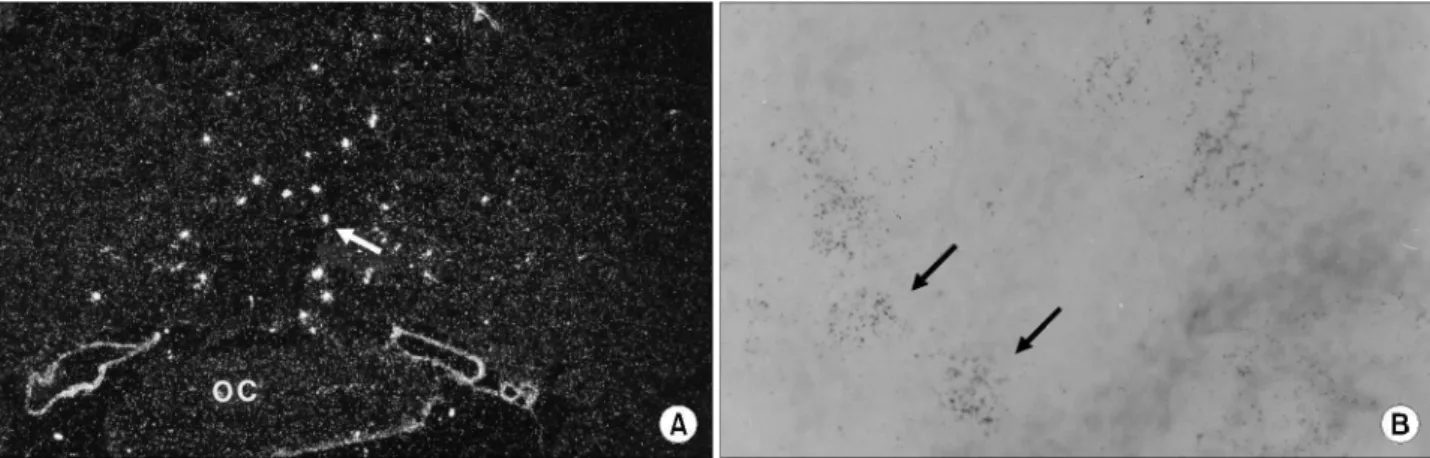
백서의 뇌조직 (대조군)에서 GnRH mRNA는 제3 ventricle (Fig. 1A)에서 발견되었으며 GnRH 수용체 mRNA는 anteri- or 뇌하수체 (Fig. 1B)에서 발견되었다.
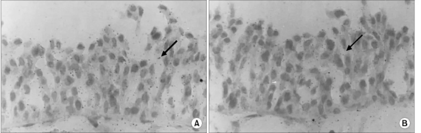
정상 인체 방광의 상피세포에서 GnRH (Fig. 2A)와 GnRH
Fig. 1. Expressions of GnRH and GnRH receptor mRNA in the preoptic area of the rat brain and anterior pituitary. The white spots around the 3rd ventricle (A) in the dark field microscopic view indicate the expression of GnRH mRNA. GnRH receptor mRNA signals are detected in the anterior pituitary (B). The signals are in clusters in both views. Arrows: positive signals. OC: optic chiasma. GnRH:
gonadotropin releasing hormone.
수용체 (Fig. 2B) mRNA가 발현하였으나, 각 세포 내에서 신 호의 숫자는 각각 일정하지 않았다. 대부분이 핵부근에서 신호를 발현하였으나, 일부에서는 세포질에서도 발견되었다.
2. GnRH와 GnRH 수용체에 대한 면역조직화학검사 면역조직화학염색에서, GnRH와 GnRH 수용체 단백은 정 상 인체 방광상피조직 (Fig. 3A, B)과 실험견 방광상피세포 (Fig. 3C, D)에서 모두에서 확인되었으나, 일부 세포에서는 확인되지 않았다. 이 연구결과 정상 방광상피세포는 GnRH
와 GnRH 수용체를 생산하였다. 염색의 정도와 위치는 각 세포에 따라 약간씩 상이하였다. 정상 방광점막에서 기저 막 부근 세포에 비하여 표면의 세포가 강하게 염색되었다.
3. GnRH의 세포증식 및 세포주기에 대한 역할 Charcoal stripped 혈청을 첨가한 배양액에 4종류의 서로 상이한 농도의 GnRH를 투여한 개 방광상피세포 (dog blad- der epithelial cell)를 이용하여 GnRH의 세포증식에 관한 결 과를 연구한 결과, GnRH 투여는 대조군에 비하여 의미 있 Fig. 2. Expressions of the GnRH (A) and GnRH receptors (B) mRNA signals from human bladder tissue. The signal numbers are different to each other, with some cells not expressing the signal. Arrow: mRNA signal. GnRH: gonadotropin releasing hormone.
Fig. 3. The expressions of the GnRH (A, C) and GnRH receptor (B, D) proteins in human normal bladder mucosa tissue (A, B) and primary cultured dog bladder mucosal epithelia (C, D). Brown color staining indicates positive staining for GnRHs and GnRH receptors. GnRH:
gonadotropin releasing hormone.
는 세포증식의 효과가 없었다 (Table 1). GnRH를 동일한 조 건으로 투여하면서 배양한 세포들에 대한 세포주기 변화에 대한 실험에서도 GnRH가 세포주기의 변화에 영향을 미치 지 못했다 (Table 2).
고 찰
방광에 대한 호르몬기능에 관한 연구는 많지 않으나, 남 성 스테로이드 호르몬은 방광종양의 재건16,17및 종양의 방 광점막상피의 성장 및 분화18와 관련 있으며, 일부 방광점막 은 이소적 골형성 (ectopic bone formation)을 유도19,20한다고
알려져 있다. Shen 등21은 방광암의 병기 및 분화도 증가와 방광의 estrogen 수용체의 증가가 관련 있음을 보고하였다.
GnRH는 시상하부-뇌하수체 생식의 축에서 중추역할을 하며, GnRH 는 시상하부에서만 생산되고 그의 수용체는 뇌 하수체에만 존재하는 것으로 여겨왔으나, 시상하부외 GnRH 및 GnRH 유사물질 또는 GnRH 수용체들은 고환13및 전립 선22에서 발견되었으나 이들의 역할은 아직 분명하지 않다.
조직 또는 세포의 분화 및 증식과 관련하여 호르몬을 포함 한 다양한 성장요소들이 관여하고 있음을 비추어 이들 시 상하부외 GnRH가 비뇨기계 장기에서 어떤 미지의 역할은 하고 있을 것이다.
저자들의 실험결과, 정상 방광점막상피는 GnRH 및 GnRH 수용체를 생산하였다. 그러나 모든 방광점막상피 조직 또 는 세포가 양성반응을 보이지 않았다는 점은 본 연구의 실 험계가 소유한 기본적인 전제 및 문제점들, 즉 이 실험의 전제조건은 내분비 (endocrine) 기전은 근본적으로 배제된 실험모델이라는 것과 모든 실험이 서로 매우 근접한 조직 에서 수행되었으나 동일 조직에서 수행되지 않았다는 것은 시상하부 외 방광점막상피 GnRH와 뇌하수체외 방광 GnRH 수용체 사이의 미지의 작용기전 연구에서 중요한 고 려사항일 것이다.
저자들의 연구결과 (Fig. 2, 3)에서, 방광점막상피 대부분 의 세포에서 GnRH 및 GnRH 수용체를 발견함으로써 유추 한 방광점막상피의 시상하부외 GnRH의 역할은, GnRH 수 용체와 관련하여, paracrine 효과 또는 autocrine 효과였다. 이 러한 유추아래 시상하부 외 방광 GnRH (extrahypothalamic bladder GnRH)가 방광점막상피세포의 증식에 관여하는지 를 알아보고자 저희들이 실시한 2가지 실험, GnRH가 제외 된 혈청 (charcoal stripped serum)을 사용한 배양액에 외부로 부터 4종류의 상이한 농도의 GnRH를 초대배양세포에 투여 후 실시한 세포증식 및 합성기를 포함한 세포주기변화를 관찰한 실험에서 의미 있는 결과를 확인하고자 하였으나, 두 실험 모두에서 의미 있는 결과를 확인하지 못했다. 이는 외부로부터 투여된 GnRH는 방광점막상피세포의 증식에 관여하지 않는다고 저자들은 추정하고 있다.
이상의 연구에서 GnRH와 GnRH 수용체가 방광점막에 존재하는 것은 밝혔으나, 이들이 동일 세포에 존재하는지 또는 그들이 방광점막상피와 관련되어 어떤 상호 간의 역 할을 하는지는 밝히지 못했다. 동시에, GnRH는 방광점막세 포의 증식을 유도하지 않았다. 그러나, 본 실험모델의 성격 상 이들 시상하부외 방광 GnRH와 뇌하추체외 방광 GnRH 수용체 사이에는 미지의 paracrine 또는 autocrine 작용기전 을 소유할 것으로 추정한다.
Table 1. The proliferation of primary cultured dog bladder mucosal cells, with treatment of exogenous GnRH
Experimental group (concentration of GnRH) Control
10-3M 10-5M 10-7M 0M
41.2± 40.46± 40.25± 39.84± 39.53±
3.59 3.47 3.25 3.92 3.18
Count: x104cells.The control was cultured in media that sup- plemented with normal serum. The experimental groups were cultured in media supplemented with charcoal stripped serum and four different concentrations of GnRH (0, 10-3, 10-5 and 10-7M). There was no significant difference (p<0.05) in pro- liferation between the control and experimental groups. Among the experimental groups, there was no significant difference in proliferation with any of the four different supplemented con- centrations of exogenous GnRH. GnRH: gonadotropin releasing hormone.
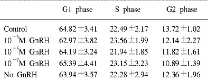
Table 2. Changes in the proportion of cell cycle phases after 6 days of culture, with supplementation of different concentrations of GnRH, in primary cultured dog bladder mucosal cells (%)
G1 phase S phase G2 phase Control 64.82±3.41 22.49±2.17 13.72±1.02 10-3M GnRH 62.97±3.82 23.56±1.99 12.14±2.27 10-5M GnRH 64.19±3.24 21.94±1.85 11.82±1.61 10-7M GnRH 65.39±4.41 23.15±3.23 10.89±1.39 No GnRH 63.94±3.57 22.28±2.94 12.36±1.96 In each test, 10,000 cells were tested. The control was cells grown in media supplemented with normal serum. The experimental groups were supplemented with charcoal stripped serum and four different concentrations of exogenous GnRH.
No GnRH: experimental group without GnRH supplementation.
GnRH: gonadotropin releasing hormone.
결 론
정상 방광점막상피세포에서 GnRH 및 GnRH 수용체가 생산되는지를 확인하고 GnRH의 방광상피세포의 증식에 대한 역할을 알아보고자 실험한 결과, 인체 및 실험견의 방 광상피세포 모두에서 시상하부 외 방광점막상피 GnRH 및 GnRH 수용체를 생산하였으나 GnRH는 방광상피세포에 의 미 있는 증식효과를 보이지 않았다. 저자들은 시상하부 외 방광 GnRH와 뇌하수체 외 방광 GnRH 수용체 사이에는 autocrine 또는 paracrine 관계가 있을 것으로 결론지었으며, 방광 GnRH의 역할에 대해서는 추가적인 연구가 필요하리 라 생각한다.
REFERENCES
1. Morisue K, Yamanaka N, Eto H, Kamidonu S, Nishimura R.
Ectopic production of HCG beta by bladder carcinoma in vitro and in vivo. Nippon Hinyokika Gakkai Zasshi 1996;87:643-9 2. Moreville M, Fritz RW, Mulholland SG. Enhancement of the bladder defense mechanism by an exogenous agent. J Urol 1983;130:607-9
3. Vaidyanathan S, McCreavy DT, McDicken IW, Soni BM, Mansour P, Wlodarski B, et al. Immunohistochemical study of parathyroid hormone-related protein in vesical transitional epithelium of patients with spinal cord injury. Spinal Cord 1999;37:760-4
4. Yamamoto M, Harm SC, Grasser WA, Thiede MA. Para- thyroid hormone-related protein in the rat urinary bladder: a smooth muscle relaxant produced locally in response to mechanical stretch. Proc Nat Acad Sci USA 1992;89:5326-30 5. Grammatico D, Grignon DJ, Eberwein P, Shepherd RR, Hearn SA, Walton JC. Transitional cell carcinoma of the renal pelvis with choriocarcinomatous differentiation. Immunohistochemi- cal and immunoelectron microscopic assessment of human chorionic gonadotropin production by transitional cell carci- noma of the urinary bladder. Cancer 1993;71:1835-41 6. Yoshimoto Y, Wolfsen AF, Odell WD. Human chorionic
gonadotrophin-like substance in nonendocrine tissues of nor- mal subjects. Science 1977;197:575-7
7. Heyderman E, Chapman DV, Richardson TC, Calvert I, Rosen SW. Human chorionic gonadotropin and human placental lac- togen in extragonadal tumors. An immununoperoxidase study of ten non-germ cell neoplasms. Cancer 1985;56:2674- 82 8. Saez S, Martin PM. Evidence of estrogen receptors in the
trigone area of the human urinary bladder. J Steroid Biochem 1981;15:317-20
9. Bodker A, Bruun J, Balslev E, Iversen HG, Meyhoff HH, Andersson KE. Estrogen receptors in the human male prostatic
urethra and prostate in prostatic cancer and benign prostatic hyperplasia. Scand J Urol Nephrol 1999;33:237-42
10. Silverman AJ, Krey LC. The luteinizing hormone releasing hormone (LHRH) neuronal networks of the guinea pig brain.
I. Intra-and extra hypothalamic projections. Brain Res 1978;
157:233-46
11. Naor Z, Clayton RN, Catt KJ. Characterization of gona- dotropin- releasing hormone receptors in cultured rat pituitary cells. Endocrinology 1980;107:1144-52
12. Gibbons JM, Mitnick M, Chieffo V. In vitro biosynthesis of TSH- and LH-releasing factors by the human placenta. Am J Obstet Gynecol 1975;121:127-31
13. Bahk JY, Hyun JS, Chung SH, Lee H, Kim MO, Lee BH, et al. Stage specific identification of the expression of GnRH mRNA and localization of the GnRH receptor in mature rat and adult human testis. J Urol 1995;154:1958-61
14. Bahk JY, Hyun JS, Kim MO, Kown IK, Park KD, Kim YH.
Study for the ureteral reconstruction with tissue engineering using poly (glycolide/e-caprolactone scaffold -1. Korean J Urol 2001;42:371-8
15. Duello TM, Tsai SJ, Van Ess PJ. In situ demonstration and characterization of progonadotropin-releasing hormone mes- senger ribonucleic acid in first trimester human placentas.
Endocrinology 1993;133:2617-23
16. Shibata MA, Shirai T, Asakawa E, Hirose M, Fukushima S.
Inhibition by dehydroepiandrosterone of butylated hydroxy- anisole (BHA) promotion of rat-bladder carcinogenesis and enhancement of BHA-induced forestomach hyperplasia. Int J Cancer 1993;53:819-23
17. Shirai T, Yamamoto A, Iwasaki S, Tamano S, Masui T.
Induction of invasive carcinomas of the seminal vesicles and coagulating glands of F344 rats by administration of N- methylnitrosourea or N-nitrosobis (2-oxopropyl)amine and fol- lowed by testosterone propionate with or without high-fat diet.
Carcinogenesis 1991;12:2169-73
18. Gunha GR, Lung B. The importance of stroma in morpho- genesis and functional activity of urogenital epithelium. In Vitro 1979;15:50-71
19. Huggins CB. The formation of bone under the influence of epithelium of the urinary tract. Clin Orthop Relat Res 1968;
59:7-19
20. Johnson FR, McMinn MR. Transitional epithelium and osteo- genesis. J Anat 1956;90:106-16
21. Shen SS, Smith CL, Hsieh JT, Yu J, Kim IY, Jian W, et al.
Expression of estrogen receptors-α and -β in bladder cancer cell line and human bladder cancer tissues. Cancer 2006;106:
2610-6
22. Bahk JY, Hyun JS, Lee H, Kim MO, Cho GJ, Lee BH, et al. Expression of Gonadotropin releasing hormone (GnRH) and GnRH receptor mRNA in prostate cancer cells and effect of GnRH on the proliferation of prostate cancer cells. Urol Res 1998;26:259-64