39
Introduction
Steroid cell tumors are rare sex cord-stromal tumors of the ovary. These tumors were previously classified as
“lipid” or “lipoid” cell tumors. The term “steroid cell tumors”
was initiated by Scully in 1979. Steroid cell tumors account for less than 0.1% of all ovarian tumors. There are three steroid cell tumor subtypes: steroid cell tumor not otherwise specified (NOS), stromal luteoma and Leydig cell tumor.
Steroid cell tumors, NOS, account for approximately 60%
of steroid cell tumors. The proportion of tumors that are clinically malignant has ranged from 25% to 43%. Therefore, steroid cell tumors, NOS, are managed surgically like other ovarian stromal tumors.1 Additionally, a gonadotropin releasing hormone agonist (GnRHa) could be used as
postoperative adjuvant therapy.2 This report describes a case of a steroid cell tumor, NOS, in an adult woman who presented with metrorrhagia and hirsutism.
Case Report
A 35-year-old Korean woman (gravida 5, para 2) visited hospital with the complaint of metrorrhagia. The patient reported that she had been suffering from metrorrhagia since 7 months ago. Her past medical history revealed left salpingectomy due to ectopic pregnancy. She was 154 cm in height and 70 kg in weight. Her body mass index was 29.5 and she was overweight. Physical examination revealed increased pubic hair and normal external genitalia.
Received: January 3, 2014 Revised: February 11, 2014 Accepted: February 11, 2014
Address for Correspondence: Seung-Ho Lee, Department of Obstetrics and Gynecology, Gachon University Gil Medical Center, 21, Namdong-daero 774beon-gil, Namdong-gu, Incheon 405-760, Korea
Tel: +82-32-460-3251, Fax: +82-32-460-3290, E-mail: [email protected]
Case Report
pISSN: 2288-6478, eISSN: 2288-6761 http://dx.doi.org/10.6118/jmm.2014.20.1.39 Journal of Menopausal Medicine 2014;20:39-42
Copyright © 2014 by The Korean Society of Meno pause
This is an Open Access article distributed under the terms of the Creative Commons Attribution Non-Commercial License (http://creativecommons.org/licenses/by-nc/3.0/).
A Case of Ovarian Steroid Cell Tumor, Not Otherwise Spe- cified, Treated with Surgery and Gonadotropin Releasing Hormone Agonist
Dong-Hae Chung, M.D.1, Seung-Ho Lee, M.D.2, Kwang-Beom Lee, M.D.2
Departments of 1Pathology, 2Obstetrics and Gynecology, Gachon University Gil Medical Center, Incheon, Korea
Steroid cell tumors account for less than 0.1% of all ovarian tumors. There are three steroid cell tumor subtypes: steroid cell tumor not otherwise specified (NOS), stromal luteoma and Leydig cell tumor. Steroid cell tumor, NOS, is the most common type and has malignant potential. This report describes a case of an ovarian steroid cell tumor, NOS. A 35-year-old woman visited hospital with the complaint of metrorrhagia. Physical examination revealed increased pubic hair. Transvaginal ultrasound indentified a 4.9 × 3.4 cm, well-circumscribed and solid left ovarian tumor. After laparoscopic left oophorectomy, the tumor was revealed as an ovarian steroid cell tumor, NOS. During the laparoscopic surgery, tumor ruptured. Complete surgical staging was performed and no evidence of metastasis was found. Gonadotropin releasing hormone agonist was administered monthly for 6 months. The patient has had no evidence of recurrence for 43 months. (J Menopausal Med 2014;20:39-42)
Key Words: Gonadotropins, Ovarian neoplasms, Ovary, Sex cord-gonadal stromal tumors, Steroids
Journal of Menopausal Medicine 2014;20:39-42
40 http://dx.doi.org/10.6118/jmm.2014.20.1.39
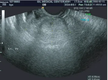
Transvaginal ultrasound indentified a 4.9 × 3.4 cm, well-circumscribed and solid left ovarian tumor (Fig. 1).
Laboratory analyses showed normal blood counts and normal serum values of cancer antigen 125 (CA-125). Serum value of dehydroepiandrosterone-sulfate (DHEA-S) was 548.9 μg/
dl (reference value, 35-430 μg/dL).
Considering transvaginal ultrasound and laboratory analyses, laparoscopic left oophorectomy was performed.
During the operation, the left ovary ruptured due to adhesion caused by previous operation.
Grossly, cut surface was vaguely lobulated, bright
yellow and soft. No hemorrhage or necrosis was noted.
Microscopically, the tumor consisted of irregular cords and nests of large rounded to polygonal cells having abundant clear vacuolated cytoplasm and centrally located monotonous nuclei with prominent nucleoli (Fig. 2). No cellular atypia was noted and mitotic figures were rare (less than 1 per 10 high-power fields). It was diagnosed as steroid cell tumor, NOS.
Two weeks postoperatively, a computerized tomography (CT) scan and endometrial biopsy were performed and no remarkable findings were found.
As she had completed childbearing, complete surgical staging including peritoneal washing, total abdominal hysterectomy, right salpingo-oophorectomy, appendectomy, bilateral pelvic lymph node dissection and infracolic omentectomy was performed after 4 weeks from the first laparoscopic surgery. Microscopically, no evidence of metastasis was found. As the left ovary rupture at initial laparoscopic surgery, the patient was treated with GnRHa monthly for 6 months. After 43 months, she has had no evidence of recurrence.
Discussion
Steroid cell tumors were previously classified as lipid or lipoid cell tumors. This prior nomenclature was misleading Fig. 1. Preoperative transvaginal ultrasonography. Four point nine
times three point four cm sized solid tumor at left ovary.
Fig. 2. Microscopic photography of steroid cell tumor, not otherwise specified. (A) The tumor consists of irregular cords and nests of large rounded to polygonal cells (H&E, x100). (B) The cytoplasm of the tumor cells show variable-sized clear vacuoles, representing fat material.
Nuclei are monotonous, round and centrally located with prominent nucleoli (H&E, x400).
41
Dong-Hae Chung, et al. An Ovarian Steroid Cell Tumor
http://dx.doi.org/10.6118/jmm.2014.20.1.39 in that some tumors have little or no lipid present. The
term steroid cell tumor more accurately describes the morphological appearance of these tumors and provides insight into their clinical manifestations. Among three steroid cell tumor subtypes, steroid cell tumor, NOS, is the most common type and associated with androgenic changes in approximately half of cases. These tumors usually occur in younger individuals (mean age, 43 years) and, in contrast to other steroid cell tumors, occasionally occur before puberty.3 Hirsutism and virilization are the most common symptoms occurring in 56-77% of patients. In our patient, increased pubic hair was found and serum value of DHEAS was elevated.
Hormones other than testosterone can be elevated in steroid cell tumors, NOS. Estradiol secretion by these tumors is not uncommon, occurring in 6-23% of patients.
This excess estrogen production can result in menorrhagia and postmenopausal bleeding; in addition, endometrial adenocarcinoma has been described.4 Our patient presented metrorrhagia, but serum value of estradiol and endometrial biopsy were normal. Steroid cell tumors, NOS, have also been associated with Cushing’s syndrome in 6-10% of cases, but approximately 25% of steroid cell tumors, NOS, do not produce hormones.5
The proportion of tumors that are clinically malignant has ranged from 25% to 43%. Extraovarian spread of tumor is noted at the time of operation in 20% of the patients. The following pathologic features indicate malignancy : two or more mitotic figures per 10 high-power fields associated with 92% malignancy, necrosis with 86% of malignancy; a
diameter of >7 cm with 78% of malignancy; hemorrhage with 77% of malignancy; and grade 2 or 3 nuclear atypia with 64% of malignancy.5 Pathologically, biopsy revealed a benign tumor in our patient.
There are few collected series in the literature. In addition, since most of these tumors are diagnosed in an early stage and do not recur or metastasize, even less is known about their response to therapy. Although the clinical course of steroid cell tumors is not exactly known, it is recommended that ovarian steroid cell tumors, NOS, are managed surgically like other ovarian stromal tumors. Conservative surgery with unilateral oophorectomy and proper staging should be performed in women with stage I disease who desire future fertility. Complete surgical staging including total abdominal hysterectomy and bilateral salpingo- oophorectomy is an accepted treatment for older women who do not want to preserve their fertility,6 as was the case here.
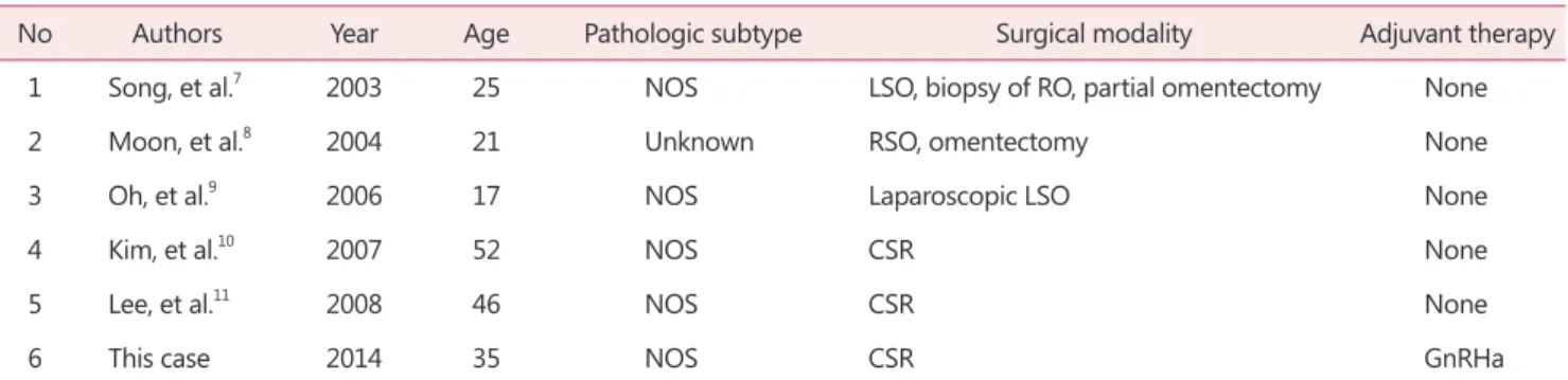
We could find 5 recently reported cases of ovarian steroid cell tumor in Korea.7~11 Conservative surgery was performed in 3 patients aged under 25 years old. Complete surgical staging was performed in 2 patients who completed childbearing (Table 1). 4 patients were premenopausal women7~10 and 1 patient was a postmenopausal woman with elevated CA-125.11 So, steroid cell tumor should be considered in postmenopausal women having ovarian tumors. Increasing use of ultrasound has resulted in increasing detection of ovarian cysts in postmenopausal women. When these cysts remain stable on follow-up, ovarian rete cyst should be considered.12
The clinical course of steroid cell tumors of the ovary
Table 1. Review of the recently reported cases of ovarian steroid cell tumor in Korea including this one
No Authors Year Age Pathologic subtype Surgical modality Adjuvant therapy
1 Song, et al.7 2003 25 NOS LSO, biopsy of RO, partial omentectomy None
2 Moon, et al.8 2004 21 Unknown RSO, omentectomy None
3 Oh, et al.9 2006 17 NOS Laparoscopic LSO None
4 Kim, et al.10 2007 52 NOS CSR None
5 Lee, et al.11 2008 46 NOS CSR None
6 This case 2014 35 NOS CSR GnRHa
NOS: not otherwise specified, LSO: left salpingo-oophorectomy, RO: right ovary, RSO: right salpingo-oophorectomy, CSR: complete surgi- cal staging, GnRHa: gonadotropin releasing hormone agonist
Journal of Menopausal Medicine 2014;20:39-42
42 http://dx.doi.org/10.6118/jmm.2014.20.1.39
is unknown, because there are few collected series in the literature. Too few cases exist to have good information as to the role of adjuvant therapy. Gonadotropin has been known to suppress androgen secretion by ovarian virilizing tumor. Pascal et al.13 suggested that various ovarian androgen-secreting tumors, as well as hyperthecosis, were not autonomous but apparently depended upon continuous gonadotropin stimulation. Imai et al.14 also noted a direct suppressive effect of GnRHa on ovarian steroidogenesis.
Wang et al.2 suggested that GnRHa might be an alternative choice as adjuvant therapy for managing recurrence of ovarian steroid cell tumor. As the tumor ruptured during the first laparoscopic surgery and we could not expect the clinical course, we administered GnRHa in our patient to prevent recurrence on the basis of these evidences.
As far as we know, there is no previously reported case of ovarian steroid cell tumor treated with complete surgical staging followed by GnRHa in Korea (Table 1). Our patient has had no evidence of recurrence for 43 months. GnRHa should be considered as one of adjuvant therapy in ovarian steroid cell tumors.
References
1. Reedy MB, Richards WE, Ueland F, Uy K, Lee EY, Bryant C, et al. Ovarian steroid cell tumors, not otherwise specified:
a case report and literature review. Gynecol Oncol 1999; 75:
293-7.
2. Wang PH, Chao HT, Lee WL. Use of a long-acting gonadotropin-releasing hormone agonist for treatment of steroid cell tumors of the ovary. Fertil Steril 1998; 69: 353- 5.
3. Wang PH, Chao HT, Lee RC, Lai CR, Lee WL, Kwok CF, et al. Steroid cell tumors of the ovary: clinical, ultrasonic, and MRI diagnosis-a case report. Eur J Radiol 1998; 26: 269-
73.
4. Luk WT, Lee N, Chang TC, Chu KK. Lipid cell tumor of the ovary associated with endometrial adenocarcinoma-a case report. Changgeng Yi Xue Za Zhi 1989; 12: 244-8.
5. Hayes MC, Scully RE. Ovarian steroid cell tumors (not otherwise specified). A clinicopathological analysis of 63 cases. Am J Surg Pathol 1987; 11: 835-45.
6. Law KS, Chang TM, Tung JN. Fertility-sparing treatment of a primary retroperitoneal mucinous cystadenocarcinoma.
BJOG 2006; 113: 612-4.
7. Song MK, Lee YY, Lee KY, Lee A, Rha JG, Ryu KS, et al.
A case of steroid cell tumor, not otherwise specified, with massive ascites. Korean J Obstet Gynecol 2003; 46: 2551- 5.
8. Moon GS, Cho KH, Kim EJ, Chung JY, Nam ES, Cho SJ, et al. A case of lipid cell tumor of the ovary. Korean J Obstet Gynecol 2004; 47: 585-8.
9. Oh JR, Cho SH, Yoon DK, Cho HB, Lee KE, Lee MB, et al.
Laparoscopic management of asymptomatic ovarian steroid cell tumors, not otherwise specified: A case report and literature review. Korean J Obstet Gynecol 2006; 49: 933- 8.
10. Kim YT, Kim SW, Yoon BS, Kim SH, Kim JH, Kim JW, et al. An ovarian steroid cell tumor causing virilization and massive ascites. Yonsei Med J 2007; 48: 142-6.
11. Lee JS, Kim BR, Lee HC, Lym BI, Kim HG, Moon HB. A case of ovarian steroid cell tumor, not otherwise specified with hypertension, obesity, ascites and elevated CA 125.
Korean J Obstet Gynecol 2008; 51: 1164-9.
12. Park J, Kim TH, Lee HH, Lee W, Chung SH. Ovarian rete cyst in a post-menopausal woman: a case report. J Korean Soc Menopause 2012; 18: 67-9.
13. Pascale MM, Pugeat M, Roberts M, Rousset H, Déchaud H, Dutrieux-Berger N, et al. Androgen suppressive effect of GnRH agonist in ovarian hyperthecosis and virilizing tumours. Clin Endocrinol (Oxf) 1994; 41: 571-6.
14. Imai A, Iida K, Tamaya T. Direct action of gonadotropin- releasing hormone (LH-RH) analogue on ovary: an alternative acting mechanism of buserelin. Arch Gynecol Obstet 1991; 248: 117-21.