INTRODUCTION
Nasopharyngeal cancer (NPC), a malignancy of the head and
neck, is rare in most regions worldwide; however; it is a rela- tively common form of cancer in Southern China, Southeast Asia, the Arctic, and the Middle East/North Africa.1,2 Even th- ough its complex etiology is not completely understood, eth- nicity, environmental factors, and Epstein-Barr virus infection are some of the risk factors associated with NPC.1 NPC is highly responsive to radiotherapy and/or chemotherapy, while sur- gical extirpation3 of NPC is difficult. Since NPC is highly inva- sive and metastatic and differs from other head and neck can- cers, surgical treatment is only attempted to salvage residual or recurrent disease with limited success rate.4,5 Further, even though the results of primary curative chemoradiation show good survival, 4–23% of patients still suffer from residual, re- current, and metastatic disease.3,6 Therefore, considerable ef- fort is being invested into developing an alternative approach for NPC treatment.
Tolfenamic Acid Inhibits the Proliferation, Migration, and Invasion of Nasopharyngeal Carcinoma:
Involvement of p38-Mediated Down-Regulation of Slug
Tatsanachat Jittreetat
1,2, Yoo Seob Shin
1,3, Hye Sook Hwang
1, Bok-Soon Lee
1,3, Yeon Soo Kim
1, Phakdee Sannikorn
2, and Chul-Ho Kim
1,31Department of Otolaryngology, School of Medicine, Ajou University, Suwon, Korea;
2Center of Excellent in Otorhinolaryngology, Head and Neck Surgery, Rajavithi Hospital, Bangkok, Thailand;
3Department of Molecular Science and Technology, Ajou University, Suwon, Korea.
Purpose: Tolfenamic acid (TA), a non-steroidal anti-inflammatory drug, is known to exhibit antitumor effects in various cancers apart from nasopharyngeal cancer (NPC). NPC exhibits high invasiveness, as well as metastatic potential, and patients continue to suffer from residual, recurrent, or metastatic disease even after chemoradiation therapy. Therefore, new treatment strategies are needed for NPC. In this study, we investigated the efficacy and molecular mechanisms of TA in NPC treatment.
Materials and Methods: TA-induced cell death was detected by cell viability assay in the NPC cell lines, HNE1 and HONE1.
Wound healing assay, invasion assay, and Western blot analysis were used to evaluate the antitumor effects of TA in NPC cell lines.
Results: Treatment with TA suppressed the migration and invasion of HNE1 and HONE1 cells. Hepatocyte growth factor en- hanced the proliferation, migration, and invasion abilities of NPC cells. This enhancement was successfully inhibited by TA treat- ment. Treatment with TA increased phosphorylation of p38, and the inhibition of p38 with SB203580 reversed the cytotoxic, anti- invasive, and anti-migratory effects of TA treatment in NPC cell lines. Moreover, inhibition of p38 also reversed the decrease in expression of Slug that was induced by TA treatment.
Conclusion: In conclusion, the activation of p38 plays a role in mediating TA-induced cytotoxicity and inhibition of invasion and migration via down-regulation of Slug.
Key Words: Tolfenamic acid, nasopharyngeal cancer, p38 mitogen-activated protein kinase, Slug Yonsei Med J 2016 May;57(3):588-598
http://dx.doi.org/10.3349/ymj.2016.57.3.588 pISSN: 0513-5796 · eISSN: 1976-2437
Received: August 10, 2015 Revised: August 22, 2015 Accepted: August 23, 2015
Co-corresponding authors: Dr. Chul-Ho Kim, Departments of Otolaryngology &
Molecular Science and Technology, Ajou University School of Medicine, 164 World cup-ro, Yeongtong-gu, Suwon 16499, Korea.
Tel: 82-31-219-5269, Fax: 82-31-219-5264, E-mail: [email protected] and Dr. Phakdee Sannikorn, Center of Excellent in Otorhinolaryngology, Head and Neck Surgery, Rajavithi Hospital, 2 Phayathai Road, Ratchathewi District, Bangkok 10400, Thailand.
Tel: 66-2-354-8108-9, Fax: 66-2-354-8100, E-mail: [email protected]
•The authors have no financial conflicts of interest.
© Copyright: Yonsei University College of Medicine 2016
This is an Open Access article distributed under the terms of the Creative Com- mons Attribution Non-Commercial License (http://creativecommons.org/licenses/
by-nc/3.0) which permits unrestricted non-commercial use, distribution, and repro- duction in any medium, provided the original work is properly cited.
cells were pre-treated with hepatocyte growth factor (HGF) (10 ng/mL), followed by treatment with TA (0, 10, 30, 50, 75, or 100 μM). After 24 h of incubation, cells were washed and sta- ined with 0.02% crystal violet (Sigma-Aldrich). The images of the wound area were captured with an Olympus SC 35 camera (Tokyo, Japan) connected to an inverted microscope. The results are presented as percentage closure area of tumor cell lines compared to the control.
Invasion assay
The invasion assay was carried out as previously described.12 Briefly, 24-well Transwell filters with an 8-μm pore size and filters coated with collagen were used. Both HNE1 and HONE1 cells (2×10⁴) in the upper chamber were pretreated with TA (0, 10, 50, or 100 μM) with or without 10 ng/mL HGF. Both inserts and lower wells were treated with vehicle control (dimethyl sulfoxide, DMSO), TA, and HGF. The chambers were incubat- ed for 24 h at 37°C in an atmosphere containing 5% CO2. After 24 h, the cells in the insert were gently removed using a cotton swab. Cells on the lower surface of the filter were fixed and stained by hematoxylin and eosin staining solutions. The number of invading cells was counted in four representative fields per membrane by using light microscopy at 40× magni- fication with Image J program (National Institutes of Health, Bethesda, MD, USA). Invasion rate is presented as a percent- age compared to control.
Western blot analysis
After cells were treated with TA (0, 10, 30, or 50 μM), total pro- teins were extracted using the ProteoExtract® Subcellular Pro- teome Extraction Kit (Calbiochem), following the manufac- turer’s instructions. Protein concentrations were measured using the BCA assay (Pierce, Rockford, IL, USA). The proteins were separated by electrophoresis on 12% and 10% sodium dodecyl sulfate polyacrylamide (SDS) gels. An equal amount of protein (10 μg) was loaded in each lane. After electrophore- sis, the proteins were transferred onto polyvinylidene difluo- ride (PVDF) membranes. The membrane was blocked in Tris- buffered saline tween-20 (TBST) containing 5% non-fat milk for 1 h, and were then incubated overnight at 4°C with prima- ry antibodies (1:1000 dilution). All primary antibodies were purchased from Cell Signaling Technology (Danvers, MA, USA). After washing the membrane extensively, incubation with horseradish peroxidase-conjugated secondary antibody (1:1000 dilution, Cell Signaling Technology) was performed for 1 h at room temperature. Protein bands on the blots were visualized using ECL Plus Western Blot detection reagents (Amersham Pharmacia Biotech, Piscataway, NJ, USA).
Statistical analysis
All values are expressed as mean±standard deviation, and sta- tistical analyses were performed using Student’s t-test and one- way ANOVA (SPSS, version 17, SPSS Inc., Chicago, IL, USA). p- Chemoprevention has recently come into the spotlight in
cancer treatment not only for interrupting the carcinogenic process but also for inhibiting the recurrence of lesions.7 Cancer chemoprevention inhibits, delays, or reverses the process of carcinogenesis. The effectiveness of chemopreventive therapy depends on the efficacy, safety, availability, and cost-effective- ness of these drugs.8,9 Tolfenamic acid (TA) is a popular non- steroidal anti-inflammatory drug (NSAID), which is proven safe and is inexpensive for use in clinical settings, especially for the treatment of migraine. Further, besides its original anti-inflam- matory effects, TA shows excellent anti-tumor effects in various cancers, including colorectal, thyroid, prostate, ovarian, lung, breast, and esophageal cancers.8-10 However, the effectiveness and possible mechanism of action of TA in treatment of NPC has not been studied. We hypothesized that TA is a promising chemopreventive agent for NPC. In this investigation, we inves- tigated whether TA has anti-proliferative and anti-progression effects on NPC cell lines, especially via p38 mitogen-activated protein kinase (p38) mediated-down-regulation of Slug.
MATERIALS AND METHODS
Cell lines, culture conditions, and reagents
HNE1, HONE1, and HaCaT cell lines were obtained from the Korean Cell Line Bank (Seoul, Korea). HNE1 cell line was main- tained in Dulbecco’s modified Eagle’s medium (DMEM) (Gib- co, Carlsbad, CA, USA) supplemented with 10% fetal bovine serum (FBS). HONE1 cell line was maintained in RPMI1640 medium (Gibco) supplemented with 10% FBS. Cells were in- cubated at 37°C in an atmosphere containing 5% CO2. TA (Sig- ma-Aldrich, St. Louis, MO, USA). SB203580 (Calbiochem, La Jolla, CA, USA, No. 559389) [p38 mitogen-activated protein ki- nase (MAPK) inhibitor] was dissolved according to the manu- facturer’s instructions for use in in vitro studies.
Cell viability assay
The viability of the cells after treatment with various concen- trations (0, 5, 10, 20, 30, 50, 75, 100, 150, or 200 μM) of TA was determined by 3-(4,5-dimethylthiazol-2-yl)-2,5-diphenyltet- razolium bromide (MTT; Sigma-Aldrich) assay. Briefly, MTT solution was added to 40 mL of cell suspension for 4 h, and the insoluble formazan product was dissolved in 100 μL of dimeth- ylsulfoxide. The optical density of each culture well was mea- sured at 540 nm using a microplate reader (Bio-Tek, Winooski, VT, USA). The results are represented as percentages relative to untreated control cells.
Wound healing assay
Cell migration was measured using the wound-healing assay as previously described.11 Briefly, cells were grown to confluent monolayers, and then wounded by scratching the surface as uniformly as possible with a 1-mL pipette tip. HNE1 and HONE1
value<0.05 was considered statistically significant [p<0.05 (*), p<0.01 (**), p<0.001 (***)].
RESULTS
TA inhibits proliferation of HNE1 and HONE1 cells
As shown in Fig. 1, 24 h and 48 h after treatment with TA, cell vi- ability significantly decreased in both NPC cell lines. We exam- ined the effects of different concentrations of TA on both HNE1and HONE1 cells and found that TA significantly inhibited NPC cell growth in a dose- and time-dependent manner (Fig. 1A). In terms of general toxicity of TA, cytotoxicity was not caused by TA treatment with a cytotoxic concentration to NPC cells (Fig. 1B).
TA decreased HGF induced migration as well as invasion of NPC cells
HGF is a potent enhancer of invasion and progression of vari- ous head and neck carcinoma cells. To analyze the suppressive effect of TA on the migration of NPC cell lines, we performed
Fig. 1. Effects of tolfenamic acid on proliferation of nasopharyngeal cancer cell lines and a normal keratinocyte cell line. Cells were seeded in 96-well tissue-culture plates, at a density of 2×10³ cells/well in a final volume of 100 μL of medium, and were allowed to attach for 24 h. The cells were then treated with ten different concentrations of tolfenamic acid (TA) for 24 h and 48 h. Cell proliferation was estimated by the MTT assay. (A) Nasopharyn- geal cancer cell lines, (B) HaCaT cell lines. Values are represented as mean±SD from three independent experiments. *p<0.05 and ***p<0.001 com- pared to the control group. MTT, 3-(4,5-dimethylthiazol-2-yl)-2,5-diphenyltetrazolium bromide.
1.2 1 0.8 0.6 0.4 0.2 0
0 5 10 20 30 50 75 100 150 200 HNE1
24 h
TA
(μM)
Cell viability
***
***
*** *** *** ***
*** ***
1.2 1 0.8 0.6 0.4 0.2 0
0 5 10 20 30 50 75 100 150 200 HONE1
24 h
TA
(μM)
Cell viability
*** ***
*** ***
***
*** *** *** ***
1.2 1 0.8 0.6 0.4 0.2 0
0 5 10 20 30 50 75 100 150 200 HONE1
48 h
TA
(μM)
Cell viability
*** ***
*** ***
***
*** *** *** ***
1.2 1 0.8 0.6 0.4 0.2 0
0 5 10 20 30 50 75 100 150 200 HNE1
48 h
TA
(μM)
Cell viability
*
***
*** ***
*** ***
*** ***
A
125
100
75
50
25
0
0 5 10 30 50 70 100 150 200 HaCaT
TA
(μM)
Cell viability (% of control)
***
***
***
B
Fig. 2. Effect of tolfenamic acid (TA) on the migratory and invasive abilities of nasopharyngeal cancer cells. The migration capability after HGF/(TA) treatment was investigated using wound healing assay. (A) HNE1 and HONE1 cells were either treated with 10 ng/mL HGF along with TA (0, 10, 30, 50, 75, 100 μM) or left untreated. Confluent monolayers of HNE1 were wounded by scratching the surface as uniformly as possible with a 1-mL pipette tip.
TA treatment significantly inhibited HGF-induced enhancement of cell migration in a dose-dependent manner. *p<0.05, **p<0.01, ***p<0.001. HGF, he- patocyte growth factor.
HNE1
Control
HGF (10 ng/mL)
TA
0 10 30 50 75 100 (μM)
HONE1
Control
HGF (10 ng/mL)
TA
0 10 30 50 75 100 (μM)
140 120 100 80 60 40 20
0 Control Control
HGF 10 ng/mL
TA10 TA30 TA50 TA75 TA100 TA10 TA30 TA50 TA75 TA100 (μM)
Migration distance (%)
** ** *** ***
*** **
** **
140 120 100 80 60 40 20
0 Control Control
HGF 10 ng/mL
TA10 TA30 TA50 TA75 TA100 TA10 TA30 TA50 TA75 TA100 (μM)
Migration distance (%)
** ***
*** *** ***
* ** ** *** ***
A
Fig. 2. Effect of tolfenamic acid (TA) on the migratory and invasive abilities of nasopharyngeal cancer cells. The migration capability after HGF/(TA) treatment was investigated using wound healing assay. (B) HNE1 and HONE1 cells (2×10⁴) in the upper chamber were pretreated with TA (0, 10, 30, 50, 75, 100 μM) and then either treated with 10 ng/mL HGF or left untreated. After 24 h, the number of invading cells was counted in four representative fields per membrane. Co-treatment with TA and HGF significantly inhibited cell invasion compared to treatment with HGF alone. The data represent the mean±SD of three independent experiments. *p<0.05, **p<0.01, ***p<0.001. HGF, hepatocyte growth factor.
B
HNE1
Control
HGF (10 ng/mL)
TA
0 10 50 100 (μM)
HONE1
Control
HGF (10 ng/mL)
TA
0 10 50 100 (μM)
350 300 250 200 150 100 50 0
HGF 10 ng/mL
TA0 TA10 TA50 TA100 TA0 TA10 TA50 TA100 (μM)
Invasion rate (%) **
***
**
*
***
140 120 100 80 60 40 20 0
HGF 10 ng/mL
TA0 TA10 TA50 TA100 TA0 TA10 TA50 TA100 (μM)
Invasion rate (%)
***
***
*** ***
**
***
a wound-healing assay. As shown in Fig. 2A, treatment with TA suppressed the migration of HNE1 and HONE1 cells. HGF enhanced the proliferation and migration abilities of the NPC cells, and this enhancement was successfully inhibited by TA treatment (Fig. 2A). We further addressed the effect of TA on invasion of NPC cells using the Transwell invasion assay. Co- treatment of cells with TA and HGF significantly inhibited cell invasion, compared to that observed with treatment with HGF alone, especially in HNE1 cells (Fig. 2B). These results indicat- ed that TA inhibits HGF-induced NPC cell migration and in- vasion in a dose-dependent manner.
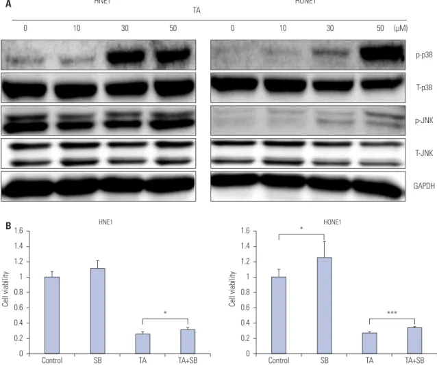
TA induced cell death of NPC cells via the p38 MAPK pathway
Next, we evaluated TA induced-changes at the protein level in NPC cells. To determine whether MAPK pathway mediated the observed cytotoxic, anti-invasive, and anti-migratory responses upon TA treatment, we determined the phosphorylation lev- els of p38 and JNK. A representative western blot is shown in
Fig. 3A. An increase in phosphorylation of p38 as well as JNK was observed (Fig. 3A) post TA treatment. Since the increase in phosphorylation of p38 signaling was more significant than that of JNK, we decided to focus on the p38-mediated signaling pathways. Treatment with the p38 inhibitor SB203580 for 48 h caused a significant increase in the viability of both NPC cell lines (Fig. 3B).
Suppression of p38 attenuates TA-induced inhibition of migration and invasion in NPC cells
As is shown in Fig. 4A, TA-induced inhibition of migration in HGF-treated NPC cells was partially reversed upon p38 inhi- bition. Similarly, attenuation of invasion following TA treatment was also restored upon SB203580 treatment (Fig. 4B).
The activation of Slug, which is a member of the Snail super- family and plays pivotal role in migration and invasion by in- ducing epithelial-mesenchymal transition (EMT),13-16 was eval- uated by western blot analysis. TA treatment decreased the phosphorylation of Slug, and p38 inhibition reversed the effect
Fig. 3. Effect of tolfenamic acid (TA) on the MAPK pathway in nasopharyngeal cancer cells. (A) Immunoblot of TA-treated cells stained with antibodies against T-p38, p-p38, T-JNK, and p-JNK. Western blot analysis showed that TA-enhanced phosphorylation of p38 and JNK in HNE1 and HONE1 cells. (B) HNE1 and HONE1 cells were treated with HGF (10 ng/mL), SB203580 (0.5 μM), and TA (50 μM) alone or in combination, as indicated. Cell viability assay was performed to investigate the cytotoxic effect of TA in nasopharyngeal cancer cells in the absence or presence of SB203580. The data represent the mean±SD of three independent experiments. *p<0.05, ***p<0.001 by one-way ANOVA. MAPK, mitogen-activated protein kinase.
HNE1
0 10 30 50 0 10 30 50 (μM)
HONE1
p-p38
T-p38
p-JNK
T-JNK
GAPDH A TA
B1.6 1.4 1.2 1 0.8 0.6 0.4 0.2 0
Cell viability
Control SB TA TA+SB
HNE1
*
1.6 1.4 1.2 1 0.8 0.6 0.4 0.2 0
Cell viability
Control SB TA TA+SB
HONE1
***
*
Fig. 4. Effect of a p38 inhibitor on anti-migratory and anti-invasive abilities of tolfenamic acid (TA) in nasopharyngeal cancer cells. HNE1 and HONE1 cells were treated with HGF (10 ng/mL), SB203580 (0.5 μM), and TA (50 μM) alone or in combination, as indicated. (A) Wound healing and (B) invasion assays were used to determine the anti-migratory and anti-invasive abilities of TA in nasopharyngeal cancer cells in the absence or presence of SB203580. The data represent the mean±SD of three independent experiments. *p<0.05, **p<0.01, ***p<0.001 by one-way ANOVA. DMSO, dimethyl sulfoxide; HGF, hepatocyte growth factor.
A HNE1
HGF (10 ng/mL)
TA (50 μM) - - + +
DMSO + + - -
SB203580 - + - +
Control
HONE1
HGF (10 ng/mL)
TA (50 μM) - - + +
DMSO + + - -
SB203580 - + - +
Control
200
150
100
50
0
HGF 10 ng/mL
Control SB TA TA+SB Control SB TA TA+SB
Migration distance (%)
**
**
200
150
100
50
0
HGF 10 ng/mL
Control SB TA TA+SB Control SB TA TA+SB
Migration distance (%)
*
*
***
Fig. 4. Effect of a p38 inhibitor on anti-migratory and anti-invasive abilities of tolfenamic acid (TA) in nasopharyngeal cancer cells. HNE1 and HONE1 cells were treated with HGF (10 ng/mL), SB203580 (0.5 μM), and TA (50 μM) alone or in combination, as indicated. (A) Wound healing and (B) invasion assays were used to determine the anti-migratory and anti-invasive abilities of TA in nasopharyngeal cancer cells in the absence or presence of SB203580. The data represent the mean±SD of three independent experiments. *p<0.05, **p<0.01, ***p<0.001 by one-way ANOVA. DMSO, dimethyl sulfoxide; HGF, hepatocyte growth factor.
B HNE1
HGF (10 ng/mL)
TA (50 μM) - - + +
DMSO + + - -
SB203580 - + - +
Control
HONE1
HGF (10 ng/mL)
TA (50 μM) - - + +
DMSO + + - -
SB203580 - + - +
Control
160 140 120 100 80 60 40 20 0
HGF 10 ng/mL
Control SB TA TA+SB Control SB TA TA+SB
Invasion rate (%)
*
160 140 120 100 80 60 40 20 0
HGF 10 ng/mL
Control SB TA TA+SB Control SB TA TA+SB
Invasion rate (%)
*
of TA in both NPC cell lines (Fig. 5). These results suggest that anti-tumor effects of TA in NPC are a result of p38 MAPK me- diated-down-regulation of Slug.
DISCUSSION
Despite the high response rate of NPC to radiotherapy and che- motherapy, a considerable number of patients eventually re- lapse.17 As an alternative, various biologic chemotherapeutics have been developed that target pathways mediating cell pro- liferation, angiogenesis, migration, and invasion.18,19 However, these are not effective molecular targets against NPC, and the life expectancy of patients with recurrent or metastatic NPC remains low. Several studies combining biologic chemothera- peutics against specific molecular targets with conventional chemotherapeutics have shown a median overall survival of only 8–12 months; therefore, the value of adding biologic agents remains unclear.20,21 Thus, development of new treatment strat- egies for NPC is needed.
Chemoprevention is an unexplored treatment modality in the field of cancer research. Cancer chemoprevention aims to delay, prevent, or reverse the development of cancers, by long- term intake of a natural or synthetic biological agent.7 A poten- tial target of chemoprevention is down-regulation of chronic inflammatory responses, which may contribute to the preven- tion of cancer initiation.7 Several preclinical and epidemiologi- cal investigations have shown strong evidence that NSAIDs reduce the overall risk of developing various cancers.10 To de-
velop strategies for treatment of NPC, understanding the me- chanisms of action of NSAIDs, rather than depending on his- torical epidemiological observations, is needed.
In this study, we investigated the efficacy of TA in NPC treat- ment and the molecular mechanism of TA in inhibition of NPC.
TA-induced cell death was detected by cell viability assay in the NPC cell lines HNE1 and HONE1. TA successfully inhibit- ed HGF-induced migration and invasion of NPC cell lines. TA mediated its effects on NPC cell lines via the p38 MAPK and JNK pathways. We observed increased phosphorylation of p38, and the inhibition of p38 with SB203580 reversed the cy- totoxic, anti-invasive, and anti-migratory effects of TA treat- ment in NPC cell lines. Moreover, inhibition of p38 also re- versed a TA treatment-induced decrease in Slug. This suggests that TA-induced anti-tumor effects in NPC cell lines might be attributed to a p38-induced down-regulation of Slug, while inhibition of p38 could reverse these effects.
Activation of p38 MAPK by various external stimuli plays an important role in biological responses, such as inflammation, cell proliferation, regulation of apoptosis, and survival.22-27 p38 MAPK is involved in tumor growth and progression in many types of human cancers, including colorectal, lung, breast, and thyroid carcinomas,28,29 as well as head and neck squamous cell carcinomas.30,31 Further, p38 MAPK signaling is associated with sensitivity to chemotherapeutics.27 Activation of non-ste- roidal anti-inflammatory drug activated gene-1 (NAG-1) via p38 MAPK pathway is responsible for rottlerin-induced apop- tosis in the HT29 colon carcinoma cell line.32 Our group previ- ously reported that TA induces apoptosis and growth inhibition
Fig. 5. Effect of p38 inhibition on Slug expression in tolfenamic acid (TA)-treated nasopharyngeal cancer cells. HNE1 and HONE1 cells were treated with TA (50 μM) and SB203580 (0.5 μM) alone or in combination, as indicated. Immunoblot of treated cells stained with antibodies against (A) p-p38 (30 min) and (B) Slug (3 h). Western blot analysis showed that TA reduced phosphorylation of p38 and Slug, while this effect was reversed by SB203580 treatment. DMSO, dimethyl sulfoxide.
HNE1 HONE1
+ + - -
DMSO DMSO + + - -
- + - +
SB203580 SB203580 - + - +
- - + +
TA (50 μM) TA (50 μM) - - + +
A
p-p38 p-p38
HSP60 HSP60
HNE1 HONE1
+ + - -
DMSO DMSO + + - -
- + - +
SB203580 SB203580 - + - +
- - + +
TA (50 μM) TA (50 μM) - - + +
B
Slug Slug
HSP60 HSP60
via NAG-1 expression in head and neck, as well as anaplastic thyroid, cancers.8,9 In this study, TA treatment-induced activa- tion of p38, while inhibition of p38 attenuated the effect of TA on cytotoxicity, as well as inhibition of invasion and migration, in NPC cell lines.
Slug is a member of the Snail superfamily, which is a family of zinc-finger transcription factors involved in the pathogene- sis of EMT. Elevated expression of Snail enhances cell invasion and migration by down-regulating epithelial markers and up- regulating mesenchymal markers. Expression of Slug is close- ly associated with tumor recurrence, metastasis, and poor sur- vival in various types of cancers.33-36 Various pathways regulate Slug, such as receptor tyrosine kinases activated by HGF, FGF, or EGF or the RAS-MAPK or PI3K-Akt pathway.37,38 HGF is also strongly involved in transcriptional regulation of Snail. HGF- mediated MAPK activation enhances the expression of Snail.39 Inhibition of p38 MAPK is also reported to reverse EMT in ad- vanced tumor phenotype. In the present study, we observed that inhibition of p38 with SB203580 reversed a TA treatment- induced decrease in Slug. Consequently, re-expression of Slug upon p38 inhibition led to the recovery of cell viability, inva- siveness, and migratory abilities in NPC cell lines.
In conclusion, these data demonstrate that TA induces p38- mediated cell death and inhibition of invasion and migration in NPC cells. The TA-induced p38 activation leads to down-reg- ulation of Slug. Further, p38 inhibition with SB203580 reversed the anti-tumor effects of TA treatment in NPC cell lines. These findings suggest that activation of p38 plays a role in mediat- ing TA-induced cytotoxicity, as well as inhibition of invasion and migration, via down-regulation of Slug.
ACKNOWLEDGEMENTS
This research was supported by the Bio & Medical Technology Development Program (2012M3A9B2052870) and Basic Sci- ence Research Program through the National Research Foun- dation of Korea (NRF) funded by the Ministry of Science, ICT and future Planning (2015R1A2A1A01002968).
REFERENCES
1. Oh JK, Weiderpass E. Infection and cancer: global distribution and burden of diseases. Ann Glob Health 2014;80:384-92.
2. Li K, Lin GZ, Shen JC, Zhou Q. Time trends of nasopharyngeal carcinoma in urban Guangzhou over a 12-year period (2000-2011):
declines in both incidence and mortality. Asian Pac J Cancer Prev 2014;15:9899-903.
3. Lee AW, Sze WM, Au JS, Leung SF, Leung TW, Chua DT, et al. Treat- ment results for nasopharyngeal carcinoma in the modern era: the Hong Kong experience. Int J Radiat Oncol Biol Phys 2005;61:1107-16.
4. Na’ara S, Amit M, Billan S, Cohen JT, Gil Z. Outcome of patients undergoing salvage surgery for recurrent nasopharyngeal carci- noma: a meta-analysis. Ann Surg Oncol 2014;21:3056-62.
5. Chan JY, Tsang RK, Wei WI. Morbidities after maxillary swing na- sopharyngectomy for recurrent nasopharyngeal carcinoma. Head
Neck 2015;37:487-92.
6. Li JX, Huang SM, Jiang XH, Ouyang B, Han F, Liu S, et al. Local failure patterns for patients with nasopharyngeal carcinoma after intensity-modulated radiotherapy. Radiat Oncol 2014;9:87.
7. Steward WP, Brown K. Cancer chemoprevention: a rapidly evolving field. Br J Cancer 2013;109:1-7.
8. Chang JW, Kang SU, Choi JW, Shin YS, Baek SJ, Lee SH, et al. Tolfe- namic acid induces apoptosis and growth inhibition in anaplastic thyroid cancer: involvement of nonsteroidal anti-inflammatory drug-activated gene-1 expression and intracellular reactive oxygen species generation. Free Radic Biol Med 2014;67:115-30.
9. Kang SU, Shin YS, Hwang HS, Baek SJ, Lee SH, Kim CH. Tolfenamic acid induces apoptosis and growth inhibition in head and neck cancer: involvement of NAG-1 expression. PLoS One 2012;7:
e34988.
10. Jeong JB, Choi J, Baek SJ, Lee SH. Reactive oxygen species medi- ate tolfenamic acid-induced apoptosis in human colorectal can- cer cells. Arch Biochem Biophys 2013;537:168-75.
11. Shin HA, Shin YS, Kang SU, Kim JH, Oh YT, Park KH, et al. Radio- protective effect of epicatechin in cultured human fibroblasts and zebrafish. J Radiat Res 2014;55:32-40.
12. Lee BS, Kang S, Kim KA, Song YJ, Cheong KH, Cha HY, et al. Met degradation by SAIT301, a Met monoclonal antibody, reduces the invasion and migration of nasopharyngeal cancer cells via inhibi- tion of EGR-1 expression. Cell Death Dis 2014;5:e1159.
13. Choi MJ, Cho KH, Lee S, Bae YJ, Jeong KJ, Rha SY, et al. hTERT mediates norepinephrine-induced Slug expression and ovarian cancer aggressiveness. Oncogene 2015;34:3402-12.
14. Zhao X, Sun B, Sun D, Liu T, Che N, Gu Q, et al. Slug promotes he- patocellular cancer cell progression by increasing sox2 and nanog expression. Oncol Rep 2015;33:149-56.
15. Merikallio H, T TT, Pääkkö P, Mäkitaro R, Kaarteenaho R, Lehtonen S, et al. Slug is associated with poor survival in squamous cell carci- noma of the lung. Int J Clin Exp Pathol 2014;7:5846-54.
16. Wang N, Dong CR, Jiang R, Tang C, Yang L, Jiang QF, et al. Overex- pression of HIF-1α, metallothionein and SLUG is associated with high TNM stage and lymph node metastasis in papillary thyroid carcinoma. Int J Clin Exp Pathol 2013;7:322-30.
17. Chang ET, Adami HO. The enigmatic epidemiology of nasopharyn- geal carcinoma. Cancer Epidemiol Biomarkers Prev 2006;15:1765- 77.
18. Tsang J, Lee VH, Kwong DL. Novel therapy for nasopharyngeal carcinoma--where are we. Oral Oncol 2014;50:798-801.
19. Chiang AK, Mak NK, Ng WT. Translational research in nasopha- ryngeal carcinoma. Oral Oncol 2014;50:345-52.
20. You B, Le Tourneau C, Chen EX, Wang L, Jarvi A, Bharadwaj RR, et al. A Phase II trial of erlotinib as maintenance treatment after gem- citabine plus platinum-based chemotherapy in patients with re- current and/or metastatic nasopharyngeal carcinoma. Am J Clin Oncol 2012;35:255-60.
21. Chan AT, Hsu MM, Goh BC, Hui EP, Liu TW, Millward MJ, et al.
Multicenter, phase II study of cetuximab in combination with car- boplatin in patients with recurrent or metastatic nasopharyngeal carcinoma. J Clin Oncol 2005;23:3568-76.
22. Kim HS, Lee JH, Park HS, Lee GS, Kim HW, Ha KT, et al. Schizan- dra chinensis extracts induce apoptosis in human gastric cancer cells via JNK/p38 MAPK activation and the ROS-mediated/mito- chondria-dependent pathway. Pharm Biol 2015;53:212-9.
23. Zhao B, Li X. Altholactone induces reactive oxygen species-medi- ated apoptosis in bladder cancer T24 cells through mitochondrial dysfunction, MAPK-p38 activation and Akt suppression. Oncol Rep 2014;31:2769-75.
24. Kang N, Wang MM, Wang YH, Zhang ZN, Cao HR, Lv YH, et al.
Tetrahydrocurcumin induces G2/M cell cycle arrest and apopto- sis involving p38 MAPK activation in human breast cancer cells.
Food Chem Toxicol 2014;67:193-200.
25. Wagner EF, Nebreda AR. Signal integration by JNK and p38 MAPK pathways in cancer development. Nat Rev Cancer 2009;9:537-49.
26. Chiacchiera F, Simone C. Signal-dependent regulation of gene expression as a target for cancer treatment: inhibiting p38alpha in colorectal tumors. Cancer Lett 2008;265:16-26.
27. Sui X, Kong N, Ye L, Han W, Zhou J, Zhang Q, et al. p38 and JNK MAPK pathways control the balance of apoptosis and autophagy in response to chemotherapeutic agents. Cancer Lett 2014;344:174-9.
28. Matrone A, Grossi V, Chiacchiera F, Fina E, Cappellari M, Carin- gella AM, et al. p38alpha is required for ovarian cancer cell me- tabolism and survival. Int J Gynecol Cancer 2010;20:203-11.
29. Grossi V, Simone C. Special Agents Hunting Down Women Silent Killer: The Emerging Role of the p38α Kinase. J Oncol 2012;2012:
382159.
30. Bakin AV, Rinehart C, Tomlinson AK, Arteaga CL. p38 mitogen-ac- tivated protein kinase is required for TGFbeta-mediated fibroblas- tic transdifferentiation and cell migration. J Cell Sci 2002;115(Pt 15):3193-206.
31. Hsieh YH, Wu TT, Huang CY, Hsieh YS, Hwang JM, Liu JY. p38 mi- togen-activated protein kinase pathway is involved in protein ki- nase Calpha-regulated invasion in human hepatocellular carci- noma cells. Cancer Res 2007;67:4320-7.
32. Lim JH, Woo SM, Min KJ, Park EJ, Jang JH, Seo BR, et al. Rottlerin
induces apoptosis of HT29 colon carcinoma cells through NAG-1 upregulation via an ERK and p38 MAPK-dependent and PKC δ-independent mechanism. Chem Biol Interact 2012;197:1-7.
33. Sharma-Walia N, Patel K, Chandran K, Marginean A, Bottero V, Kerur N, et al. COX-2/PGE2: molecular ambassadors of Kaposi’s sarcoma-associated herpes virus oncoprotein-v-FLIP. Oncogene- sis 2012;1:e5.
34. Hemavathy K, Ashraf SI, Ip YT. Snail/slug family of repressors:
slowly going into the fast lane of development and cancer. Gene 2000;257:1-12.
35. Peinado H, Olmeda D, Cano A. Snail, Zeb and bHLH factors in tumour progression: an alliance against the epithelial phenotype?
Nat Rev Cancer 2007;7:415-28.
36. Moody SE, Perez D, Pan TC, Sarkisian CJ, Portocarrero CP, Sterner CJ, et al. The transcriptional repressor Snail promotes mammary tumor recurrence. Cancer Cell 2005;8:197-209.
37. Ciruna B, Rossant J. FGF signaling regulates mesoderm cell fate specification and morphogenetic movement at the primitive streak.
Dev Cell 2001;1:37-49.
38. Lu Z, Ghosh S, Wang Z, Hunter T. Downregulation of caveolin-1 function by EGF leads to the loss of E-cadherin, increased tran- scriptional activity of beta-catenin, and enhanced tumor cell inva- sion. Cancer Cell 2003;4:499-515.
39. Wang Y, Shi J, Chai K, Ying X, Zhou BP. The Role of Snail in EMT and Tumorigenesis. Curr Cancer Drug Targets 2013;13:963-72.