A Case of Acquired Brown Syndrome after Surgical Repair of a Medial Orbital Wall Fracture
4
0
0
전체 글
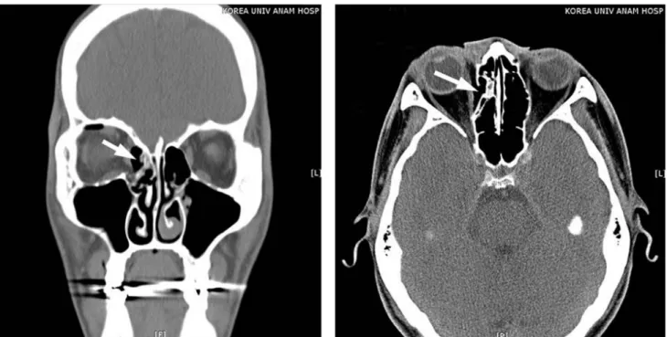
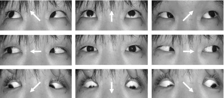
(2) Korean J Ophthalmol Vol.24, No.1, 2010. Fig. 1. A computerized tomography scan was performed before surgery and showed a medial orbital wall fracture (arrow).. Fig. 2. Postoperative computerized tomography scan of the orbits showed the right superior oblique muscle was entrapped between the autografted bone fragment and posterior margin of the fracture (arrow).. at the department of plastic surgery 3 days after the injury (Fig. 2). Immediately after surgery, the patient complained of newly- developed diplopia. The patient visited the ophthalmologic department 2 months after the surgery. A prism cover test at distance fixation revealed 4 prism diopters (PD) of right hypotropia in the primary position, 20 PD hypotropia in left gaze, and orthophoric in right gaze. Eye movement of the right eye was markedly limited on elevation in adduction while elevation in abduction 54. was normal (Fig. 3). Fundus examination showed intorsion of the right eye (Fig. 4). Forced duction test of the right eye demonstrated restricted elevation in adduction. A postoperative CT scan of the orbits showed the right superior oblique muscle was entrapped between the autografted bone fragment and posterior margin of the fracture (Fig. 2)..
(3) IH Seo, et al. Acquired Brown syndrome. Fig. 3. After repair of the blow-out fracture, limitation of elevation in adduction of the right eye developed (arrow) while elevation in abduction was normal.. Fig. 4. Fundus examination showed intorsion of the right eye.. Discussion Parks and Brown [1] first reported on an unusual motility disorder characterized by limited elevation when the eye is in adduction. At first, Brown attributed the limited elevation to a short or tight anterior superior oblique tendon sheath and termed this newly discovered syndrome the “superior oblique tendon sheath syndrome”. Later, Brown divided the syndrome into true sheath and simulated sheath syndromes based on whether the congenital short anterior sheath of the superior oblique tendon is the only cause of the symptom or not [9]. The clinical features of true and simulated sheath. syndromes are similar, but true sheath syndrome is always congenital as well as constant, and does not show spontaneous recovery. The true sheath syndrome was further subdivided into typical and atypical forms [10]. A classification of Brown's syndrome can be made on the basis of onset, and congenital versus acquired. Acquired Brown syndrome is associated with limited elevation in adduction that occurs after infancy. In certain cases, acquired Brown syndrome is idiopathic. In other instances, Brown syndrome can be associated with a systemic disease or local orbital pathology [2]. Inflammatory diseases, such as rheumatoid arthritis or 55.
(4) Korean J Ophthalmol Vol.24, No.1, 2010. systemic lupus erythematosus, can cause acquired Brown syndrome [11-14]. Local inflammation of the superior oblique tendon and trochlea can occur as an idiopathic form without systemic disease [15, 16]. Scarring in the area of the trochlea from trauma or from periocular or sinus surgery can also produce Brown syndrome [17, 18]. There are numerous case reports of postoperative Brown syndrome. Dobler et al. [19] reported Brown syndrome after implantation of a double plate Molteno valve in the superior nasal position. Coats et al. [6] reported Brown ® syndrome after Ahmed valve implantation. Ball et al. [4] reported a case of Brown syndrome after Baevelt implantation. Munoz and Rosenbaum [5] observed Brown syndrome after retinal detachment surgery. Brown syndrome related to surgical repair of an orbital wall fracture has also been reported. Lauer et al. reported Brown syndrome diagnosed following repair of an orbital roof fracture [7]. Hwang and Lim [20] reported Brown syndrome after reconstruction of a medial wall fracture and observed the tendon and muscle fiber near the trochlea were hypertrophied. Surgical or accidental trauma to the area of the trochlea has been reported to cause acquired Brown syndrome. In medial orbital wall reconstruction surgery, the superior oblique muscle is easily damaged because of its close proximity. The superior oblique muscle and the tendon can be injured during an operation, or adhesion can occur post-operatively. Edema or hemorrhage can also induce Brown syndrome [21]. In the present case study reported, the authors could confirm by CT scan of the orbits that the right superior oblique muscle was entrapped between the autograft bone fragment and posterior margin of the fracture (Fig. 2). The indications for Brown syndrome surgery include hypotropia in the primary position, diplopia, anomalous head posture, and downshoot during adduction [22]. There are several surgical procedures for the treatment of Brown's syndrome including superior oblique tenotomy, tenectomy and elongation of the superior oblique muscle [23]. In the present case study, removal of the bone graft was difficult, and even if successfully performed, the symptoms due to adhesion probably would not improve. As hypotropia at the primary gaze was small and the patient did not want surgical management, the authors decided to manage the patient conservatively. In reconstruction of medial wall blow-out fracture, there is a risk of external ocular muscle entrapment. The muscle entrapment can occur when the implant is inserted without identifying the margin of fracture clearly. Although superior oblique entrapment is not common, the surgeon should always consider the possibility of Brown syndrome if the implant is placed without securing the superior and posterior margin of the orbital fracture site, at which the superior oblique may strangulate. Surgeons should also perform an intraoperative forced duction test after inserting the implant to ensure all extraocular mus56. cles are released.. References 1. Parks MM, Brown M. Superior oblique tendon sheath syndrome of Brown. Am J Ophthalmol 1975;79:82-6. 2. von Noorden GK. Binocular vision and ocular motility: theory and management of strabismus. 3rd ed. St. Louis: Mosby; 1985. p. 380-1. 3. Wright KW. Brown’s syndrome: diagnosis and management. Trans Am Ophthalmol Soc 1999;97:1023-109. 4. Ball SF, Ellis GS Jr, Herrington RG, Liang K. Brown’s superior oblique tendon syndrome after Baerveldt glaucoma implant. Arch Ophthalmol 1992;110:1368-9. 5. Muñoz M, Rosenbaum AL. Long-term strabismus complications following retinal detachment surgery. J Pediatr Ophthalmol Strabismus 1987;24:309-14. 6. Coats DK, Paysse EA, Orenga-Nania S. Acquired PseudoBrown’s syndrome immediately following Ahmed valve glaucoma implant. Ophthalmic Surg laser 1999;30:396-7. 7. Lauer SA, Sauer H, Pak SM. Brown’s syndrome diagnosed following repair of an orbital roof fracture: a case report. J Craniomaxillofac Trauma 1998;4:20-2. 8. Zipf RF, Trokel SL. Simulated superior oblique tendon sheath syndrome following orbital floor fracture. Am J Ophthalmol 1973;75:700-5. 9. Brown HW. True and simulated superior oblique tendon sheath syndromes. Doc Ophthalmol 1973;34:123-36. 10. Wilson ME, Eustis HS Jr, Parks MM. Brown's syndrome. Surv Ophthalmol 1989;34:153-72. 11. Hickling P, Beck M. Brown's syndrome: an unusual ocular complication of rheumatoid arthritis. Ann Rheum Dis 1991;50:66. 12. Killian Pj, McClain B, Lawless OJ. Brown's syndrome: an unusual manifestation of rheumatoid arthritis. Arthritis Rheum 1977;20:1080-4. 13. Walters G, Bradbury JA. Brown's syndrome: an important cause of diplopia in systemic lupus erythematosus. Ann Rheum Dis 1995;54:934. 14. Whitefield L, Isenberg DA, Brazier DJ, Forbes J. Acquired Brown's syndrome in systemic lupus erythematosus. Br J Rheumatol 1995;34:1092-4. 15. Hermann JS. Acquired Brown's syndrome of inflammatory origin: response to locally injected steroids. Arch Ophthalmol 1978;96:1228-32. 16. Moore AT, Morin JD. Bilateral acquired inflammatory Brown's syndrome. J Pediatr Ophthalmol Strabismus 1985;22:26-30. 17. Przewoska-Błaszczykowa R, Bieganowski L, Goszczyński W. Treatment of Brown's syndrome of traumatic etiology. Klin Oczna 1981;83:191-2. 18. Rosenbaum AL, Astle WF. Superior oblique and inferior rectus muscle injury following frontal and intranasal sinus surgery. J Pediatr Ophthalmol Strabismus 1985;22:194-202. 19. Dobler AA, Sondhi N, Cantor LB, Ku S. Acquired Brown’s syndrome after double-plate Molteno implant. Am J Ophthalmol 1993;116:641-2. 20. Hwang JU, Lim HT. Acquired simulated Brown syndrome following surgical repair of medial orbital wall fracture. Korean J Ophthalmol 2005;19:80-3. 21. Wilson ME, Eustis HS Jr, Parks MM. Brown’s syndrome. Surv Ophthalmol 1989;34:153-72. 22. Crawford JS. Surgical treatment of true Brown’s syndrome. Am J Ophthalmol 1976;81:289-95. 23. Von Noorden GK. Olivier P. Superior oblique tenectomy in Brown syndrome. Ophthalmology 1982;89:303-9..
(5)
수치
관련 문서
Therefore depending on the size of the fracture in the veneering porcelain, intraoral repair methods using surface treatment and composite resins may
In the efficiency review of the Trombe wall system through the experiment, if the Trombe wall was applied in the building structure, the Trombe wall had the
Purpose: Calcaneal fracture is a rare fracture, which accounts for about 2% of all fractures, but is one of the most common fractures in the ankle bone.. There is
The third was combination of levator palpebrae, superior rectus, inferior oblique muscle weakness(n=5) and the fourth was the third group plus medial rectus
• Increase in the elevation of a feature causes its position to be displaced radially outward from the principal point. • When a vertical feature is photographed,
It considers the energy use of the different components that are involved in the distribution and viewing of video content: data centres and content delivery networks
After first field tests, we expect electric passenger drones or eVTOL aircraft (short for electric vertical take-off and landing) to start providing commercial mobility
1 John Owen, Justification by Faith Alone, in The Works of John Owen, ed. John Bolt, trans. Scott Clark, "Do This and Live: Christ's Active Obedience as the
