pISSN: 1738-222X eISSN: 2093-8047
Revision and update on clinical practice guideline for liver cirrhosis
Ki Tae Suk1, Soon Koo Baik2*, Jung Hwan Yoon3*, Jae Youn Cheong4, Yong Han Paik5, Chang Hyeong Lee6, Young Seok Kim7, Jin Woo Lee8, Dong Joon Kim1, Sung Won Cho4, Seong Gyu Hwang9, Joo Hyun Sohn10, Moon Young Kim2, Young Bae Kim11, Jae Geun Kim12,
Yong Kyun Cho5, Moon Seok Choi5, Hyung Joon Kim13, Hyun Woong Lee13, Seung Up Kim14, Ja Kyung Kim14, Jin Young Choi15, Dae Won Jun16, Won Young Tak17, Byung Seok Lee18, Byoung Kuk Jang19, Woo Jin Chung19, Hong Soo Kim20, Jae Young Jang21, Soung Won Jeong21,
Sang Gyune Kim7, Oh Sang Kwon22, Young Kul Jung22, Won Hyeok Choe23, June Sung Lee24, In Hee Kim25, Jae Jun Shim26, Gab Jin Cheon27, Si Hyun Bae28, Yeon Seok Seo29,
Dae Hee Choi30, and Se Jin Jang31 (random order)
1Department of Internal Medicine, Hallym University College of Medicine, Chuncheon; 2Department of Internal Medicine and Cell Therapy and Tissue Engineering Center, Yonsei University Wonju College of Medicine, Wonju;
3Department of Internal Medicine and Liver Research Institute, Seoul National University College of Medicine, Seoul; 4Department of Internal Medicine, Ajou University College of Medicine, Suwon; 5Department of Internal Medicine, Sungkyunkwan University College of Medicine, Seoul; 6Department of Internal Medicine, Catholic University of Daegu College of Medicine, Daegu; 7Department of Internal Medicine, Soonchunhyang University Hospital Bucheon, Soonchunhyang University College of Medicine, Bucheon; 8Department of Internal Medicine, Inha University College of Medicine, Incheon; 9Department of Internal Medicine, Cha University College of Medicine, Seongnam; 10Department of Internal Medicine, Hanyang University Guri Hospital, Hanyang University
College of Medicine, Guri; Departments of 11Pathology and 12Radiology, Ajou University College of Medicine, Suwon; 13Department of Internal Medicine, Chung-Ang University College of Medicine, Seoul; Departments of
14Internal Medicine and 15Radiology, Yonsei University College of Medicine, Seoul; 16Department of Internal Medicine, Hanyang University Seoul Hospital, Hanyang University College of Medicine, Seoul; 17Department of Internal Medicine, Kyungpook National University College of Medicine, Daegu; 18Department of Internal Medicine,
Chungnam National University College of Medicine, Daejeon; 19Department of Internal Medicine, Keimyung University College of Medicine, Daegu; 20Department of Internal Medicine, Soonchunhyang University Hospital
Cheonan, Soonchunhyang University College of Medicine, Cheonan; 21Department of Internal Medicine, Soonchunhyang University Hospital Seoul, Soonchunhyang University College of Medicine, Seoul; 22Department of
Internal Medicine, Gachon University of Medicine and Science, Incheon; 23Department of Internal Medicine, Konkuk University College of Medicine, Seoul; 24Department of Internal Medicine, Inje University College of Medicine, Goyang; 25Department of Internal Medicine, Chonbuk National University College of Medicine, Jeonju;
26Department of Internal Medicine, Kyung Hee University College of Medicine, Seoul; 27Department of Internal Medicine, Ulsan University College of Medicine, Gangneung; 28Department of Internal Medicine, The Catholic University of Korea College of Medicine, Seoul; 29Department of Internal Medicine, Korea University College of
Medicine, Seoul; 30Department of Internal Medicine, Kangwon National University College of Medicine, Chuncheon; 31Department of Preventive Medicine, Yonsei University Wonju College of Medicine, Wonju, Korea Keywords: Liver cirrhosis; Clinical practice guideline
Received February 12, 2012; Accepted March 5, 2012
Abbreviations: CHB, chronic hepatitis B; CHC, chronic hepatitis C; EVL, endoscopic variceal ligation; EVO, endoscopic variceal obturation; GOV, gastroesophageal varices; IGV, isolated gastric varices; LC, liver cirrhosis; LOLA, L-ornithine-L-aspartate; PMN, polymorphonuclear leukocyte;
SBP, spontaneous bacterial peritonitis; TIPS, transjugular intrahepatic portosystemic shunt
*Co-corresponding author:
Soon Koo Baik
Division of Gastroenterology & Hepatology, Department of Internal Medicine and Cell Therapy and Tissue Engineering Center, Yonsei University Wonju College of Medicine, 162 Ilsan-ro, Wonju 220-701, Korea
Tel. +82-33-741-1229, Fax. +82-33-741-1228, E-mail; [email protected] Jung Hwan Yoon
Department of Internal Medicine and Liver Research Institute, Seoul National University College of Medicine, 101 Daehak-ro, Jongno-gu, Seoul 110-744, Korea
Tel. +82-2-2072-2731, Fax. +82-2-743-6701, E-mail; [email protected] Copyright Ⓒ 2012 by The Korean Association for the Study of the Liver
This is an Open Access article distributed under the terms of the Creative Commons Attribution Non-Commercial License (http://creativecommons.org/licenses/by-nc/3.0/) which permits unrestricted non-commercial use, distribution, and reproduction in any medium, provided the original work is properly cited.
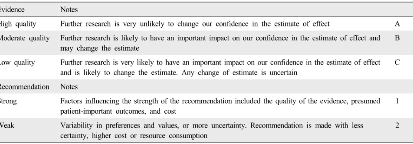
Table 1. Grading evidence and recommendations
Evidence Notes
High quality Further research is very unlikely to change our confidence in the estimate of effect A Moderate quality Further research is likely to have an important impact on our confidence in the estimate of effect and
may change the estimate
B
Low quality Further research is very likely to have an important impact on our confidence in the estimate of effect and is likely to change the estimate. Any change of estimate is uncertain
C
Recommendation Notes
Strong Factors influencing the strength of the recommendation included the quality of the evidence, presumed patient-important outcomes, and cost
1
Weak Variability in preferences and values, or more uncertainty. Recommendation is made with less certainty, higher cost or resource consumption
2
INTRODUCTION
Liver cirrhosis (LC) is a disease with a high rate of prevalence and one of the most common causes of mortality in the Republic of Korea (hereafter "Korea"). In Korea, the main etiologies of LC have been found to be chronic hepatitis B (CHB), alcohol, and chronic hepatitis C (CHC).
In patients with complications such as ascites, variceal bleeding, and encephalopathy, the 5-year survival rates were 32%, 21%, and 40%, respectively, reflecting the poor prognosis of patients with LC. Consequently, a clinical practice guideline appropriate for the medical milieu of Korea is important for both patients and clinicians.
In 2005, the Korean Association for the Study of the Liver established a guideline for the treatment of LC that is now widely used. However, it is currently necessary to revise and update the clinical practice guideline based on new evidence over the past 6 years regarding the diagnosis, treatment, and prevention of LC. Therefore, the Korean Association for the Study of the Liver undertook a revision and update of the clinical practice guideline co-organized by the Liver Cirrhosis Clinical Research Center. This guideline was based on an interdisciplinary (hepatology, radiology, pathology, and preventive medicine) approach. A panel of experts selected by the Korean Association for the Study of the Liver and Liver Cirrhosis Clinical Research Center met several times to discuss and write this guideline during 2005-2011. This guideline was written in light of published studies retrieved from MEDLINE, EMBASE, and Cochrane Library. The panel aimed to address 5 subjects: diagnosis of LC, anti- fibrotic therapy for LC, variceal bleeding, ascites, and hepatic encephalopathy.
The evidence and recommendations made in this guideline have been graded according to the GRADE (Grading of Recommendations Assessment Development and Evaluation) system. The strength of evidence has been classified into 3 levels: A (high-quality evidence), B (moderate-quality evidence), and C (low-quality evidence).
The strength of recommendation has been classified into 2 categories: strong and weak (Table 1). Where there was no clear evidence, the recommendations were based on the consensus expert opinion(s) in literature and that of the writing committee.
1. Diagnosis of LC
LC is a pathologically defined disease, and is clinically classified as compensated and decompensated LC. Decom- pensated LC includes cases with ascites, variceal bleeding, hepatic encephalopathy, or jaundice. Image studies for diagnosing LC are CT, abdominal ultrasound, and MRI.
Typical findings of these images are nodular liver surface, splenomegaly, and the presence of intra-abdominal collateral vessels, which mean increasing portal venous pressure.
Although there are not established criteria for the diagnosis of compensated LC, imaging studies may be helpful for the diagnosis of LC b y integrating laboratory findings such as albumin, bilirubin, or prothrombin time and platelet values.
1-1. Diagnostic approach-patient history, physical examination, and laboratory tests
When dealing with patients with LC, evaluation of the cause, severity, and stage is the first step. In patients with chronic liver disease, history taking (drug use, blood transfusion, or alcohol use), physical examination (jaundice, ascites,
Recommendations
1. What test is needed for the diagnosis of LC in patients with chronic liver disease?
- Find evidence for LC by history taking and physical examination. (A1)
- In patients with chronic liver disease, to find LC, the following tests are recommended and the findings in parentheses should be checked. (A1)
(1) Complete blood count with platelet count (thrombocytopenia)
(2) Liver function test (hypoalbuminemia) (3) Prolonged prothrombin time
(4) Imaging studies (nodularity of liver surface and findings of LC)
(5) Endoscopy (presence of varices)
- The cause of LC should be examined including hepatitis B virus and hepatitis C virus tests. (A1)
- The severity of LC could be assessed by Child-Pugh classification with score. (B1)
2. Which findings are compatible with the diagnosis of LC in the imaging studies?
- For the diagnosis of LC, compatible findings of imaging studies (abdominal ultrasound, CT, and MRI) are morphologic changes (nodularity of liver surface, atrophy of the right lobe, hypertrophy of the left and caudate lobe, expansion of the periportal space, and intrahepatic nodule), ascites, and presence of portal hypertension (collateral vessel or splenomegaly). (B1)
spider angioma, hepatomegaly, or splenomegaly), and symptom such as fatigue from hepatitis should be assessed. In patients with LC, a whole blood test including platelet count, liver function test (albumin, aspartate aminotransferase, alanine aminotransferase, alkaline phosphatase, and gamma glutamyl transpeptidase), prothrombin time, abdominal ultrasound, abdominal CT, and endoscopy should be carried out to confirm the presence or absence of cirrhosis. In addition, laboratory tests for hepatitis B or C virus infection are needed for the evaluation of its cause. Generally, the Child- Pugh score is used to assess the severity of LC. In clinical practice for the diagnosis of LC, findings of portal hyper- tension such as ascites, hepatic encephalopathy, or varices, imaging findings, and laboratory findings are common diagnostic tools. Recently, it was found that nodularity of the liver surface, a platelet count of less than 100,000/mm3, albumin less than 3.5 g/dL, and an international normalized ratio of 1.3 or more are related to the presence of LC.
Presence of one condition of these findings showed a specificity of 90.42% and a sensitivity of 61.11%.1
1-2. Imaging modalities for the diagnosis of LC Abdominal ultrasound is safe and less expensive, and it can be easily used as a screening test for the diagnosis of LC.
Abdominal ultrasound confirms the size of the liver, echogenicity of the liver parenchyme, morphological changes of the liver, ascites, and thrombus of the portal vein.
Typical ultrasound finding of LC is a coarse echo pattern by fibrosis and regeneration. This finding ranges from mild coarsening to visible nodules. The accuracy of CT in diagnosing LC is 66-95%.2-6 The most important CT finding for the diagnosis of LC is nodularity caused by regenerative nodules, fibrosis scars, and lobar non-uniform atrophy and hypertrophy. Nodularity of the liver surface showed a specificity of 95%.2-4,7 Additionally, other radiologic findings such as splenomegaly, blunt angle, morphological changes (nodularity of liver surface, atrophy of right lobe, hypertrophy of left and caudate lobe, expansion of periportal space, and intrahepatic nodule), velocity of portal flow, shape of hepatic vein waveform (Doppler test),2,4,8 and caudate lobe/right lobe ratio (CT or MRI) are useful indices.9 If the caudate lobe/right lobe ratio is above 0.65, it can indicate LC, and its sensitivity, specificity, and accuracy reach 84%, 100%, and 94%, respectively.10
1-3. Pathological diagnosis of liver cirrhosis The gold standard for confirming the diagnosis of LC is liver biopsy, but it is invasive and susceptible to a sampling error and inter-observer discrepancy. Therefore, liver biopsy has not been widely used in clinical practice. Liver biopsy can be carried out selectively for evaluating activity of the underlying disease and fibrosis. Particularly, when cirrhosis is diagnosed by clinical findings and imaging studies, but not compatible with laboratory data, a liver biopsy is especially helpful for diagnosing LC.11
1-4. Others
The serum markers for liver fibrosis directly or indirectly reflect extracellular matrix metabolism. However, their clinical utility in the diagnosis of LC has not been verified.12 FibroScan has been introduced to measure liver elasticity noninvasively and is an objective diagnostic tool for the diagnosis of LC. However, there is no exact guideline on how to apply it clinically, and there is some variation in its diagnostic accuracy for the diagnosis of LC. Therefore, the practical use of FibroScan is limited.
3. When should liver biopsy be performed in patients with chronic liver disease for the diagnosis of LC?
- Liver biopsy should be selectively performed to detect the cause of LC and assess the disease activity and fibrosis.
Liver biopsy is necessary on patients in whom the diagnosis remains unclear from the conventional test (B1)
2. Anti-fibrotic therapy for LC
LC is a pathological condition of liver fibrosis generated by continuous scar formation and recovery due to chronic liver injury, eventually leading to the development of regenerative nodules around the fibrotic scar.13 If liver injury is chronically repeated, damaged liver cells cannot regenerate and instead migrate to the extracellular matrix such as collagen.14 The mechanism of liver fibrosis is variable, depending on causes such as alcohol, hepatitis virus, or bile acids. Hepatic stellate cells, which are located in the space of Disse, play a major role in liver fibrosis.14 Histological changes associated with LC have long been believed to be irreversible. However, in patients receiving antiviral therapy for CHB, liver fibrosis has been observed to be significantly improved.15 Similarly, the primary approach to treating LC is treatment of the underlying disease. The second approach in the treatment of LC is anti-fibrotic therapy that targets the liver fibrosis-generating mechanism irrespective of the cause of LC. In order to develop effective treatments for liver fibrosis, the molecular pathogenesis and treatment of liver fibrosis have been exhaustively investigated over the last 2 decades. Subsequently, a number of substances or drugs with anti-fibrotic effects in animal experiments have been found; however, most of these have not been verified for use in human beings.
2-1. Chronic hepatitis B
Antiviral therapy that regulates inflammation via the inhibition of viral proliferation is recommended for improving liver fibrosis.15-21 There were no significant differences in the degree of improvement in fibrosis according to antiviral drugs.22-24 As liver fibrosis progresses, the rate of improvement by antiviral treatment increases.18,25 Therefore, in patients with advanced liver fibrosis, antiviral treatment is needed in the case with viral proliferation. In patients with LC due to CHB, antiviral therapy with a sufficient treatment period can be recommended to reduce liver fibrosis from virus proliferation. However, in the case of drug-resistant viral infection, the benefits of antiviral treatment are reduced.19,22,26
The extent of liver fibrosis was reduced by 49% when treated with interferon α or peginterferon α.20 In patients with HBeAg-positive CHB, hepatic fibrosis improved in 39% of patients after initial treatment with entecavir, whereas in HBeAg-negative CHB, improvement was observed in 36% of patients.23,27 With 1-year tenofovir treatment, inflam- mation improved without aggravation in 74% of HBeAg- positive CHB patients and in 71% of HBeAg-negative CHB patients.21,24,28
2-2. Chronic hepatitis C
Interferon treatment for CHC reduces the hepatic necrotic inflammatory reaction; particularly, in patients with a sustained virological response, the reductions in serum hepatitis C virus RNA and intrahepatic necrotic inflammatory reactions are correlated.29 In particular, antiviral treatment should be actively considered for patients presenting with genotype 2 or 3 and a Child-Pugh score <7. In patients with a Child-Pugh score of 7-9, antiviral treatment can be considered with careful attention to hepatic dysfunction.30-34
2-3. Alcohol
Abstinence is the most important treatment of alcoholic liver disease.35 Abstinence not only improves fibrosis in hepatic tissue but also reduces portal pressure and inhibits progression to LC. Ultimately, the survival rate of patients in all stages of alcoholic liver disease increases following abstinence, with improvements being observed in up to 66%
of patients.35 Sustained drinking increases the risk of variceal bleeding by portal hypertension and is closely related with mortality.
In severe alcoholic hepatitis, steroid and pentoxifylline therapy have been recommended by several studies. However, the effect of these drugs on fibrosis has not been confirmed;
therefore, these medications are not recommended in the treatment of fibrosis as anti-fibrotic therapy.
2-4. Nonalcoholic fatty liver
Treatment of nonalcoholic fatty liver comprises improving insulin resistance, removing risk factors of metabolic syndrome, lifestyle modifications, drug therapy, and operative treatment.36-52 Weight loss appears to help in liver steatosis as well as relieves hepatic inflammation and fibrosis.36-39 Therefore, the first choice of therapy in nonalcoholic fatty liver disease is weight loss through diet therapy and lifestyle
Recommendations
1. What is anti-fibrotic therapy according to the causes of LC?
- In patients with LC, it is recommended that the cause of disease should be treated to improve liver fibrosis. (A1) - In patients with LC due to CHB, antiviral therapy with a
sufficient treatment period can be recommended to reduce liver fibrosis from virus proliferation. (B1)
- In patients with LC due to CHC, antiviral therapy with a combination of peginterferon and ribavirin can be recommended, if the normal range of liver function with viral proliferation. In addition, monitoring for side effects is needed. (B1)
- In patients with alcoholic LC, strict abstinence is recommended to prevent worsening of disease. (A1) - In patients with nonalcoholic fatty liver disease, losing
weight, diet therapy, and exercise can be recommended.
(B1)
- In patients with primary biliary cirrhosis, ursodeoxycholic acid is recommended with a dose of 13-15 mg/kg/day.
(A1)
Recommendations
1. What is the role of endoscopy in patients with LC?
- It is recommended that all patients undergo endoscopy when they are first diagnosed with LC in order to evaluate the presence of esophageal varix and the risk of bleeding. (A1)
- Esophageal varices are classified into small (F1) and large (F2 and F3). Also, red color sign should be checked during endoscopy. (B1)
- For patients with compensated LC should be considered for endoscopy every 2 to 3 years, and patients with decompensated LC every 1 to 2 years in order to evaluate the occurrence and progression of varices. (B1)
- Patients with compensated LC with small varices not using nonselective beta-blocker, should be considered for endoscopy every 2 years to evaluate the progression of varices. (B1)
- The frequency of endoscopy can be adjusted according to the cause and progression of LC.
modifications, with more than 7% weight loss being the recommended target.39 For nonalcoholic fatty liver disease, clinical trials using multiple drugs have currently being attempted. However, data regarding its effectiveness, safety, and dosing period remain limited.40
2-5. Primary biliary cirrhosis
Ursodeoxycholic acid has been proven to improve primary biliary cirrhosis and is the only drug recognized for its treatment.53-56 Ursodeoxycholic acid reduces serum bilirubin levels, which is an important prognostic factor, and delays the progression and deterioration of fibrosis55,56 13-15 mg/kg/day is the most effective dose in terms of biological effect and cost-effectiveness. Improvements of liver function tests appear within 6-9 months of therapy in more than 90% of patients.57 Ursodeoxycholic acid treatment is not known to improve fatigue, itching, bone diseases, or autoimmune reactions associated with primary biliary cirrhosis.57
3. Variceal bleeding
Esophageal variceal bleeding is found in approximately 40% of patients with Child-Pugh A class LC, and in approximately 80% of patients with Child-Pugh C class LC.58 The risk of variceal bleeding depends on the size of varices, red color sign, and the Child-Pugh classification.
Variceal bleeding due to portal hypertension can be prevented when the value of hepatic venous pressure gradient is lower
than 12 mmHg or when it is decreased to lower than 20% of the baseline measurement.59
3-1. Diagnosis
It is recommended that all patients undergo endoscopy when they are first diagnosed with LC in order to evaluate the presence of esophageal varix and the risk of bleeding.60 Esophageal varices are classified based on the diameter of varices and their morphology as follows: varices with the findings of larger than 5 mm61 or F2-F3 (beaded varices - crystalled varices) are defined as large and varices with the findings of smaller than 5 mm or F1 (straight varices) are defined as small.62 Treatment to prevent esophageal variceal bleeding should be performed when large varices are present. For patients with compensated LC should be considered for endoscopy every 2 to 3 years, and patients with decompensated LC every 1 to 2 years in order to evaluate the occurrence and progression of varix.63 Patients with compensated LC with small varices not using nonselective beta-blocker, should be considered for endoscopy every 2 years to evaluate the progression of varix.
3-2. Acute esophageal variceal bleeding 3-2-1. Definition and diagnosis
Acute variceal bleeding is defined as hematemesis within last 24 hours of presentation, and/or ongoing melena, with the last melenic stool within last 24 hours before the hospital visit in a known or suspected case of portal hypertension.
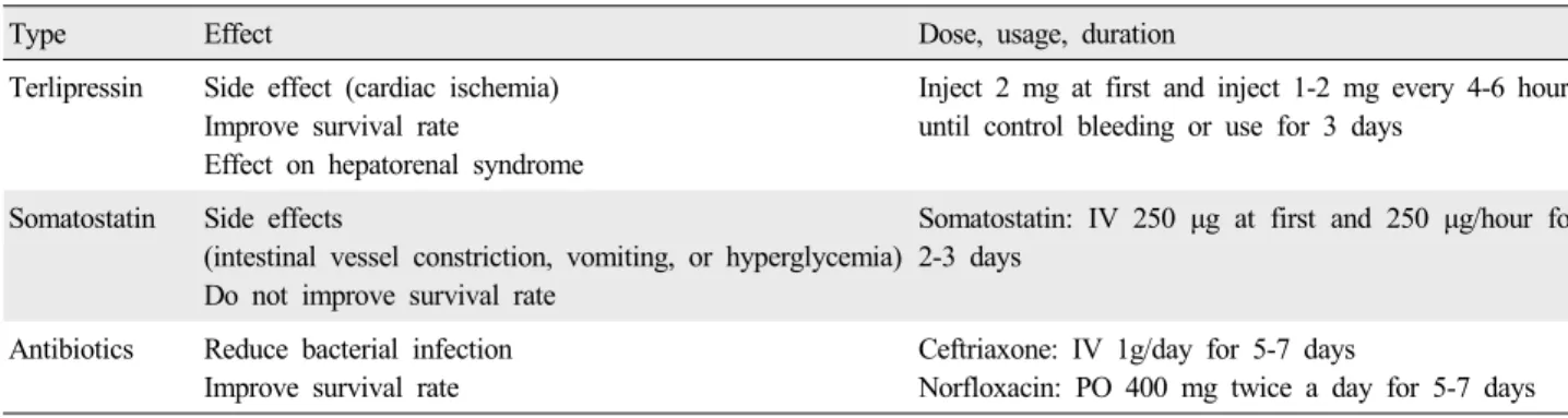
Table 2. Drugs for acute esophageal variceal bleeding
Type Effect Dose, usage, duration
Terlipressin Side effect (cardiac ischemia) Improve survival rate
Effect on hepatorenal syndrome
Inject 2 mg at first and inject 1-2 mg every 4-6 hours until control bleeding or use for 3 days
Somatostatin Side effects
(intestinal vessel constriction, vomiting, or hyperglycemia) Do not improve survival rate
Somatostatin: IV 250 μg at first and 250 μg/hour for 2-3 days
Antibiotics Reduce bacterial infection Improve survival rate
Ceftriaxone: IV 1g/day for 5-7 days
Norfloxacin: PO 400 mg twice a day for 5-7 days Recommendations
2. How should acute variceal bleeding be treated?
- It is recommended that patients with acute variceal bleeding initially be administered vasoconstrictor and antibiotic treatment. (A1)
- Endoscopic treatment is recommended for patients with acute variceal bleeding. (A1)
- TIPS can be recommended if drugs and endoscopic therapy have failed or endoscopic treatment is impossible. (B1) Endoscopy is the most definite diagnostic method for identifying acute esophageal variceal bleeding. Variceal bleeding can be diagnosed as follows: direct visualization of blood issuing from esophageal varices, presence of signs of recent bleeds on varices such as white nipple sign or overlying clot, or presence of blood in the stomach in the absence of another source of bleeding.64
3-2-2. Treatment
The first line of therapy includes vasoconstrictors such as terlipressin and somatostatin.65 Antibiotics are recommended for patients admitted in the hospital.66 Antibiotic therapy is recommended for 5-7 days to prevent sepsis and rebleeding following endoscopy (Table 2).67
Medication and endoscopic therapy are recommended in patients with acute variceal bleeding for the first time.68 If active bleeding is present, banding of the culprit vessel or that just below the ooze should be performed endoscopically.
After ligation of the active bleeding site, even in the absence of active bleeding, banding should be performed starting from just above the gastro-esophageal junction (5-10 mm) in a sequential manner up to 5 cm. Endoscopic injection sclerotherapy is recommended if endoscopic variceal ligation is technically impossible or has failed.
Balloon tamponade can be used as a rescue therapy if active variceal bleeding cannot be controlled. Transjugular intrahepatic portosystemic shunt (TIPS) is recommended if medication and endoscopic therapy have failed or if endoscopic therapy is impossible.
3-3. Prevention
3-3-1. Prevention of first bleeding in patients without esophageal varices
Nonselective beta-blockers are not recommended in patients without esophageal varices.69,70
3-3-2. Prevention of first bleeding in patients with small esophageal varices
Nonselective beta-blockers do not lower the incidence of bleeding in patients with small esophageal varices.71 However, patients with a high risk of bleeding (Child-Pugh class B/C or endoscopic red color sign), are considered for nonselective beta-blocker therapy.70 The dose of nonselec- tive beta-blockers is adjusted for a reduction in the resting heart rate by 25%, to 55 beats/minute, or until the occurrence of side effects. In Koreans, the mean adjusted dose of propranolol is 160 mg/day.72,73
3-3-3. Prevention of first bleeding in patients with large esophageal varices
In large esophageal varices, endoscopic variceal ligation (EVL) and nonselective beta-blockers are effective in preventing the first bleeding occurrence.74,75 In patients with large varices (F2 or F3), which have never been observed to bleed, nonselective beta-blockers or EVL is recommended.
Repeated EVL is recommended until the disappearance of esophageal varices.76 Low doses of carvedilol have been shown to lower the frequency of variceal bleeding
Recommendations
3. How can a first variceal bleeding be prevented in patients with LC?
- In patients without varices, a nonselective beta-blockers are not recommended for the purpose of preventing the formation of varix and first bleeding of esophageal varix.
(B1)
- Nonselective beta-blockers should be considered for patients with small varices which have never bled but have a high risk of bleeding (Child-Pugh class B/C or red color sign on endoscopy). (B1)
- In patients with large varices (F2 or F3) in which bleeding has never been observed, nonselective beta-blockers or EVL are recommended. (A1)
4. How should nonselective beta-blockers be administered to patients with LC?
- Nonselective beta-blockers are adjusted at the dose of reduction in resting heart rate by 25% or 55 beats/minute, or until the side effects occur. (B1)
Recommendations
5. How to prevent recurrence of variceal bleeding?
- Patients who experience acute variceal bleeding need treatment to prevent rebleeding. (A1)
- EVL alone or in combination with nonselective beta-blockers should be considered for the prevention of rebleeding. (B1)
- TIPS should be considered as a rescue therapy in Child- Pugh A/B patients in whom other therapies have failed.
(B1)
- Liver transplantation should be considered for patients who meet indications for liver transplantation. (B1) with lesser side effects as compared with EVL. However,
additional research is needed before they can be routinely used.66,77 Combination therapy with EVL and nonselective beta-blockers for the prevention of first bleeding has shown no differences as compared with monotherapy.78
3-4. Prevention of variceal rebleeding 3-4-1. Definition and diagnosis
Variceal rebleeding is defined as bleeding after 5 days of recovery from acute variceal bleeding.70 It is diagnosed similar to acute variceal bleeding.
3-4-2. Prevention
Nonselective beta-blockers alone or combination therapy with isosorbide mononitrate are known to be effective in the prevention of rebleeding.79 EVL shows better outcomes than endoscopic injection sclerotherapy in endoscopic therapy for prevention of rebleeding.80 Rebleeding has been reported in 32% of patients after EVL. Combination therapy of EVL and nonselective beta-blockers has been shown to have better outcomes than EVL alone.81,82
TIPS shows a significantly low rebleeding rate as compared with endoscopic therapy. However, it shows a significantly high incidence of hepatic encephalopathy. TIPS is not recommended as a first-line therapy for esophageal variceal bleeding and is recommended only as a rescue therapy when combination therapy fails.83 Liver transplantation should be considered for patients who meet indications for liver
transplantation.84,85
If the hepatic venous pressure gradient is reduced to less than 12 mmHg or to more than 20% reduction in baseline levels by medical therapy, the incidence rate of rebleeding is low (10%).86 In such cases, further endoscopic therapy may not be required.
3-5. Gastric varices 3-5-1. Definition and diagnosis
Gastric varices are enlarged submucosal veins of the stomach that cause critical upper gastrointestinal bleeding.
Gastric varices occur in approximately 20% of patients with portal hypertension, and the bleeding rate in 2 years is known to be 25%.87 Diagnosis is performed by endoscopy.
Endoscopic ultrasound can also be helpful.88
3-5-2. Classification
Gastroesophageal varices are classified depending on whether esophageal varices are extended along lesser curvature (gastroesophageal varices, GOV1) or gastric fundus (GOV2). Gastric varices alone are classified as varices located in the fundus (isolated gastric varices, IGV1) and any other regions, i.e., stomach or duodenum (IGV2).87
3-5-3. Treatment and prevention
EVL is recommended for the treatment of GOV1.
Endoscopic variceal obturation (EVO) can be an alter- native.89-93 EVO involves the injection of tissue adhesives such as N-butyl-2-cyanoacrylate (histoacrylⓇ) into the varices via endoscopy and can be recommended in treating GOV2 and IGV.91,94-96 Endoscopic injection sclerotherapy is not recommended in treating GOV2 or IGV1 because of its low rate of hemostasis, high rate of rebleeding, and other complications.97-100 If endoscopic treatment is not possible,
Recommendations
6. How should gastric variceal bleeding be treated?
- In gastric variceal bleeding, EVL or EVO are considered for GOV1 accompanying esophageal varices extended along the lesser curvature. (B1)
- EVO is preferable in patients with GOV2 or IGV1. If endoscopic treatment is not possible, TIPS can be used. If gastric varix is accompanied with gastrorenal shunt, balloon-occluded retrograde transvenous obliteration can be considered. (B1)
- Surgery such as distal splenorenal shunt or vascular shunt should be considered for patients (Child-Pugh A/B) and liver transplantation for patients (Child-Pugh B/C) who are not eligible for endoscopic treatment.
Recommendations
7. How should portal hypertensive gastropathy be treated?
- If chronic bleeding due to portal hypertensive gastropathy presents, nonselective beta-blockers can be used. (B1)
Recommendations
1. How should ascites of LC be diagnosed?
- Paracentesis should be performed when Grade 2 or 3 ascites occurs, when there is clinical suspicion of infection, or when there are complications of LC such as encephalopathy or kidney dysfunction. (A1)
- When the initial paracentesis is performed, a total cell count and differential, albumin, and total protein tests should be performed. A culture of ascitic fluid in blood culture bottles at the bedside is recommended. (A1) - If serum-ascites albumin gradient is greater than or equal
to 1.1 g/dL, it indicates ascites by portal hypertension. (B1) TIPS can be performed.101,102 If a gastrorenal shunt is
present, balloon-occluded retrograde transvenous obliteration can be performed.103-106 Surgery, such as distal splenorenal shunting or vascular shunting, can be attempted if endoscopic treatment is not possible in case of Child-Pugh A/B patients, while liver transplantation is considered in the case of Child-Pugh B/C patients.107
Empirical use of nonselective beta-blockers, balloon- occluded retrograde transvenous obliteration, and EVO can prevent first gastric variceal bleeding, while EVO, TIPS, and balloon-occluded retrograde transvenous obliteration are considered to prevent gastric variceal rebleeding.70,108
3-6. Portal hypertensive gastropathy 3-6-1. Diagnosis
Portal hypertensive gastropathy is diagnosed when gastric mucosal changes of snake-skin appearance (or mosaic pattern) are found on endoscopy in patients with portal hypertension.109 When gastric mucosal changes alone are found, this is diagnosed as a mild form. When red or dark brown viscous changes with changes in gastric mucosa are found, it is considered severe.72 Severe portal hypertensive gastropathy causes more chronic bleeding than does the mild form.110
3-6-2. Treatment
In chronic bleeding due to portal hypertensive gastropathy, the goal of treatment is lowering the portal pressure with nonselective beta-blockers. In addition, iron supplementation is recommended.111 Despite these treatments, if repeated transfusions are needed for the treatment of chronic anemia, TIPS can be considered.112
4. Ascites
Ascites is the most common complication in LC, occurring in 60% of the patients with compensated LC within 10 years.113 Ascites appears in 2/3 of the patients who required admission due to LC,114 and 60% of the patients requiring paracentesis is because of LC.115
4-1. Diagnosis
Abdominal ultrasound can diagnose ascites with only 100 mL.116 Ascites is classified by the amount of fluid as follows:
Grade 1 is diagnosed through imaging study, Grade 2 through inspection and physical examination, Grade 3 based on marked abdominal expansion. Paracentesis is the most useful and simple diagnostic tool in Grade 2 or 3 ascites.
Furthermore, paracentesis should be performed in cirrhotic patients with fever, abdominal pain, bleeding, encephalopathy, hypotension, or kidney dysfunction to assess for spontaneous bacterial peritonitis (SBP), since 10-27% of the patients with ascites have SBP.117 The main purpose of paracentesis is to discriminate the cause of ascites. Therefore, screening tests should include total cell count and differential, albumin, and total protein. Blood cell count is the most useful test for diagnosing SBP. For differential diagnosis of ascites, a serum-ascites albumin gradient can be calculated. If the serum-ascites albumin gradient is is greater than of equal to 1.1 g/dL, ascites is ascribed to portal hypertension with an accuracy of 97%.
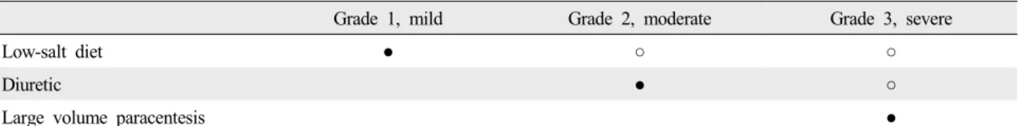
Table 3. Treatment of ascites depending on the grade
Grade 1, mild Grade 2, moderate Grade 3, severe
Low-salt diet ● ○ ○
Diuretic ● ○
Large volume paracentesis ●
Grade 1, a small amount of ascites detected in the ultrasound test; Grade 2, ascites of the amount which distends abdomen symmetrically; Grade 3, ascites of the large volume which distends abdomen; ●, major treatment; ○, recommended.
4-2. Treatment
Although controversial,118 a low salt diet is considered effective for controlling ascites and shortening hospitalization.
Less than 5 g/day of salt (sodium for 2 g, 88 mEq) is recommended. When plasma sodium is lower than 120-125 mEq/L, water intake should be restricted to 1-1.5 L/day.119
4-2-1. Medications
If severe ascites is present, diuretic therapy should be used for negative sodium balance.120,121 Secondary hyperaldo- steronism in patients with LC induces reabsorption of sodium and water in the distal renal tubule and collecting tubule, consequently causing hypokalemia. Aldosterone antagonists inhibit this mechanism, and hence is mainly used for controlling ascites in patients with LC. Spirono- lactone has a long half-life but has a slow onset of action, and therefore requires 3-4 days to achieve a stable concentration. It is initiated at a dose of 50-100 mg/day, with a maximum dose of 400 mg/day. Side effects include hyperkalemia, gynecomastia, mastalgia, hyposexuality, and erectile dysfunction.122
Loop diuretics operate by blocking Na-K-2Cl receptors in the thick ascending limb of Henle’s loop. They are mostly used along with aldosterone antagonists in patients with LC.
Hypokalemia may occur as a side effect, but via such mechanism, hyperkalemia caused by the aldosterone anta- gonist can be corrected. The starting dose is 20-40 mg/day, with the maximum dose being 160 mg/day. Monotherapy with loop diuretics is less effective than aldosterone anta- gonist monotherapy. Spironolactone monotherapy can be used initially on patients with ascites.123 Combination therapy of an aldosterone antagonist and a loop diuretic is recommended at first in a ratio of 100:40, which stabilizes plasma potassium levels. Combination therapy shows a faster effect in controlling ascites and lowers the possibility of hyperkalemia as compared to aldosterone monotherapy.124
When peripheral edema is present, the rate of weight loss
should be no greater than 1 kg/day. For patients without edema, 0.5 kg/day of weight loss is ideal.120,125 If there is no weight loss even with 5 g/day of salt intake, the diet and diuretic dose should be evaluated by examining the amount of urinary sodium excretion per day.120,126 Patients with low salt intake should not excrete more than 78 mEq of urinal sodium in 24 hours. In such patients, if urinary sodium excretion is more than 78 mEq, low salt intake is judged not to be followed. When the excretion is less than 78 mEq, the diuretic is considered to be inadequate and the dose needs to be increased. Spot urine Na/K ratio of more than 1 repre- sents 24 hours urine sodium excretion more than 78 mEq.127 It is important to check the weight loss, vital sign, changes in consciousness level, and the level of plasma sodium, potassium, and creatinine during diuretic administration.
When plasma sodium is more than 126 mEq/L, diuretics can be used without the restriction of water intake. However, when plasma sodium is less than 125 mEq/L, the physician should consider cessation or reduction in the dose of the diuretic and restriction of water intake. If plasma sodium is less than 120 mEq/L, the diuretic and water intake should be stopped and a plasma expander such as albumin should be administered. If plasma sodium is less than 125 mEq/L with kidney dysfunction, the diuretic should be stopped and a plasma expander should be administered.128 If plasma potassium is less than 3.5 mEq/L, the dose of loop diuretic should be reduced or stopped. If plasma potassium is more than 5.5 mEq/L, the dose of aldosterone antagonist should be reduced. And if plasma potassium is more than 6.0 mEq/L, the aldosterone antagonist should be ceased.120
4-2-2. Therapeutic paracentesis (reference to intractable ascites section)
Therapeutic paracentesis is an effective treatment for tension-type ascites, because it relieves the symptoms more quickly than diuretics and shortens hospitalization (Table 3).129
Recommendations
2. How should ascites of LC be treated?
- Patients with cirrhotic ascites should be advised to take in less than 5 g of salt a day. (B1)
- When the serum sodium is normal, restriction of water intake is not necessary. (B1)
- Bed rest is not recommended for the treatment of ascites.
(B1)
- The first-choice diuretic for patients with cirrhotic ascites is aldosterone antagonist. (A1) Loop diuretics can be used along with aldosterone antagonist. (B1) Spironolactone can be used with a starting dose of 50-100 mg/day up to 400 mg/day. To increase the diuretic effects and maintain a normal serum potassium level, 20-40 mg of furosemide should be used with spironolactone (40:100) beginning at the initial stage.
- When peripheral edema is present, the rate of weight loss should be recommended up to 1 kg/day. For patients without edema, 0.5 kg/day of weight loss should be recommended. (A1)
- In cases of severe hyponatremia, kidney dysfunction, encephalopathy, or severe muscle spasms, diuretics should be stopped. (B1)
- In cases of hypokalemia, loop diuretic should be reduced or stopped, and if hyperkalemia occurs, the dose of aldosterone antagonist should be adjusted. (B1)
- Therapeutic large volume paracentesis is recommended as the first-line treatment for tension-type ascites. (A1)
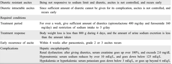
Table 4. Definition and diagnosis of refractory ascites206
Diuretic resistant ascites Being not responsive to sodium limit and diuretic, ascites is not controlled, and recurs early Diuretic intractable ascites Since sufficient amount of diuretic cannot be given for its complication, ascites is not controlled, and
recurs early Required conditions
Treatment period For over a week, give sufficient amount of diuretics (spironolactone 400 mg/day and furosemide 160 mg/day) and restriction of sodium intake to 5 g/day
Treatment response Body weight loss is less than 800 g during 4 days, and the amount of urine sodium excretion is less than the amount taken
Early recurrence of ascite Within 4 weeks after paracentesis, grade 2 or 3 ascites recurs Complications Hepatic encephalopathy
Renal dysfunction: after giving diuretics, serum creatinine goes up over 100%, and exceeds 2.0 mg/dL Hyponatremia: serum sodium reduces by over 10 mEq/L, and goes down below 125 mEq/L Hypokalemia or hyperkalemia: serum potassium goes down below 3 mEq/L, or goes up beyond 6 mEq/L 4-3. Refractory ascites and hyponatremia
4-3-1. Refractory ascites
Refractory ascites130 is defined as fluid overload that (1) is not controlled despite restriction of sodium intake and the maximum dose of diuretics, and (2) recurs rapidly after paracentesis. There are 2 types of refractory ascites-diuretic resistant and diuretic intractable (Table 4).
Large-volume paracentesis is not considered for every patient as a first-line treatment. It can be selectively performed in occasions in which the patient has difficulties eating or breathing because of abdominal distension. After relieving the symptoms by paracentesis, the maintenance treatment should be administered. In the case of large- volume paracentesis over 5 L, an infusion of 8-10 g of albumin per L is recommended. In the case of paracentesis less than 5 L, although the occurrence of circulatory dysfunction is not frequent, an infusion of volume expander can be considered. Albumin can also be used for this purpose.125,131 Medications such as midodrine,132 norad- renaline,133 or terlipressin134 can be used. Compared with repetitive paracenteses, TIPS is an effective method to prevent the recurrence of ascites and the occurrence hepatorenal syndrome. However, hepatic encephalopathy occurs in 30-50% of cases after TIPS. As 21% of the patients with refractory ascites die within 6 months and the median survival period is also less than 1 year, they should be considered for liver transplantation.135
4-3-2. Hyponatremia
The diagnostic criterion for hyponatremia is less than 130 mEq/L.119 A hypervolemic state can be corrected to normal by a negative water balance. Eventually, dilutional hyponatremia can be improved.136 Restriction of water intake can prevent a decrease in the serum sodium level.125 Hypertonic sodium injection can worsen ascites and edema.137 Plasma expanders can be useful in the treatment of hyponatremia.138 Vaptan,139 a selective vasopressin 2 receptor antagonist of arginine vasopressin, leads to the excretion of
Recommendations
3. How should intractable ascites be treated?
- Repetitive large volume paracentesis is recommended in patients with refractory ascites. (A1)
- In the case of large volume paracentesis, 8-10 g/L (albumin/
ascites) is recommended for the prevention of postparacentesis circulation dysfunction. (A1)
- TIPS can be used for the treatment of refractory ascites. (B1) - Because of poor prognosis, liver transplantation is
recommended in patients with refractory ascites. (A1) - If the serum sodium concentration is less than 120-125
mEq/L, restriction of fluid intake to 1-1.5 L/day is recom- mended. (A1)
- Albumin or vaptan can be used in severe dilutional hypo- natremia (<125 mEq/L). (B2)
Table 5. New International Ascites Club's diagnostic criteria of hepatorenal syndrome 1) Cirrhosis with ascites
2) Serum creatinine >133 mmol/L (1.5 mg/dL)
3) No improvement of serum creatinine (decrease to a level of 133 mmol/L) after at least 2 days with diuretic withdrawal and volume expansion with albumin; the recommended dose of albumin is 1 g/kg of body weight per day up to a maximum of 100 g/day 4) Absence of shock
5) No current or recent treatment with nephrotoxic drugs
6) Absence of parenchymal kidney disease as indicated by proteinuria >500 mg/day, microhematuria (>50 red blood cells/high-power field), and/or abnormal renal ultrasound.
solute-free water. This agent can be used in the treatment of hyponatremia caused by inappropriate antidiuretic hormone secretion, heart failure, or liver cirrhosis. In the United States and Europe, tolvaptan and conivaptan are approved for the treatment of severe hyponatremia (<125 mEq/L).
4-4. Hepatorenal syndrome 4-4-1. Definition and diagnosis
Renal failure in LC occurs in 2 forms. First, type 1 hepatorenal syndrome is a rapid progressive acute renal dysfunction which occurs due to the strong contraction of the renal vasculature. Type 2 hepatorenal syndrome is a relatively slow process with a rather moderate renal dysfunction. The most important mechanism involved in the occurrence of hepatorenal syndrome is decreased effective blood volume due to the dilation of the splanchnic and peripheral circulation. This situation activates the sympathetic nervous system as well as the renin-angiotensin system and causes functional renal disorder.140-142
In 1994, the International Ascites Club announced the diagnostic criteria for hepatorenal syndrome; in 2007, they revised these criteria to providing clearer diagnostic methods and including infectious diseases (Table 5).130,143
4-4-2. Treatment
Dilatation of splanchnic artery and decrease in effective arterial blood volume arethe primary mechanisms of hepatorenal syndrome. The effect of albumin monotherapy is not sufficient in this situation.144,145 Vasoconstrictors have been used in the treatment of the hepatorenal syndrome, and combination therapy with vasoconstrictors and albumin is effective in hepatorenal syndrome. The combination therapy of terlipressin and albumin improves renal function in 60-75% of the patients.144,146-152 Maintenance therapy is possible up to 15 days or until the serum creatinine levels decrease (<1.5 mg/dL). Further, long-term usage (approximately 2 months) can be considered as a bridging therapy prior to liver transplantation.153,154 The combination therapy of noradrenaline and albumin can also improve renal function in hepatorenal syndrome.155,156 Combination therapy of midodrine and octreotide or triple therapy of midodrine, octreotide, and albumin also meaningfully improve renal function in patients with hepatorenal syndrome.157-160
After administration of terlipressin, hepatic venous pressure gradient is known to decrease while the renal blood flow increases.161,162 Therefore, it can also be used for treatment in patients with secondary renal insufficiency due to variceal bleeding.147,163,164
Although TIPS157,165,166
reduces the serum creatinine levels in most patients with hepatorenal syndrome, the effect is slower than the combined use of terlipressin and albumin.
In a group that used molecular adsorbent recirculating system167 in the treatment of hepatorenal syndrome, laboratory findings (serum creatinine, bilirubin, and prothrombin time) and 30-days survival rate were improved compared to the group that underwent intermittent dialysis and drug therapy. Continuous arterio-venous hemofiltration and continuous veno-venous hemofiltration can be considered to minimize the changes in blood pressure.168,169
Recommendations
4. How should hepatorenal syndrome be treated?
- In type 1 hepatorenal syndrome, combination therapy of terlipressin and albumin can improve renal function. (A1) - In type 1 hepatorenal syndrome, combination therapy of
midodrine, octreotide, and albumin can be considered.
(B2)
- The best treatment for type 1 hepatorenal syndrome is liver transplantation. (A1)
- In high risk patients who have ascites accompanied by SBP, the use of albumin can decrease the incidence of hepatorenal syndrome. (A1)
Recommendations
5. How should SBP be diagnosed and treated?
- If SBP is suspected and the result of paracentesis show a PMN of more than 250/mm3, empirical antibiotic therapy should be started immediately without results of ascitic fluid culture. (A1)
- Third-generation cephalosporine is recommended as an initial antibiotic. (A1)
- The patient with symptoms or signs of infection should receive empirical antibiotics while awaiting of ascitic fluid culture even if the number of PMN is less than 250/mm3. (A1)
- If the secondary bactrieal peritonitisis suspected, an imaging study such as a CT should be performed (A1), and additional examinations such as those for total protein, LDH, glucose, or gram staining can be performed.
(B1)
- If the patient has a history of SBP, gastrointestinal bleeding, or protein in ascites less than 1.5 g/dL, although there is no gastrointestinal bleeding, prophylactic antibiotics should be considered because the chance of SBP is high. (B1) 4-4-3. Prevention
Hepatorenal syndrome can be prevented by inhibiting the decrease of plasma volume. Diuretics and lactulose should be used carefully to prevent excessive fluid loss.132,133,170,171
In patients with SBP, the use of albumin and antibiotics reduce the incidence of hepatorenal syndrome.172,173 The use of oral norfloxacin has been shown to reduce the occurrence of hepatorenal syndrome and increase 3-month survival rates in patients with low serum protein (<1.5 g/dL) or renal insufficiency (creatinine ≥1.2 mg/dL, or blood urea nitrogen
≥25 mg/dL, or serum Na ≤130 mEq/L).174 Administration of pentoxifylline improved survival rates as compared to corticosteroids in severe acute alcoholic hepatitis patients (Maddrey’s discriminant factor ≥32).175-177
4-5. Spontaneous bacterial peritonitis 4-5-1. Definition and diagnosis
SBP is bacterial infection of ascites, without an evident intra-abdominal, surgically treatable source of infection. It occurs in 10-30% of the patients with cirrhotic ascites and recurs in 70% of the patients within 1 year even if treated.126,178
SBP can be diagnosed in patients with findings of ascitic polymorphonuclear leukocyte (PMN) ≥250/mm3and bacteria in the ascitic culture without an evident intra-abdominal infection. If ascites fluid contains red blood cells, PMN is calculated by subtracting 1/mm3 per red blood cell 750 /mm3. Culture-negative neutrocytic ascites is a condition where ascitic PMN ≥250/mm3 but no cultured bacteria are observed. These patients show a clinical course mostly similar to patients with SBP. Empiric antibiotics treatment is recommended. Monomicrobial non-neutrocytic bacterascites is a condition with ascitic PMN <250/mm3, but cultured single-strain bacteria being observed.
4-5-2. Treatment
Administration of third generation cephalosporin is recommended. Cefotaxime is recommended at a dose of 2 g every 6-8 hours by intravenous injection. Subsequently, selective antibiotic therapy based on the result of ascites culture should be administered for 5-10 days.179 Treatment duration varies according to the symptoms and/or results of antibiotic sensitivity. Albumin during the treatment of SBP, especially in patients with renal dysfunction, can be helpful.173
4-5-3. Prevention
If a patient presentsacute gastrointestinal bleeding, administration of norfloxacin at a dose of 400 mg twice daily for 7 days orally or ceftriaxone at a dose of 1 g daily for 7 days intravenously is effective in preventing SBP. In patients with a concentration of protein in ascites fluid less than 1.5 g/dL or bilirubin in plasma is more than 2.5 mg/dL, administration of norfloxacin at a dose of 400 mg daily for longer than 6 months orally is effective in prolonging the survival.
5. Hepatic encephalopathy
5-1. Definition
Hepatic encephalopathy is a neuropsychiatric syndrome that follows liver dysfunction. Patients with hepatic encephalopathy can show various neurological illnesses
Table 6. Classification of hepatic encephalopathy180
HE type Nomenclature Subcategory Subdivisions
A Encephalopathy associated with acute liver failure
B Encephalopathy associated with portal-systemic bypass and no intrinsic hepatocellular disease
C Encepahlopathy associated with cirrhosis and portal hypertension/or systemic shunts
Episodic HE Precipitated Spontaneous Recurrent Persistent HE Mild
Severe
Treatment-dependent Minimal HE
HE, hepatic encephalopathy.
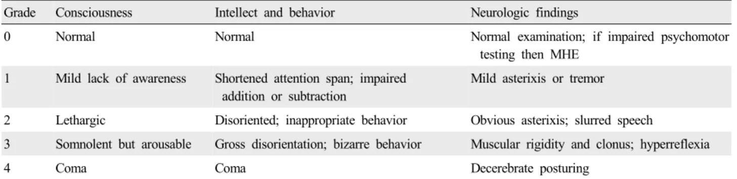
Table 7. West-Haven criteria for hepatic encephalopathy183
Grade Consciousness Intellect and behavior Neurologic findings
0 Normal Normal Normal examination; if impaired psychomotor
testing then MHE 1 Mild lack of awareness Shortened attention span; impaired
addition or subtraction
Mild asterixis or tremor
2 Lethargic Disoriented; inappropriate behavior Obvious asterixis; slurred speech
3 Somnolent but arousable Gross disorientation; bizarre behavior Muscular rigidity and clonus; hyperreflexia
4 Coma Coma Decerebrate posturing
MHE, minimal hepatic encephalopathy.
such as cognition and orientation disorders. Hepatic encephalopathy is classified into 3 groups according to the causative liver disease (Table 6).180,181
5-2. Diagnosis
Hepatic encephalopathy is generally accompanied by advanced liver disease; therefore, muscle weakness, jaundice, ascites, palmar erythema, edema, spider telangiectasias, and fetor hepaticus can be noted on physical examination.
Clinicians should check for gastrointestinal hemorrhage, uremia, use of anti-psychotics or diuretics, protein hyper- ingestion, infection, constipation, dehydration, electrolyte imbalance, etc.182 Common symptoms include concentration disorders, sleep disorders, and movement disorders, including lethargy or coma. The severity of hepatic encephalopathy can be evaluated using the West Haven criteria (Table 7).183
Venous levels of ammonia are not helpful because they are not proportional to the severity of hepatic encephalopathy and some patients with sever hepatic encephalopathy have
normal venous ammonia levels.184
Brain MRI is considered better than brain CT in the diagnosis of brain edema accompanying hepatic failure, but this is not true for hepatic encephalopathy. Brain CT is useful when differentiating between organic causes of neuropsychiatric disorders such as intracranial hemorrhage.185
5-3. Treatment
The goal of treatment is to prevent secondary damage caused by decreased consciousness, normalize the patient’s state of consciousness, prevent recurrence, and to improve the prognosis and quality of life by eliminating the social and economic restrictions caused by hepatic encephalopathy.
The precipitating factor can be identified in more than 80%
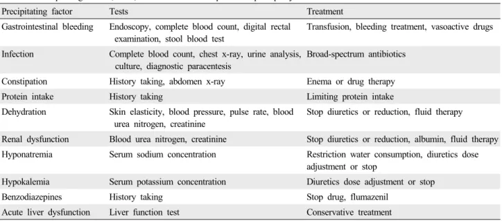
of patients with hepatic encephalopathy.182 The currently known precipitating factors of hepatic encephalopathy and the corresponding tests and treatments are shown in Table 8.186
Table 8. Precipitating factors, tests, and treatment of hepatic encephalopathy186
Precipitating factor Tests Treatment
Gastrointestinal bleeding Endoscopy, complete blood count, digital rectal examination, stool blood test
Transfusion, bleeding treatment, vasoactive drugs
Infection Complete blood count, chest x-ray, urine analysis, culture, diagnostic paracentesis
Broad-spectrum antibiotics
Constipation History taking, abdomen x-ray Enema or drug therapy
Protein intake History taking Limiting protein intake
Dehydration Skin elasticity, blood pressure, pulse rate, blood urea nitrogen, creatinine
Stop diuretics or reduction, fluid therapy
Renal dysfunction Blood urea nitrogen, creatinine Stop diuretics or reduction, albumin, fluid therapy Hyponatremia Serum sodium concentration Restriction water consumption, diuretics dose
adjustment or stop
Hypokalemia Serum potassium concentration Diuretics dose adjustment or stop
Benzodiazepines History taking Stop drug, flumazenil
Acute liver dysfunction Liver function test Conservative treatment
5-3-1. Medications
The primary treatment of hepatic encephalopathy is nonabsorbable disaccharides such as lactulose (β-galactosido- fructose) or lactitiol (β-galctoside sorbitol). These treatments lead to the recovery of 70-90% of patients with hepatic encephalopathy. Although nonabsorbable disaccha- rides have been reported to have no significant effect on hepatic encephalopathy,187 randomized controlled studies have indicated a positive effect of lactulose in the treatment and prevention of hepatic encephalopathy.188,189 Enema with nonabsorbable disaccharides can be used until the cons- ciousness is recovered. After consciousness is restored, nonabsorbable disaccharides (15-45 mL, orally 2-4 times/day) should be recommended for loose stool defecation 2-3 times a day.
Antibiotics such as neomycin, metronidazole, and rifaximin that are not absorbed by the intestine, affect urea-producing bacteria and reduce the generation of ammonia, thereby improving hepatic encephalopathy.
Neomycin and metronidazole are not recommended as atreatment of hepatic encephalopathy because of their side effects such as intestinal malabsorption, nephrotoxicity, and ototoxicity for neomycin and peripheral neuropathy for metronidazole.190 Rifaximin maintains high levels of concentration in the intestine because it is not absorbed by the intestine and remains in an active form until it is excreted. It has a broad antimicrobial activity on aerobic and anaerobic gram-positive and gram-negative bacteria. It has been proven effective and safe in hepatic encephalopathy,191-193
and recently has been focused upon as a first-line treatment for hepatic encephalopathy with a maximum dose of 1,200 mg/day.
Ornithine and aspartate are important substrates used to metabolize ammonia to urea and glutamine. L-ornithine- L-aspartate (LOLA) can therefore be administered to patients with hepatic encephalopathy for reducing blood ammonia levels, with subsequent improvements in hepatic encephalopathy. LOLA is available in oral and injection forms, both of which are available in Korea currently.
5-3-2. Liver transplantation
Liver transplantation is indicated in patients with severe hepatic encephalopathy, who do not respond to the above treatments. Patient with acute liver failure who shows hepatic encephalopathy are also considered for liver transplantation because of the poor prognosis.194
5-3-3. Prevention of relapse
Because the recurrence rate of hepatic encephalopathy is 50-70%,189,195,196 therapy for the prevention of recurrence should be considered. Lactulose189 or rifaximin196 have been used for the prevention of recurrence.
5-4. Minimal hepatic encephalopathy 5-4-1. Definition and diagnosis
Minimal hepatic encephalopathy is a mild form of hepatic encephalopathy that is defined as a cognitive dysfunction presenting a abnormal psychometric tests without clinical
Recommendations
1. What are the precipitating factors of hepatic encephalopathy?
- Precipitating factors of hepatic encephalopathy are gastrointestinal bleeding, infection, constipation, excessive intake of protein, dehydration, renal function disorder, electrolyte imbalance, psychoactive medication, and acute hepatic injury. (A1)
2. How should hepatic encephalopathy be treated?
- Nonabsorbent disaccharides (ex. lactulose, lactitol) (A1) and rifaximin (B1) are recommended for treating patients with hepatic encephalopathy. Nonabsorbable disaccharides can be used to adjust the bowel movement-loose stool (2-3 times/day), and rifaximin 1,200 mg should be given
orally in 2-3 divided doses for 1-3 weeks.
- A lactulose enema is recommended in severe hepatic encephalopathy (West Haven grade ≥III). (A1) - LOLA can be used in patients with hepatic
encephalopathy, and LOLA of 20 g can be injected daily for 1-2 weeks or LOLA of 6 g can be given orally 3 times per day for 1-2 weeks. (B2)
- Flumazenil can be used in patients with hepatic encephalopathy caused by benzodiazepine for the improvement of consciousness. (B2)
- In patients who do not respond to treatment or acute liver injury with hepatic encephalopathy, liver transplantation is recommended. (A1)
- In patients with a history of hepatic encephalopathy, nonabsorbable disaccharide can be used until patients have loose stools 2-3 times a day (A1), or 600 mg of rifaximin can be used twice a day (B1).
3. How protein be supplied to patients with hepatic encephal- opathy?
- Protein intake should be restricted in patients with initial hepatic encephalopathy, and gradually can be increased according to the patient’s condition. (B1)
- Oral branched-chain amino acids can be used as a protein source in case of worsening or recurrence of hepatic encephalopathy due to as a consequence of high protein intake. (B2)
4. Is it necessary to examine and treat minimal hepatic encephalopathy for patients with LC?
- In patients with LC, if there are any symptoms of low cognitive function, tests and treatment for minimal hepatic encephalopathy can be considered. (B1)
symptoms.180 Overall, 22-74% of patients with non-fulminant hepatic encephalopathy have minimal hepatic encephalo- pathy,197 and its frequency is proportional to patient age and severity of liver disease.197 It is impossible to diagnose minimal hepatic encephalopathy on clinical examination alone. Only mild disturbances in cognitive and psychomotor functions can be observed. Patients with minimal hepatic encephalopathy exhibit disability in most functional behaviors such as social connection, alertness, emotional behavior, sleep, work, and leisure.188,198
Mini-Mental State Examination can be useful in differen- tiating West Haven criteria 0 and stage 1-2, and if the score is 23 or lower, it suggests that the patient has overt hepatic encephalopathy causing a cognitive disorder and is not a primary target for psychometric tests.199 Since there are still no decisive diagnostic tests, psychometric hepatic encephalopathy score battery is recommended as a standard method consisting of number connection test-A, number connection test-B, line drawing test, serial dotting test, and digit symbol test, and its benefits have been proven in studies from Spain, Germany, India, and Korea.181,200-202
5-4-2. Treatment
Cognition and health-related quality of life improve significantly in the treatment group compared to placebo group.188 It has been reported that microviral agents (e.g., probiotics, synbiotics, etc.) improve minimal encephalopathy by changing intestinal normal flora and suppressing the production of ammonia.203 Even though reports are displayed that LOLA204 and acetyl L-carnitine205 improved minimal encephalopathy, there is no evidence regarding its effectiveness.
Acknowledgements
This research was supported by a grant from the Ministry of Health and Welfare, Republic of Korea (A102065) and the Korean Association for the Study of the Liver (KASL).
The Korean version of this guideline is available on the KASL web site (http://www.kasl.org/). This version is revision and update of the clinical practice guideline established by the Korean Association for the Study of the Liver (KASL) in 2005.
Conflict of interest
Authors attest that there are no commercial associations that might be a conflict of interest in relation to the sub- mitted manuscript.