ORIGINAL ARTICLE
위 정맥류 출혈에 대한 내시경적 정맥류 폐색술, 내시경적 정맥류 결찰술, 역행성 경정맥 위정맥류 폐색술 간의 치료효과 비교
민슬기, 김상균, 김영석, 배준용, 이종찬, 이세환, 김홍수, 정승원, 장재영, 문종호, 이문성, 김부성
순천향대학교 의과대학 내과학교실
Comparison among Endoscopic Variceal Obliteration, Endoscopic Band Ligation, and Balloon-occluded Retrograde Transvenous Obliteration for Treatment of Gastric Variceal Bleeding
Seul Ki Min, Sang Gyune Kim, Young Seok Kim, Jun Yong Bae, Jong Chan Lee, Sae Hwan Lee, Hong Soo Kim, Soung Won Jeong, Jae Young Jang, Jong Ho Moon, Moon Sung Lee and Boo Sung Kim
Department of Internal Medicine, Soon Chun Hyang University School of Medicine, Bucheon, Korea
Background/Aims: Endoscopic variceal obliteration (EVO), endoscopic variceal ligation (EVL), and balloon-occluded retrograde transvenous obliteration (BRTO) are used to manage gastric variceal bleeding. We compared the re-bleeding rates and survival times of these modalities.
Methods: The study enrolled 103 patients with suspected gastric variceal bleeding between July 2001 and May 2009. For the management of gastric variceal bleeding, 52 patients underwent EVO; 36, EVL; and 15, BRTO. We evaluated their laboratory results and vital signs, and calculated the Child score, Child classification, and Model for End-stage Liver Disease score.
Rebleeding was defined as new-onset hematemesis, hematochezia, melena, or endoscopically proven bleeding. Time-to-rebleeding and survival time were examined by Kaplan-Meyer analysis. A value of p<0.05 indicated statistical significance.
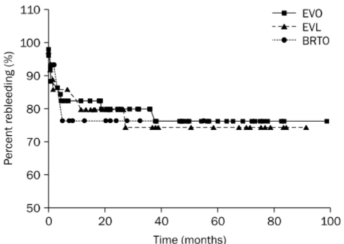
Results: There were no significant differences in baseline characteristics among the three groups. The overall follow-up period averaged 65.13 months. During follow-up, rebleeding occurred in 17 patients (11 EVO, 5 EVL, and 1 BRTO). The times-to-rebleeding were 63.59, 75.79, and 51.41 months for EVO, EVL, and BRTO, respectively, and did not differ significantly (p=0.515). The median survival times were 77.42, 70.14, and 42.79 months, respectively, and also were not different significantly (p=0.978).
Conclusions: There were no significant differences in the time-to-rebleeding or survival time among EVO, EVL, and BRTO. Further prospective, large-scale studies are needed. (Korean J Gastroenterol 2011;57:302-308)
Key Words: Esophageal and gastric varices; Gastrointestinal hemorrhage; Liver cirrhosis; Treatment
Received August 24, 2010. Revised December 7, 2010. Accepted December 7, 2010.
CC This is an open access article distributed under the terms of the Creative Commons Attribution Non-Commercial License (http://creativecommons.org/licenses/
by-nc/3.0) which permits unrestricted non-commercial use, distribution, and reproduction in any medium, provided the original work is properly cited.
교신저자: 김영석, 420-767, 부천시 원미구 중동 1174, 순천향대학교 의과대학 내과학교실
Correspondence to: Young Seok Kim, Department of Internal Medicine, Soon Chun Hyang University College of Medicine, 1174, Jung-dong, Wonmi-gu, Bucheon 420-767, Korea. Tel: +82-32-621-6546, Fax: +82-32-621-5016, E-mail: [email protected]
Financial support: None. Conflict of interest: None.
서 론
위정맥류는 간경변 환자의 약 20-70%에서 발생하고,1-4 일 단 출혈이 발생하면 자연적인 지혈이나 시술 후 지혈 모두 어렵다.3 따라서 사망률은 식도정맥류에 비해 높아 약 45-55%까지 이르며, 이는 측부 순환정맥의 혈류가 풍부하여
출혈량이 많고, 식도정맥류에 비해 내시경 시술로 지혈이 더 어려우며, 재출혈률이 높기 때문이다.3,5-7 위정맥류 출혈시 초 치료로는 내시경 정맥류 결찰술(endoscopic variceal liga- tion, EVL), HistoacrylⓇ (n-butyl-2-cyanoacrylate)을 이용 한 내시경적 정맥류 폐색술(endoscopic variceal oblitera- tion, EVO), 그리고 경경정맥 간내문맥전신 단락술(transju-
gular intrahepatic portosystemic shunt, TIPS) 등 여러 치 료법이 추천되어 왔다. 위정맥류 출혈의 예방에 대해서는 비 선택적 베타차단제 또는 TIPS를 제시하고 있으나 자료가 부 족하다.8,9
1986년 Soehendra 등10이 처음으로 시술을 보고한 Histo- acrylⓇ을 이용한 내시경적 위정맥류 폐색술은 비교적 안전하 고 효과적인 위정맥류 출혈의 지혈법으로 현재 일부 지역을 제외한 전 세계에서 널리 사용되고 있다.11,12 하지만 안정성에 대한 논란이 남아있으며 재출혈률과 합병증 및 장기 효과에 관한 연구는 많지 않다. 한편 1996년 Kanagawa 등13에 의해 예방적 시술로 시도된 역행성 경정맥 위정맥류 폐색술 (balloon- occluded retrograde transvenous obliteration, BRTO)은 출혈성 위정맥류의 치료에도 사용되어 높은 지혈 률, 정맥류 소실률을 보여주고 있다.14-16
EVL은 많은 발전과 안전성이 입증되어 정맥류 출혈의 치 료에 널리 사용되어 온 방법으로 경화요법에 비해 낮은 재출 혈률, 합병증 및 치사율을 보여주나 정맥류의 재발이 더 빈발 한 것으로 보고되고 있다.17 현재 위정맥류 출혈의 치료에 있 어 주로 두 가지 치료법에 대한 연구는 단일군 분석 연구나 두 군 간의 비교연구가 주를 이루어 세 방법을 동시에 비교한 연구는 없는 실정으로, 세 방법을 동시에 비교할 경우 통계학 적 제한점을 가지게 된다. 이에 저자는 위정맥류 출혈 후 치료 에 대해 상기 세 치료법 간의 재출혈률, 생존기간을 비교하고 자 이번 연구를 시행하였다.
대상 및 방법
1. 대상
2001년 7월부터 2009년 5월까지 순천향대학교 부천병원 에 위정맥류 출혈을 주소로 내원한 환자들 가운데 보존적 치 료 후 48시간 이내에 재출혈의 예방목적으로 EVO를 시행받 은 환자 52명, EVL을 시행받은 환자 36명 그리고 BRTO를 시행받은 환자 15명을 대상으로 하였다.
2. 방법
1) 내시경적 정맥류 폐색술(EVO)
사용기기는 직시경인 GIF series (Olympus Co., Tokyo, Japan)와 23 G 주사침 그리고 1,650 mm의 주입도관을 사용 하였다. 위정맥류 내 HistoacrylⓇ의 주입방법은 HistoacrylⓇ 0.5 mL와 lipiodol (Guerbet, Aulnay-Sous-Bois, France) 0.5 mL를 혼합한 용액을 위정맥류 내로 신속하게 주입한 후 X-선 투시 하에 위정맥류 내 lipiodol의 위치를 확인하고 곧바 로 순수 lipiodol을 주입하면서 정맥류 주사침을 정맥류로부 터 제거하고 난 다음, 증류수로 정맥류 주입도관을 세척하였
다. 필요에 따라 위정맥류 내에 2-4차례 반복적으로 주입하였 다.
2) EVL
SaeedTM Multi-Band Ligator (Cook Medical, Winston- Salem, NC, USA)를 상부위장관 내시경 GIF series (Olym- pus Co., Tokyo, Japan)의 말단에 부착한 후 출혈가능성이 높은 정맥류 부위에 대해 결찰술을 시행하였으며 이후 주변의 정맥류에 대해 추가적인 결찰술을 시행하였다. 1회에 1-4개의 고무 밴드가 사용되었으며 위정맥류의 감소 또는 소실이 이루 어질 때까지 정기적으로 상부 소화관 내시경을 시행하였다.
3) 역행성 경정맥 위정맥류 폐색술
복부전산화단층촬영, 신동맥조영술을 통해 위신정맥단락 의 존재가 확인된 환자를 대상으로 시행하였다. 대퇴정맥을 천자하여 폐쇄풍선도관을 위신정맥단락 기시부에 위치시키고 좌측 신정맥조영술을 시행하였으며 조영제가 위정맥류 내에 4분 이상 저류하는 것을 관찰하였다. 투시 유도 하에 풍선을 팽창시켜 위신정맥단락을 폐쇄하고 50% glucose 20 mL를 주입한 후 조영제와 경화제를 1:1 비율로 혼합한 용액 10 mL 를 위정맥류에 가득찰 때까지 주입하였다.
3. 정의
위정맥류는 Sarin 등3의 분류에 따라 GOV1, GOV2, IGV1, IGV2로 나누었으며, 위 정맥류의 형태는 Hashizume 등18의 분류에 따라 F1, F2, F3로 나누었다. 위정맥류의 재출혈은 지 혈술을 시행한 이후에 임상적으로 토혈, 커피색의 토사물, 혈 변 또는 흑색변이 보이면서 동시에, 첫째, 내시경적으로 위정 맥류에서 피가 분출되거나 스며 나오는 것이 확인되었을 때, 둘째, 위정맥류 위로 혈전이 붙어 있는 것이 확인되거나 궤양 이 관찰될 때, 셋째, 임상적으로 의미가 있는 식도정맥류 또는 다른 부위에서의 출혈의 증거 없이 적색흔을 동반하고 있는 F2 이상의 위정맥류가 관찰될 때로 정의하였다.19,20
4. 통계
통계 분석은 윈도우용 SPSS 12.0 (SPSS Inc., Chicago, IL, USA)을 사용하였다. 각 치료방법에 따른 환자군 간의 임 상적 특징의 비교에는 연속변수에 대해서는 one-way ANOVA 또는 Kruskal-Wallis test를, 범주형 변수에 대해서 는 Chi-square test 또는 Fisher’s exact test를 사용하였다.
위정맥류의 출혈 및 사망 누적발생률은 Kaplan-Meyer sur- vival analysis를 이용하여 계산하였고, log-rank test를 이용 하여 비교하였다. 모든 통계 분석에서 p-value가 0.05 미만인 경우를 유의한 것으로 간주하였다.
Table 2. Analysis of Time to Rebleeding Number of patients
with rebleeding (%)
Mean time to
rebleeding (months) p-value EVO 11/52 (21.2) 63.59 (53.42-73.77) 0.515 EVL 5/36 (13.9) 75.79 (63.39-88.19)
BRTO 1/15 (6.7) 51.41 (44.55-58.27)
EVO, endoscopic variceal obliteration; EVL, endoscopic variceal ligation; BRTO, balloon-occluded retrograde transvenous oblitera- tion.
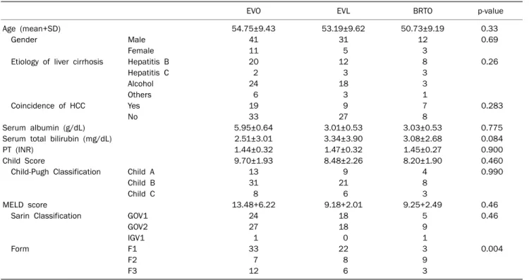
Table 1. Baseline Characteristics of Patients
EVO EVL BRTO p-value
Age (mean+SD) 54.75±9.43 53.19±9.62 50.73±9.19 0.33
Gender Male 41 31 12 0.69
Female 11 5 3
Etiology of liver cirrhosis Hepatitis B 20 12 8 0.26
Hepatitis C 2 3 3
Alcohol 24 18 3
Others 6 3 1
Coincidence of HCC Yes 19 9 7 0.283
No 33 27 8
Serum albumin (g/dL) 5.95±0.64 3.01±0.53 3.03±0.53 0.775
Serum total bilirubin (mg/dL) 2.51±3.01 3.34±3.90 3.08±2.68 0.084
PT (INR) 1.44±0.32 1.47±0.32 1.45±0.27 0.900
Child Score 9.70±1.93 8.48±2.26 8.20±1.90 0.460
Child-Pugh Classification Child A 13 9 4 0.990
Child B 31 21 8
Child C 8 6 3
MELD score 13.48+6.22 9.18+2.01 9.25+2.49 0.46
Sarin Classification GOV1 24 18 5 0.46
GOV2 27 18 9
IGV1 1 0 1
Form F1 33 22 3 0.004
F2 7 8 9
F3 12 6 3
EVO, Endoscopic variceal obliteration; EVL, endoscopic variceal ligation; BRTO, balloon-occluded retrograde transvenous obliteration; MELD, model for end stage liver disease; HCC, hepatocellular carcinoma.
결 과
1. 연령 및 성별분포
대상이 되는 총 103명 환자 중 EVO군의 평균 연령은 54.75±9.43세이었으며 남녀 각각 41명과 11명이었고, EVL 군의 평균 연령은 53.19±9.62세이었으며 남녀 각각 31명과 5명, 그리고 BRTO군은 평균 연령이 50.73±9.19세이었으며 남녀 각각 12명과 3명으로 세 집단 간에서 통계적으로 유의한 차이는 없었다(Table 1).
2. 간경변증의 원인 및 내시경 소견
EVO군의 기저 간질환으로는 만성 B형 간염 20명, 만성 C 형 간염 2명, 알코올성 간경변 24명, 원인미상 6명이었으며, EVL군은 각각 12명, 3명, 18명, 3명이었고 BRTO군은 각각 8명, 3명, 3명, 1명이었다. 통계적으로 유의한 차이는 없었다.
(p=0.243) Child-Pugh 분류상 EVO군에서 Child A는 13명, Child B는 31명, Child C는 8명이었고, EVL군에서는 각각 9명, 21명, 6명, 그리고 BRTO군에서는 각각 4명, 8명, 3명이 었다. 통계적으로 유의한 차이는 없었다(p=0.983). Sarin 분 류상 EVO군에서 GOV1는 24명, GOV2는 27명, IGV1 1명이 었고, EVL군에서는 GOV1가 18명, GOV2가 18명이었으며, BRTO군에서는 GOV1 5명, GOV2 9명, IGV1 1명이었다. 통
계적으로 유의한 차이는 없었다(p=0.437).
EVO군에서는 F1, F2, F3 각각 33명, 7명, 12명이었으며 EVL군에서는 각각 22명, 8명, 6명이었고 BRTO군에서는 각 각 3명, 9명, 3명으로 BRTO군에서 통계적으로 유의하게 정 맥류의 크기가 큰 결과를 보여주었다(p=0.004) (Table 1).
3. 위정맥류의 재출혈 및 생존기간
평균 추적기간 72.99개월간 재출혈이 발생한 환자의 수는 EVO군에서 52명 중 11명(21.2%), EVL군에서 36명 중 5명 (13.9%) 그리고 BRTO군에서 15명 중 1명(6.7%)이었다. EVO 군은 재출혈까지 평균 63.59개월(95% CI: 53.42-73.77), EVL 군은 평균 75.79개월(95% CI: 63.39-88.19), 그리고 BRTO군 은 평균 51.41개월(95% CI: 44.55-58.27)이었으며 통계적으
Table 3. Analysis of Patients’ Survival Number of
expired patients (%)
Mean survival time
(months) p-value EVO 11/52 (21.2) 77.42 (66.30-88.53) 0.978 EVL 8/36 (22.2) 70.14 (57.19-83.09)
BRTO 3/5 (20) 42.79 (30.57-55.00)
EVO, endoscopic variceal obliteration; EVL, endoscopic variceal ligation; BRTO, balloon-occluded retrograde transvenous oblitera- tion.
Fig. 2. Kaplan-Meier plots of time to survival.
EVO, endoscopic variceal obliteration; EVL, endoscopic variceal ligation; BRTO, balloon-occluded retrograde transvenous oblitera- tion.
Fig. 1. Kaplan-Meier plots of time to rebleeding.
EVO, endoscopic variceal obliteration; EVL, endoscopic variceal ligation; BRTO, balloon-occluded retrograde transvenous oblitera- tion.
로 유의한 차이는 없었다(p=0.515) (Table 2, Fig. 1).
평균 추적기간 76.49개월(95% CI: 68.29-84.69)동안 EVO 군에서는 52명 중 11명(21.2%), EVL군에서는 36명 중 8명 (22.2%), BRTO군에서는 5명 중 3명(20%)이 사망하였다.
EVO군의 생존기간은 평균 77.42개월(95% CI: 66.30-88.53 개월), EVL군은 평균 70.14개월(95% CI: 57.19-83.09개월), 그리고 BRTO군은 평균 42.79개월(95% CI: 30.57-55.00개월) 이었으며 통계적으로 유의한 차이는 없었다(p=0.978) (Table 3, Fig. 2).
고 찰
대상 간경변 환자의 약 40%에서 식도위정맥류를 가지고 있고 정맥류 발생은 매년 6% 정도 증가하며, 위정맥류 발생 빈도는 보고자마다 다양하나 간경변 환자의 20-70%에서 발 생하는 것으로 알려져왔다.1-3,18 위정맥류의 출혈은 식도정맥 류의 출혈보다는 빈도가 적으나 14-36%의 정맥류 출혈은 위 정맥류의 출혈에 의해 발생한다.21 위정맥류는 위의 전 부위에 서 발생 가능하나 60-75%가 위분문부, 나머지 25-40%가 위 기저부로 대부분 위분문부와 위기저부에서 발견된다. 위정맥
류는 식도정맥류와는 다른 혈류 공급을 받게 되는데 원칙적으 로 위정맥류는 어떤 위정맥에 의해서도 발생할 수 있으나 대 부분의 위정맥류는 좌측 위정맥과 후위정맥의 혈액공급을 받 게 된다. 좌측 위정맥에 의한 위정맥류는 주로 위분문부에 발 생하며 후위정맥 및 단위정맥에 의한 위정맥류는 주로 위기저 부에 발생하게 된다.22 대부분의 위기저부에 발생한 위정맥류 의 경우는 하위 횡경정맥으로 유출되어 좌측 신정맥과 만나 위신정맥단락(80-85%)을 형성하거나 횡경막 직하부에서 하 대정맥과 만나 위대정맥단락(10-15%)을 형성한다.23,24 위정맥 류는 또한 식도정맥류와 달리 혈류량이 많고 측부혈관이 발달 하였으며 위내측뿐만 아니라 외측에도 정맥류가 발달해 있어 치료가 힘들며 정립된 치료지침이 없는 현실이다.
위정맥류의 출혈에 대해서는 크게 내과적 치료와 외과적 치료가 있으며 내과적 치료로는 vasopressin이나 terlipre- ssin 등과 같은 혈관에 작용하는 약물이나 초기출혈을 방지하 는 비선택적 베타수용체 차단제 등의 약물치료, 내시경적 치 료, Sengstaken-Blackmore tube를 이용한 기계적인 지혈법 등이 있으며 외과적 수술 방법과 난치성 정맥류출혈의 치료에 TIPS를 시도해 볼 수 있다. 위정맥류 내시경 치료술의 적응증 에 대한 일반적인 견해는 활동 위정맥류 출혈 환자는 응급 내시경 지혈술이 요구되며, 지혈술 후 재출혈의 빈도가 높으 므로 반복 대기요법으로 정맥류 근절을 시도하고, 활동 출혈 은 없으나 위정맥류 출혈의 확실한 증거가 있는 급성 출혈 환자는 응급 및 대기 내시경 지혈술의 적응이 되며, 내시경 치료 후 잔존 및 재발 위정맥류, 정맥류 출혈의 병력이 있으면 서 적색징후나 점막미란이 관찰되는 경우 F2, F3의 위정맥류 와 단기간에 커지는 위정맥류는 예방 치료술의 적응이 될 수 있다.
HistoacrylⓇ은 혈액이나 조직수분 같은 생리적인 매개체
와 접촉했을 때 바로 중합체를 이루면서 고형화되어 정맥류를 폐쇄시키기 때문에 활동성 출혈을 멈추게 할 뿐만 아니라 정 맥류의 근절을 유도하여 재출혈을 줄이는 효과가 있다.25 이러 한 HistoacrylⓇ주입법을 다른 약제를 이용한 경화요법, 밴드 결찰술과 비교하는 많은 연구들에서 16개월 동안 재출혈은 16.7%를 보이는 등 재출혈률, 생존율에서 모두 나은 결과를
보였다.26-28 Ethanolamine oleate 등과 같은 다른 경화제는
위정맥류 내의 혈류가 빨라서 혈전을 형성하는 것이 어려웠으 나, HistoacrylⓇ을 이용한 폐색술은 혈류와 무관하게 혈전을 빠르게 형성할 수 있는 이점이 있어 다른 경화제에 비해서 높은 지혈률이 보고되고 있고, 반복적인 시술을 통해 정맥류 의 완전소실과 재출혈의 예방에도 우수한 효과가 있는 것으로 알려지고 있다.10 그러나 지혈 실패나 전신 색전증과 같은 심 각한 합병증과 내시경 기구 손상 등의 문제점29,30이 있으며 이차예방의 효과에 대해서는 논란의 여지가 있다.31,32
EVL은 정맥류를 흡인한 후에 O형 고무밴드를 이용하여 정 맥류를 기계적으로 결찰하는 방법이며 결찰된 조직은 고무밴 드에 의한 압박으로 괴사되어 궤양을 형성한 후에 섬유화를 유발하여 반흔이 만들어지는 원리를 이용하는 것으로 경화요 법에 비해 재출혈의 상대위험도의 37% 감소와 절대 위험도의 13% 감소를 보여줬다.33 또한 비선택적 베타차단제와의 병용 은 EVL 단독치료보다 우월하다는 보고도 있다.34 그러나 최근 에 HistoacrylⓇ을 이용한 폐색술이 더 효과적이라는 보고가 있어 추가적인 연구가 필요할 것으로 보인다.27
위신단락을 동반하고 간문맥압차가 높지 않은 위정맥류 출 혈 환자에서 위신단락을 통해 경화제를 주입하여 정맥류를 폐 쇄시키는 BRTO는 1996년 Kanagawa 등13에 의해 출혈이 없 는 환자에 대한 예방적 시술로 시도되어 75%의 성공률을 보 였으며, 이후 출혈성 위정맥류의 치료에도 사용되어 높은 지 혈률, 정맥류 소실률을 보였다.14,16,35 또 단락을 인위적으로 소실시키기 때문에 간문맥압이 상승하며 이로 인해 기존의 식 도정맥류가 악화하거나 새로운 식도정맥류를 생성할 수 있
다.14,16,35 그러나 BRTO는 TIPS나 EVO에 비해 재발이 드물
고 거의 대부분의 위정맥류를 소실시킬 수 있다는 장점을 가 지고 있어 위정맥류 출혈의 이차예방에 가장 적절한 치료법이 라 할 수 있다.14,16,35 BRTO는 TIPS와의 비교연구15에서 재출 혈률에서는 차이가 없었으며, 합병증이나 위정맥류 소실에서 는 통계적인 차이는 없었으나 더 나은 결과를 보여주었다. 그 러나 아직은 BRTO에 대한 장기추적결과가 부족하며 다른 치 료법과의 비교연구도 부족하므로 이에 대한 더 많은 전향적 임상연구가 필요할 것이다.
일반적으로 간정맥류 출혈의 예방에 대한 연구는 두 가지 방법을 비교하는 것이 대다수이며 BRTO에 대한 연구는 주로 TIPS와 비교되어 있고 내시경적 출혈예방법과의 비교는 더욱
적은 형편이다. 이번 연구에서는 위정맥류 출혈의 이차예방에 대한 세가지 치료법에 대해 재출혈률 및 생존율을 동시에 비 교하였다. 이는 통계상의 제1종 오류를 줄이기 위한 것으로 세 치료법을 비교하기 위해 t-test를 3회 시행할 경우 95%의 신뢰도이므로 알파값을 0.05로 설정한 의학연구에서는 0.143 으로 알파값이 증가하며, 귀무가설일 확률이 95%에서 85.7%
로 감소하여 귀무가설을 받아들일 확률이 줄어들게 된다. 이 에 세 변수를 동시에 비교하는 one-way ANOVA나 Kruskal- Wallis 검정을 시행한 이번 연구 필요성이 나타나게 되었다.
이번 연구에서 세 치료법 간의 재출혈까지의 기간 및 생존 기간을 동시에 비교해 보았으나 통계적으로 의미 있는 차이는 나타나지 않았다. 또한 두 치료법씩 다시 짝지어 사후분석을 해보아도 치료법 간에 차이는 보이지 않았다. 과거 EVO가 EVL에 비하여 더 우월하다는 여러 연구의 결과와 상반된 것 으로 이는 이번 연구가 전향적 무작위 연구가 아닌 후향적 분석이며 대상환자수가 적고, 기저 특성면에서 차이가 있기 때문일 수 있으며 세 군을 비교하는 경우는 두 군 간의 비교에 비해 통계학적인 차이가 나타날 가능성이 낮은 것도 이유로 생각된다. 그리고 EVO군과 EVL 간에 위정맥류의 크기에 대 해 통계적으로 의미 있는 차이는 보이지 않았으나 GOV1 및 F1의 정맥류가 더 많이 포함된 경향을 보였다. 이는 정맥류의 크기가 큰 경우 밴드결찰로 정맥류를 완전히 결찰하기가 어려 운 경우가 많아 EVO군에서 고위험군이 더 포함되었을 가능 성이 있다. BRTO군에는 위정맥류의 크기가 F2 또는 F3인 환 자 빈도가 다른 두 치료법에 비해 많아 세 치료법 중 고위험군 의 빈도가 가장 높음을 보여주며 이는 정맥류의 크기가 크거 나 환자와의 협조가 어려워 EVO를 시행하기 어렵거나 불가 능한 경우에 BRTO가 많이 시행되었을 가능성을 보여준다. 이 러한 선택편견의 존재로 인해 이번 연구에서 BRTO 환자군의 재출혈까지의 기간과 생존기간이 짧게 측정되었을 수 있다.
EVO군과 EVL군은 추적관찰 중 환자가 소실되는 좌측 중 도절단의 빈도가 높았으나 BRTO군에서는 사건이 발생한 시 간이 중도절단 시기보다 긴 우측 중도절단의 빈도가 높았다.
이 또한 BRTO의 효과를 과소평가하게 하는 원인의 하나로 생각할 수 있다. 이의 극복을 위해 지속적인 추적관찰을 통한 장기연구가 필요할 것으로 보인다. 세 치료법 간의 재출혈의 빈도는 BRTO가 EVO나 EVL에 비해 낮게 나타났으나 재출혈 까지의 기간은 다른 치료법에 비해 짧은 것으로 나타났다. 이 또한 BRTO군에서는 다른 치료법에 비해 우측 중도절단된 환 자가 많았기 때문으로 생각된다.
재출혈에 영향을 주는 다른 인자로는 Sarin 분류법에 따른 GOV2가 재출혈이 높은 경향이 있었으나 통계적으로 의미는 없었다. 이는 GOV2가 GOV1에 비해 정맥류가 넓게 위치하여 폐쇄가 쉽지 않았을 가능성과 GOV1의 다수가 식도정맥류의
치료에 의해 자연적으로 소실되기 때문에 재출혈률이 높은 경 향을 보였을 가능성이 있다. 그 외 위정맥류의 형태나 원인, 간기능 등은 재출혈률과 관련은 보이지 않았다.
결론으로, 위정맥류출혈의 치료 및 재출혈 예방에 있어 세 치료법 간에 의미 있는 차이는 보이지 않았으며 향후 지속적 인 추적관찰을 통한 전향적인 장기연구가 필요할 것으로 생각 된다.
요 약
목적: 위정맥류 출혈에 대한 치료는 약물적, 내시경적, 방사선 적, 그리고 수술적 치료가 제시되고 있으며 이 중에서 내시경 적 정맥류 폐색술(endoscopic variceal obliteration, EVO), 내시경적 정맥류 결찰술(endoscopic variceal ligation, EVL), 그리고 역행성 경정맥 위정맥류 폐색술(balloon- oc- cluded retrograde transvenous obliteration, BRTO) 등의 내시경 및 방사선적 치료가 우선적으로 고려되고 있다. 그러 나 아직까지 세 치료법의 간의 치료효과에 대한 명확한 비교 한 연구가 없는 실정으로 이에 저자들은 위 정맥류 출혈에 있어 EVO, EVL 그리고 BRTO 간의 치료효과를 비교하였다.
대상 및 방법: 2001년 7월부터 2009년 5월까지 순천향대학교 부천병원에 위정맥류 출혈로 내원한 환자들 가운데 치료를 시 행한 환자(EVO 52명, EVL 36명, BRTO 환자 15명)를 후향적 으로 분석하였다. 시술 전 환자의 간기능 검사 및 활력징후를 측정하였으며 Child score, model for end stage liver dis- ease (MELD) score 및 Child-Pugh Score을 기록하였다. 시 술 후 새로 발생된 토혈, 혈변, 흑색변 또는 내시경으로 확인 된 출혈이 있는 경우 재출혈로 정의하였다.
결과: 세 환자군 간에 연령, 성별, Child score, MELD score, 간암유무, 기저질환, 간기능 장애 정도, 사망원인 등에서 통계 적으로 유의한 차이는 보이지 않았다. 평균 65.13개월의 추적 기간 중 17명(13%)에서 재출혈이 발생하였으며 EVO군, EVL 군, 그리고 BRTO군에서 각각 11명(21.2%), 5명(13.9%), 1명 (6.7%)이었다. 재출혈까지의 평균기간은 각각 63.59개월, 75.79개월, 51.41개월이었으며 통계적으로 유의한 차이를 보 여주지는 않았다(p-value=0.515). 생존기간은 각 군 77.42개 월, 70.14개월, 42.79개월 이었으며 통계적으로 유의한 차이 를 보이지 않았다(p-value=0.978).
결론: 누적 지혈률 및 누적 생존율에 있어 EVO, EVL 그리고 BRTO 치료법 간에 의미 있는 통계적 효과의 차이는 보이지 않았다. 이는 이번 연구가 후향적으로 시행되어 발생한 선택 편견의 가능성과 충분한 환자군이 포함되지 못했을 가능성이 있으므로 향후 전향적인 비교 연구가 필요할 것으로 보인다.
색인단어: 식도위정맥류; 위장관출혈; 간경변; 치료
REFERENCES
1. Sarin SK, Kumar A. Gastric varices: profile, classification, and management. Am J Gastroenterol 1989;84:1244-1249.
2. Greig JD, Garden OJ, Anderson JR, Carter DC. Management of gastric variceal haemorrhage. Br J Surg 1990;77:297-299.
3. Sarin SK, Lahoti D, Saxena SP, Murthy NS, Makwana UK.
Prevalence, classification and natural history of gastric varices:
a long-term follow-up study in 568 portal hypertension patients.
Hepatology 1992;16:1343-1349.
4. Hashizume M, Sugimachi K. Classification of gastric lesions as- sociated with portal hypertension. J Gastroenterol Hepatol 1995;10:339-343.
5. Ohnishi K, Saito M, Sato S, et al. Direction of splenic venous flow assessed by pulsed Doppler flowmetry in patients with a large splenorenal shunt. Relation to spontaneous hepatic encepha- lopathy. Gastroenterology 1985;89:180-185.
6. Watanabe K, Kimura K, Matsutani S, Ohto M, Okuda K. Portal hemodynamics in patients with gastric varices. A study in 230 patients with esophageal and/or gastric varices using portal vein catheterization. Gastroenterology 1988;95:434-440.
7. Arakawa M, Masuzaki T, Okuda K. Pathomorphology of esoph- ageal and gastric varices. Semin Liver Dis 2002;22:73-82.
8. Wu CY, Yeh HZ, Chen GH. Pharmacologic efficacy in gastric vari- ceal rebleeding and survival: including multivariate analysis. J Clin Gastroenterol 2002;35:127-132.
9. Lo GH, Liang HL, Chen WC, et al. A prospective, randomized con- trolled trial of transjugular intrahepatic portosystemic shunt ver- sus cyanoacrylate injection in the prevention of gastric variceal rebleeding. Endoscopy 2007;39:679-685.
10. Soehendra N, Nam VC, Grimm H, Kempeneers I. Endoscopic ob- literation of large esophagogastric varices with bucrylate.
Endoscopy 1986;18:25-26.
11. Japanese Research Society for Portal Hypertension. The general rules for recording endoscopic findings on esophageal varices.
Jpn J Surg 1980;10:84-87.
12. Feretis C, Tabakopoulos D, Benakis P, Xenofontos M, Golematis B. Endoscopic hemostasis of esophageal and gastric variceal bleeding with Histoacryl. Endoscopy 1990;22:282-284.
13. Kanagawa H, Mima S, Kouyama H, Gotoh K, Uchida T, Okuda K.
Treatment of gastric fundal varices by balloon-occluded retro- grade transvenous obliteration. J Gastroenterol Hepatol 1996;
11:51-58.
14. Hirota S, Matsumoto S, Tomita M, Sako M, Kono M. Retrograde transvenous obliteration of gastric varices. Radiology 1999;
211:349-356.
15. Choi YH, Yoon CJ, Park JH, Chung JW, Kwon JW, Choi GM.
Balloon-occluded retrograde transvenous obliteration for gas- tric variceal bleeding: Its feasibility compared with transjugular intrahepatic portosystemic shunt. Korean J Radiol 2003;4:
109-116.
16. Baik GH, Kim DJ, Lee HG, et al. Therapeutic efficacy of bal- loon-occluded retrograde transvenous obliteration in the treat- ment of gastric varices in cirrhotic patients with gastrorenal shunt. Korean J Gastroenterol 2004;43:196-203.
17. Garcia-Pagán JC, Bosch J. Endoscopic band ligation in the treat- ment of portal hypertension. Nat Clin Pract Gastroenterol Hepatol 2005;2:526-535.
18. Hashizume M, Kitano S, Yamaga H, Koyanagi N, Sugimachi K.
Endoscopic classification of gastric varices. Gastrointest Endosc 1990;36:276-280.
19. Ramond MJ, Valla D, Mosnier JF, et al. Successful endoscopic obturation of gastric varices with butyl cyanoacrylate. Hepatolo- gy 1989;10:488-493.
20. Hou MC, Lin HC, Kuo BI, Lee FY, Schmidt CM, Lee SD. Clinical im- plications of the white nipple sign and its role in the diagnosis of esophageal variceal hemorrhage. Am J Gastroenterol 1996;
91:2103-2109.
21. Mitchell KJ, MacDougall BR, Silk DB, Williams R. A prospective reappraisal of emergency endoscopy in patients with portal hypertension. Scand J Gastroenterol 1982;17:965-968.
22. Kiyosue H, Mori H, Matsumoto S, Yamada Y, Hori Y, Okino Y.
Transcatheter obliteration of gastric varices. Part 1. Anatomic classification. Radiographics 2003;23:911-920.
23. Koito K, Namieno T, Nagakawa T, Morita K. Balloon-occluded ret- rograde transvenous obliteration for gastric varices with gastro- renal or gastrocaval collaterals. AJR Am J Roentgenol 1996;
167:1317-1320.
24. Chikamori F, Kuniyoshi N, Shibuya S, Takase Y. Correlation be- tween endoscopic and angiographic findings in patients with esophageal and isolated gastric varices. Dig Surg 2001;18:
176-181.
25. Binmoeller KF, Soehendra N. "Superglue": the answer to variceal bleeding and fundal varices? Endoscopy 1995;27:392-396.
26. Huang YH, Yeh HZ, Chen GH, et al. Endoscopic treatment of bleeding gastric varices by N-butyl-2-cyanoacrylate (Histoacryl) injection: long-term efficacy and safety. Gastrointest Endosc
2000;52:160-167.
27. Lo GH, Lai KH, Cheng JS, Chen MH, Chiang HT. A prospective, randomized trial of butyl cyanoacrylate injection versus band li- gation in the management of bleeding gastric varices.
Hepatology 2001;33:1060-1064.
28. Sarin SK, Jain AK, Jain M, Gupta R. A randomized controlled trial of cyanoacrylate versus alcohol injection in patients with iso- lated fundic varices. Am J Gastroenterol 2002;97:1010-1015.
29. Tan YM, Goh KL, Kamarulzaman A, et al. Multiple systemic emb- olisms with septicemia after gastric variceal obliteration with cyanoacrylate. Gastrointest Endosc 2002;55:276-278.
30. Roesch W, Rexroth G. Pulmonary, cerebral and coronary emboli during bucrylate injection of bleeding fundic varices. Endoscopy 1998;30:S89-S90.
31. Akahoshi T, Hashizume M, Shimabukuro R, et al. Long-term re- sults of endoscopic Histoacryl injection sclerotherapy for gastric variceal bleeding: a 10-year experience. Surgery 2002;131(1 Suppl):S176-S181.
32. Evrard S, Dumonceau JM, Delhaye M, Golstein P, Devière J, Le Moine O. Endoscopic histoacryl obliteration vs. propranolol in the prevention of esophagogastric variceal rebleeding: a randomized trial. Endoscopy 2003;35:729-735.
33. Laine L, Cook D. Endoscopic ligation compared with scle- rotherapy for treatment of esophageal variceal bleeding. A meta-analysis. Ann Intern Med 1995;123:280-287.
34. Lo GH, Lai KH, Cheng JS, et al. Endoscopic variceal ligation plus nadolol and sucralfate compared with ligation alone for the pre- vention of variceal rebleeding: a prospective, randomized trial.
Hepatology 2000;32:461-465.
35. Kim ES, Park SY, Kwon KT, et al. The clinical usefulness of bal- loon occluded retrograde transvenous obliteration in gastric variceal bleeding. Korean J Hepatol 2003;9:315-323.