ORIGINAL ARTICLE
내시경 정맥류 결찰술 실패의 위험인자 및 내시경적 구제 치료
김동현, 조은애, 전충환, 손동준, 이면재, 박창환, 조성범, 박선영, 김현수, 최성규, 류종선
전남대학교 의과대학 전남대학교병원 소화기내과
Risk Factors and On-site Rescue Treatments for Endoscopic Variceal Ligation Failure
Dong Hyun Kim*, Eunae Cho*, Chung Hwan Jun, Dong Jun Son, Myeon Jae Lee, Chang Hwan Park, Sung Bum Cho, Seon-Young Park, Hyun Soo Kim, Sung Kyu Choi and Jong Sun Rew
Division of Gastroenterology, Department of Internal Medicine, Chonnam National University Hospital, Chonnam National University Medical School, Gwangju, Korea
Background/Aims: The success rate of endoscopic variceal ligation (EVL) is about 85-94%. There is only a few studies attempting to determine the cause of EVL failure, and to date, on-site rescue treatments remains unestablished. This study aimed to elucidate the risk factors for EVL failure and the effectiveness of on-site rescue treatment.
Methods: Data of 454 patients who underwent emergency EVL at Chonnam National University Hospital were retrospectively analyzed. Enrolled patients were divided into two groups: the EVL success and EVL failure groups. EVL failures were defined as inability to ligate the varices due to poor endoscopic visual field, or failure of hemostasis after band ligation for the culprit lesion.
Results: Forty-seven patients experienced EVL failure. In the multivariate analysis, male patients, initial hypovolemic shock, active bleeding on endoscopy, and history of previous EVL were independent risk factors for EVL failure. During endoscopic procedure, we came across the common causes of EVL failure, including unsuctioned varix due to previous EVL-induced scars followed by insufficient ligation of the stigmata and inability to ligate the varix due to poor endoscopic visual field. Endoscopic variceal obturation using N-bu- tyl-2-cyanoacrylate (48.9%) was the most commonly used on-site rescue treatment method, followed by insertion of Sangstaken Blakemore tube (14.9%), and EVL retrial (12.8%). The rescue treatments successfully achieved hemostasis in 91.7% of those in the EVL failure group.
Conclusions: The risk factors of EVL failure should be considered before performing EVL, and in case of such scenario, on-site rescue treatment is needed. (Korean J Gastroenterol 2018;72:188-196)
Key Words: Esophageal and gastric varices; Treatment failure; Salvage therapy
Received September 6, 2018. Revised October 9, 2018. Accepted October 10, 2018.
CC This is an open access article distributed under the terms of the Creative Commons Attribution Non-Commercial License (http://creativecommons.org/licenses/
by-nc/4.0) which permits unrestricted non-commercial use, distribution, and reproduction in any medium, provided the original work is properly cited.
Copyright © 2018. Korean Society of Gastroenterology.
교신저자: 전충환, 61469, 광주시 동구 제봉로 42, 전남대학교 의과대학 전남대학교병원 소화기내과
Correspondence to: Chung Hwan Jun, Division of Gastroenterology, Department of Internal Medicine, Chonnam National University Hospital, Chonnam National University Medical School, 42 Jebong-ro, Dong-gu, Gwangju 61469, Korea. Tel: +82-62-220-6296, Fax: +82-62-220-8578, E-mail: [email protected], ORCID:
https://orcid.org/0000-0002-7136-8350
Financial support: This study was supported by a grant (CRI 18091-1) of Chonnam National University Hospital Biomedical Research Institute.
Conflict of interest: None.
*The first two authors equally contributed to this article.
INTRODUCTION
Acute variceal hemorrhage is considered an emergent sit- uation, with an incidence of 5-15% and a 6-week mortality rate of about 20% in patients with cirrhosis.1,2 Rebleeding is reported to occur in as many as 60% of patients, with a mortality rate of 30% within the first two years after the initial bleeding episode.3
The current approaches to manage esophageal varices or variceal hemorrhage are as follows: pharmacological therapy consisting of splanchnic vasoconstrictors; endoscopic thera- pies, such as endoscopic injection sclerotherapy (EIS) or endo- scopic variceal ligation (EVL); shunting therapy, such as trans- jugular intrahepatic portosystemic shunting (TIPS); and liver transplantation.2,4,5 Another report showed that acrylate glue injection was effective and safe for the treatment of EV bleed- ing,6 with improved outcomes compared with EIS.7
Baveno VI consensus recommended EVL as a standard en- doscopic treatment for acute esophageal variceal bleeding,8 due to its superiority compared with EIS with respect to the rebleeding rate, complications, and survival rate.9-11 However, EVL is not always successful in controlling active variceal hemorrhage. The success rate of EVL is about 85-94%,9,12-14 and Chen et al.15 have reported an EVL failure rate of 4.8%
in cases of acute EV hemorrhage.
The prognosis of patients with failed EVL is dismal; but only a few studies have addressed the risk factors for EVL failure, and currently, there is no optimal recommendation for the on-site management of EVL failure. If the risk factors for EVL failure can be identified, bleeding-related mortality can further be minimized. Therefore, this study was designed to investigate the risk factors and on-site rescue treatments for EVL failure.
SUBJECTS AND METHODS
1. Ethical considerations
The present study was conducted in accordance with the ethical guidelines of the Declaration of Helsinki. This study was approved by the Institutional Review Board of Chonnam National University Hospital (IRB No. CNUH-2018-034).
2. Patients and study protocol
This study was a retrospective case-control study. A total
of 454 patients who underwent emergency EVL to control bleeding esophageal varices at Chonnam National University Hospital between April 2004 and October 2017 were analyzed. Most patients received adequate fluid resuscitation, prophylactic antibiotics, and vasoactive drugs immediately at the time of admission. After hemostasis, most patients were given non-selective beta-blockers, and EVL sessions were re- peated until variceal obliteration – dependent on patient con- sent – according to the current guidelines. Experienced endo- scopists who performed more than 100 EVLs performed all endoscopic procedures. All endoscopic records were reviewed by three experienced endoscopists and hepatologists.
3. Endoscopic treatment and on-site rescue treatment modalities
EVL was performed using a multi-band ligator (Cook Medical Endoscopy, Limerick, Ireland) with a forward-viewing endoscope (GIF Q 260, Olympus, Tokyo, Japan). When EVL failed, on-site rescue treatments, including endoscopic variceal obturation (EVO) using n-butyl-2-cyanoacrylate (NBC), EVL retrial, insertion of a Sengstaken-Blakemore (SB) tube, TIPS, or a combination of multiple treatment modalities were performed. Combined treatment was defined as active rescue treatment with EVL retrial or EVO, followed by SB tube insertion. When EVO was performed, NBC (Histoacryl®; B. Braun Dexon, Spangenberg, Germany) was mixed with ethiodized oil (Lipiodol; Guerbert, Roissy, France), which was then injected as a bolus dose of 0.5-2 mL, depending on the amount of bleeding.
4. Definitions
EVL failures were defined in two ways: 1) the inability to ligate the varices due to poor endoscopic visual field (e.g., massive bleeding or severe belching of the patient), or 2) failure of hemostasis after band ligation for the culprit lesion. The size of EV was classified as small and straight (form 1); enlarged and tortuous (form 2); or large and coil-shaped that occupied more than one-third of the lumen (form 3).16 Bleeding-related death was defined as death within 6 weeks of the index bleed- ing episode.17 Active rescue treatment was defined as on-site EVO, EVL retrial, combination treatment, or TIPS. Non-active rescue treatment was defined as SB tube insertion or medical treatment only. Success of active rescue treatment was defined as no bleeding-related death in patients after active rescue treatments.
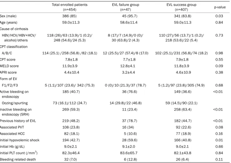
Table 1. Baseline Clinical Characteristics of the Enrolled Patients and Comparison of Characteristics between the EVL Failure and EVL Success Groups
Total enrolled patients (n=454)
EVL failure group (n=47)
EVL success group
(n=407) p-value
Sex (male) 386 (85) 45 (95.7) 341 (83.8) 0.03
Age (years) 59.0±11.3 58.6±11.4 59.0±11.3 0.84
Cause of cirrhosis HBV/HCV/HBV+HCV/
alcohol/others
118 (26)/63 (13.9)/1 (0.2)/
248 (54.6)/24 (5.3)
8 (17)/7 (14.9)/0 (0)/
30 (63.8)/2 (4.3)
110 (27)/56 (13.7)/1 (0.2)/
218 (53.6)/22 (5.4)
0.73
CPT classification
A/B/C 114 (25.1) /258 (56.8) /82 (18.1) 12 (25.5)/27 (57.4)/8 (17.0) 102 (25.1)/231 (56.8)/74 (18.2) 0.98
CPT score 7.8±1.8 7.7±1.8 7.9±1.8 0.55
MELD score 11.9±3.9 12.8±4.1 11.8±3.9 0.09
APRI score 4.4±10.4 3.2±4.4 4.6±10.9 0.38
Form of EV
F1/F2/F3 5 (1.1)/107 (23.6)/ 342 (75.3) 0 (0)/10 (21.3)/37 (78.7) 5 (1.2)/97 (23.8)/305 (74.9) 0.68 Active bleeding on
endoscopy
185 (40.7) 36 (76.6) 149 (36.6) <0.01
Oozing/spurting 73 (16.1)/112 (24.7) 14 (29.8)/22 (46.8) 59 (14.5)/90 (22.1)
Inactive bleeding on endoscopy (SRH)
269 (59.3) 11 (23.4) 258 (63.4) <0.01
Previous history of EVL 219 (48.2) 37 (78.7) 182 (44.7) <0.01
Associated PVT 108 (23.8) 16 (34) 92 (22.6) 0.08
Associated HCC 82 (18.1) 5 (10.6) 77 (18.9) 0.16
Initial hypovolemic shock 194 (42.7) 28 (59.6) 166 (40.8) 0.01
Initial Hb (g/dL) 9.0±2.1 9.1±2.0 9.0±2.1 0.66
Initial PLT count (/mm3) 82.3±46.4 83.6±65.7 82.1±43.8 0.84
Bleeding related death 32 (7.0) 6 (12.8) 26 (6.4) 0.11
Values are presented as mean±standard deviation or n (%).
EVL, esophageal variceal ligation; HBV, hepatitis B virus; HCV, hepatitis C virus; F, form; CPT, Child-Pugh-Turcotte; MELD, model for end-stage liver disease; APRI, AST to platelet ratio index; EV, esophageal varix; SRH, stigmata of recent hemorrhage; PVT, portal vein thrombosis; HCC, hepatocellular carcinoma; Hb, hemoglobin; PLT, platelet.
5. Statistical analysis
All statistical analyses were performed using SPSS ver- sion 23.0 (SPSS Inc., IBM, Chicago, IL, USA). Continuous data are shown as the mean±standard deviation, or me- dians (ranges) and categorical data as absolute and rela- tive frequencies. The continuous variables of the EVL failure and EVL success groups were analyzed using Student’s t-test. Categorical data were examined using Fisher’s exact test or x2 test with Yates’s correction. In a multivariate anal- ysis, binary logistic regression models were used to inves- tigate the risk factors associated with EVL failure. Variables with a p-value <0.05 in the univariate analysis were se- lected for possible inclusion in the multivariate analysis.
Data that were included in the regression analysis are pre- sented as the OR with 95% CI.
RESULTS
1. The baseline characteristics of the enrolled patients The cohort was comprised of 386 men (85%) and 68 women (15%). The mean age of the enrolled patients was 59.0 years (range: 27-87). EV was classified as form one in five patients (1.1%), form two in 107 (23.6%) patients, and form three in 342 (75.3%) patients. During endoscopy, ac- tive bleeding (oozing or spurting) was found in 185 patients (40.7%) and inactive bleeding (stigmata of recent hemor- rhage) in 269 patients (59.3%). Two hundred nineteen pa- tients (48.2%) had a history of EVL, 108 (23.8%) had portal vein thrombosis (PVT), and 82 (18.1%) had hepatocellular carcinoma (HCC). Initial hypovolemic shock was observed in 194 patients (42.7%). EVL failure was observed in 47 pa-
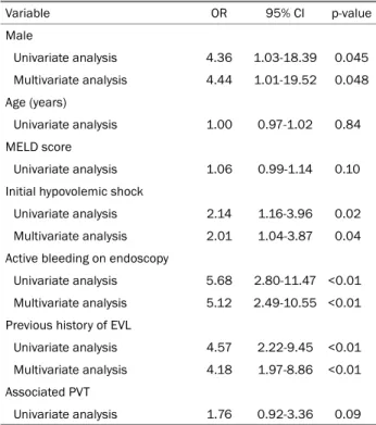
Table 2. Univariate and Multivariate Analyses of Potential Risk Factors for EVL Failure
Variable OR 95% CI p-value
Male
Univariate analysis 4.36 1.03-18.39 0.045 Multivariate analysis 4.44 1.01-19.52 0.048 Age (years)
Univariate analysis 1.00 0.97-1.02 0.84 MELD score
Univariate analysis 1.06 0.99-1.14 0.10 Initial hypovolemic shock
Univariate analysis 2.14 1.16-3.96 0.02 Multivariate analysis 2.01 1.04-3.87 0.04 Active bleeding on endoscopy
Univariate analysis 5.68 2.80-11.47 <0.01 Multivariate analysis 5.12 2.49-10.55 <0.01 Previous history of EVL
Univariate analysis 4.57 2.22-9.45 <0.01 Multivariate analysis 4.18 1.97-8.86 <0.01 Associated PVT
Univariate analysis 1.76 0.92-3.36 0.09 EVL, esophageal variceal ligation; OR, odds ratio; CI, confidence interval; MELD, model for end-stage liver disease; PVT, portal vein thrombosis.
Fig. 1. (A) Endoscopic picture of the SRH (black arrow) adjacent to the previous EVL induced ulcer scar (white arrow). (B) Insufficient ligation of the varix due to the adjacent EVL scar. (C) Insufficient ligation of the varix with SRH (black arrow) out of the EVL band. SRH, stigmata of recent hemorrhage; EVL, endoscopic variceal ligation.
tients (10.4%) and bleeding-related death was observed in 32 (7%). The baseline clinical characteristics of the enrolled patients are shown in Table 1.
2. Comparison of the characteristics between the EVL success group and EVL failure group
Of the 454 enrolled patients, 47 patients (10.4%) who expe- rienced EVL failure were classified into the “EVL failure group,”
and the other 407 patients were classified into the “EVL suc-
cess group”. Analyses of both groups are shown in Table 1.
There were a greater number of male patients (95.7% vs.
83.8%, p=0.03), patients with active bleeding on endoscopy (76.6% vs. 36.6%, p<0.01), a previous history of EVL (78.7%
vs. 44.7%, p<0.01), and initial hypovolemic shock (59.6% vs.
40.8%, p=0.01) in the EVL failure group than in the EVL suc- cess group. A high Model for End-Stage Liver Disease score was related to EVL failure without statistical significance (12.8±4.1 vs. 11.8±3.9, p=0.09). Bleeding-related death was more common in the EVL failure group than in the EVL success group, but without statistical significance (12.8% vs. 6.4%, respectively, p=0.11). The other baseline clinical character- istics were not significantly different between the two groups (Table 1).
3. Analysis of the potential risk factors for EVL failure We evaluated the potential risk factors for EVL failure. In the univariate analysis, male patients, initial hypovolemic shock, active bleeding on endoscopy, and previous history of EVL were associated with EVL failure. In the multivariate analy- sis, male (OR: 4.44, p=0.048, CI: 1.01-19.52), initial hypo- volemic shock (OR: 2.01, p=0.04, CI: 1.04-3.87), active bleed- ing on endoscopy (OR: 5.12, p<0.01, CI: 2.49-10.55), and previous history of EVL (OR: 4.18, p<0.01, CI: 1.97-8.86) were independent risk factors for EVL failure (Table 2).
4. The endoscopic findings associated with EVL failure and on-site rescue treatments
Among the 47 patients who experienced EVL failure, the most common cause of EVL failure was an unsuctioned varix due to previous EVL-induced scars (24 patients, 51.1%). In
A
A BB CC
Fig. 2. Endoscopic findings of rescue treatments for EVL failure. (A) The varix was insufficiently ligated due to previous EVL induced scar.
(B) Immediate spontaneous detachment of the EVL band and blood oozing from the varix was noted. (C) Glue was injected into the bleeding varix with immediate hemostasis. (D) In another patient, retrial of EVL achieved successful hemostasis after EVL failure (note two EVL bands over the varix, white arrows). EVL, endoscopic variceal ligation.
Fig. 3. Flow chart of the on-site rescue treatments for EVL failure. EVL, endoscopic variceal ligation; EVO, endoscopic variceal obturation;
TIPS, transjugular intrahepatic portosystemic shunt; SB tube, Sengstaken–Blakemore tube.
16 patients (34.0%), ligation was insufficient due to nearby previous EVL-induced scars, 6 patients (12.8%) had poor en- doscopic visual field, and EVL could not be performed in one patient due to hemodynamic instability (2.1%) (Fig. 1).
We performed immediate rescue therapy for patients with EVL failure. The most commonly performed rescue therapy was EVO using NBC (23 patients, 48.9%), followed by insertion of an SB tube (seven patients, 14.9%), EVL retrial (six patients,
12.8%), combination treatment (six patients, 12.8%), TIPS (one patient, 2.1%), and medical treatment (four patients, 8.5%) (Fig. 2). These immediate active rescue treatments ach- ieved successful hemostasis in 91.7% (33/36) of patients with EVL failure. The medical treatment-only group included hemodynamically stable patients with failed EVL (n=3) and one patient with a sudden change in vital signs. The success rate for rescue TIPS was 33.3% (1/3).
A
A BB CC DD
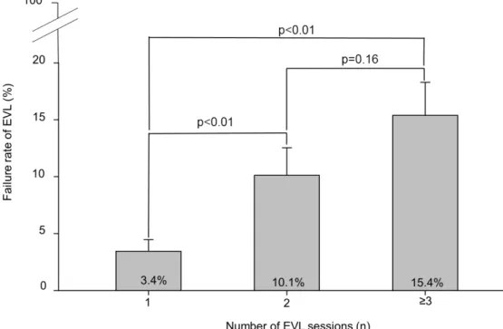
Fig. 4. Relationship between the numbers of EVL session and EVL failure. EVL, endoscopic variceal ligation.
Rebleeding from a variceal source was assessed in patients with EVL failure. The median bleeding-free survival was the longest in patients who received EVO (360 days), followed by EVL retrial (224.5 days), combination treatment (209.5 days), TIPS (209 days), and SB tube insertion (17 days).
Rebleeding occurred earliest in the medical treatment-only group (12.5 days). The median bleeding-free survival was lon- ger in the active rescue treatment group than in the non-active rescue treatment group, but without statistical significance (294.5 days vs. 17 days, respectively, p=0.2) (Fig. 3).
Complications related to rescue treatments were ulcers (21.7%), mild fever (17.4%), and bacteremia (8.7%) in the EVO group; mild fever (50.0%) and ulcers (16.7%) in the retrial EVL group; and pressure ulcers (28.6%), pneumonia (14.3%), and bacteremia (14.3%) in the SB tube insertion group.
5. The relationship between previous EVL sessions and EVL failure
The rate of EVL failure was the lowest (4.3%, 10/235) in patients without a history of previous EVL, and increased with repetitive EVL sessions (16.0%, 15/94, for the second EVL session; 17.6%, 22/125, for the third or more EVL session, p<0.01) (Fig. 4).
DISCUSSION
EVL failure is a situation that even an experienced endo- scopist can face during endoscopic procedures. However, there are limited data with respect to the risk factors of EVL failure, and no rescue treatments have been clearly evaluated. Chen et al.15 reported an EVL failure rate of 4.8%
for acute EV hemorrhage; however, they failed to discuss the cause of EVL failure and the potential rescue treatments.
Another study reported that emergent endoscopic treatment failed to achieve hemostasis in 10-20% of patients.18 Patients who did not achieve hemostasis are at an increased risk for experiencing exsanguination and death. If one endoscopic modality fails to control bleeding, it is reasonable to try a different treatment modality. However, data regarding a sec- ond on-site endoscopic treatment attempt are lacking.19
This study is unique in that the incidence rate, risk factors, and on-site rescue treatments of EVL failure were all compre- hensively evaluated. EVL failure occurred in 10.4% of patients in the present study, which is consistent with the rates that were observed in other previous studies.15,18 We found that male patients, initial hypovolemic shock, active bleeding on endoscopy, and previous EVL history increased the risk for EVL failure. However, PVT, HCC, hemoglobin level, platelet count, prothrombin time index, aspartate aminotransferase to platelet ratio index score, and Child-Pugh-Turcotte score were
not significantly associated with EVL failure.
In emergent EVL conditions, various factors, including he- modynamic instability, poor visual field due to massive blood clots, and poor cooperation by the patient may hinder EVL from being performed successfully. In our study, the most common cause of EVL failure was an unsuctioned esophageal varix due to previous EVL-induced scars. The EVL failure rate was the lowest in patients without a history of previous EVL, but increased with repetitive EVL sessions. According to these findings, we suggest that repeated EVL might increase the chance of EVL failure due to increased fibrosis of the esoph- ageal mucosa. However, further studies are needed to confirm this finding.
Regarding the on-site rescue treatments, the most common rescue therapy for patients with failed EVL was EVO. Other rescue therapies included SB tube insertion, EVL retrial, combi- nation modalities (such as EVO following insertion of an SB tube, EVL retrial following SB tube), TIPS, and medical treat- ment only. Hemostasis was achieved in 91.7% (33/36) of pa- tients with EVL failure after immediate active rescue treatment.
Active treatments using EVO, EVL, TIPS, or combination treat- ments were more effective than non-active treatments, such as insertion of an SB tube or medical treatment only. Furthermore, there was a tendency of shorter median bleeding-free survival in the non-active treatment group than in the active treatment group; however, this was without statistical significance. This finding may be explained by the small number of patients (n=47) in this study. Accordingly, on-site active rescue treat- ments should be considered in patients with EVL failure.
However, the number of failed patients was not large enough to determine which rescue treatment was the most effective, and the rescue treatment modalities that were used in the present study were not randomly assigned. Thus, the most efficacious rescue treatment in the management of EVL failure remains unknown, and large, prospective, randomized studies are warranted.
Tissue adhesives, such as NBC, have been used to manage esophageal variceal bleeding, since they promote immediate obturation of the vessel after injection of NBC.20 Cipolletta et al.20 reported that EVO using NBC initially achieved hemo- stasis at a rate of 94.2% in patients with esophageal variceal bleeding. EIS is performed by an injection of sclerosant (ethanolamine or polidocanol) into the varix. One to 2 mL of sclerosant is injected at each site, with a total of 10-15 mL
per session. EIS is reported to be effective in about 80-90%
for hemostasis in cases of EV bleeding.18 However, previous studies showed that EVO may be more efficacious and safer than EIS for treating acute esophageal variceal hemorrhage.
Moreover, EVO improved the clinical outcomes, such as in-hos- pital mortality compared to EIS.6,7 Furthermore, EIS is more time-consuming compared to EVO in emergency condition.
According to these findings, we had performed EVO for rescue treatment in EVL failure cases. In our study, EVO as an on-site rescue therapy was safe and effective for hemostasis in 21/23 patients (91.3%). To date, the efficacy and safety of EVO in patients with failed EVL have not been fully evaluated.
This study is novel in that it showed that the on-site rescue EVO effectively achieved hemostasis in patients with failed EVL.
Balloon tamponade, as a single therapy, may control the initial variceal bleeding in >80% of patients.21 However, hemo- stasis is transient, and it is associated with a high rate of complications (such as aspiration pneumonia, necrosis of the esophagus, or airway obstruction) and a mortality rate of 20%.
It is recommended that a balloon tamponade should only be used as a temporary bridge, and the treatment should not be continued for more than 24 hours.8 Recently, esophageal self-expandable metallic stents have been shown to be more effective and safer than balloon tamponade; however, this study included a small number of patients.22
A recent study showed that salvage TIPS achieved hemo- stasis in 90-100% of patients, with a rebleeding rate of 6-16%, in-hospital mortality rate of 75%, and 30 day mor- tality rate of 15%.23 Early TIPS should be considered at high risk of treatment failure according to the Baveno VI consensus. However, given that the rescue TIPS is still as- sociated with high mortality, it is related to the develop- ment of hepatic encephalopathy in one-third of patients.24 Patients with heart failure, multiple hepatic cysts, and un- controlled systemic infection are contraindicated for TIPS;
and Child-Pugh-Turcotte score >14 points, encephalopathy, PVT, and HCC are relative contraindications. However, pa- tients with contraindications for TIPS (e.g., CTP class C, PVT, and HCC) are at increased risk of recurrent EV bleeding.
Moreover, TIPS may necessitate the urgent transfer of he- modynamically unstable patients to a specialized liver cen- ter because appropriate interventional radiology expertise may not be present in every medical center. Therefore, pa-
tients who are not eligible for early TIPS may benefit from on-site rescue treatments in case of EVL failure. In the present study, the success rate of emergent rescue TIPS was 33.3%, which was lower than the rate of 84% reported in other studies.25 The reason for the low success rate might be due to the small number of patients who were in- cluded in our study and the hemodynamic instability of these patients.
The rate of bleeding-related death in the present study was 7%, which was lower than the rate of 15-20% reported in other studies.26 In the present study, there was a tendency of higher bleeding-related death in the EVL failure group (12.8%) compared with the EVL success group (6.4%). We think that active rescue treatments, through which most bleeding were successfully controlled, might have reduced bleeding-related death in the EVL failure group.
The present study has some limitations. First, this is a retro- spective study, which may lead to various biases, such as patient selection and information. Second, since this is a sin- gle-center study, the results cannot be generalized to other patient populations. Third, the number of patients with failure was not so large as to evaluate which rescue therapy is the most effective. Furthermore, the rescue treatments were not randomized and were dependent on the endoscopist’s experi- ence; therefore, there was a selection bias depending on the patient’s situation. The most effective rescue treatment in the management of patients with EVL failure remains unknown.
In conclusion, risk stratification of EVL failure should be con- ducted before performing EVL, and active on-site rescue treat- ments should be undertaken in case of EVL failure.
REFERENCES
1. Sanyal AJ, Fontana RJ, Di Bisceglie AM, et al. The prevalence and risk factors associated with esophageal varices in subjects with hepatitis C and advanced fibrosis. Gastrointest Endosc 2006;
64:855-864.
2. Garcia‐Tsao G, Sanyal AJ, Grace ND, Carey W, Practice Guidelines Committee of the American Association for the Study of Liver Diseases, Practice Parameters Committee of the American College of Gastroenterology. Prevention and management of gastroesophageal varices and variceal hemorrhage in cirrhosis.
Hepatology 2007;46:922-938.
3. Bosch J, García-Pagán JC. Prevention of variceal rebleeding.
Lancet 2003;361:952-954.
4. Saad WE, Darcy MD. Transjugular intrahepatic portosystemic shunt (TIPS) versus balloon-occluded retrograde transvenous
obliteration (BRTO) for the management of gastric varices.
Semin Intervent Radiol 2011;28:339-349.
5. Iwatsuki S, Starzl TE, Todo S, et al. Liver transplantation in the treatment of bleeding esophageal varices. Surgery 1988;104:
697-705.
6. Maluf-Filho F, Sakai P, Ishioka S, Matuguma SE. Endoscopic scle- rosis versus cyanoacrylate endoscopic injection for the first epi- sode of variceal bleeding: a prospective, controlled, and random- ized study in Child-Pugh class C patients. Endoscopy 2001;33:
421-427.
7. Sung JJ, Yeo W, Suen R, et al. Injection sclerotherapy for variceal bleeding in patients with hepatocellular carcinoma: cyanoacry- late versus sodium tetradecyl sulphate. Gastrointest Endosc 1998;47:235-239.
8. de Franchis R, Baveno VI Faculty. Expanding consensus in portal hypertension: report of the Baveno VI consensus workshop:
stratifying risk and individualizing care for portal hypertension.
J Hepatol 2015;63:743-752.
9. Stiegmann GV, Goff JS, Michaletz-Onody PA, et al. Endoscopic sclerotherapy as compared with endoscopic ligation for bleeding esophageal varices. N Engl J Med 1992;326:1527-1532.
10. Garcia-Pagán JC, Bosch J. Endoscopic band ligation in the treat- ment of portal hypertension. Nat Clin Pract Gastroenterol Hepatol 2005;2:526-535.
11. Laine L, Cook D. Endoscopic ligation compared with sclerotherapy for treatment of esophageal variceal bleeding. A meta-analysis.
Ann Intern Med 1995;123:280-287.
12. Gimson AE, Ramage JK, Panos MZ, et al. Randomised trial of vari- ceal banding ligation versus injection sclerotherapy for bleeding oesophageal varices. Lancet 1993;342:391-394.
13. Lo GH, Lai KH, Cheng JS, et al. A prospective, randomized trial of sclerotherapy versus ligation in the management of bleeding esophageal varices. Hepatology 1995;22:466-471.
14. Kim HY, Park JK, Shim JH, et al. Clinical course of esophageal vari- ces treated with endoscopic variceal ligation. Korean J Med 2005;68:498-503.
15. Chen WC, Lo GH, Tsai WL, Hsu PI, Lin CK, Lai KH. Emergency en- doscopic variceal ligation versus somatostatin for acute esoph- ageal variceal bleeding. J Chin Med Assoc 2006;69:60-67.
16. Idezuki Y. General rules for recording endoscopic findings of esophagogastric varices (1991). Japanese Society for Portal Hypertension. World J Surg 1995;19:420-422; discussion 423.
17. de Franchis R, Primignani M. Natural history of portal hyper- tension in patients with cirrhosis. Clin Liver Dis 2001;5:645-663.
18. Habib A, Sanyal AJ. Acute variceal hemorrhage. Gastrointest Endosc Clin N Am 2007;17:223-252, v.
19. Saeed ZA, Michaletz PA, Winchester CB, et al. Endoscopic variceal ligation in patients who have failed endoscopic sclerotherapy.
Gastrointest Endosc 1990;36:572-574.
20. Cipolletta L, Zambelli A, Bianco MA, et al. Acrylate glue injection for acutely bleeding oesophageal varices: a prospective cohort study. Dig Liver Dis 2009;41:729-734.
21. Avgerinos A, Armonis A. Balloon tamponade technique and effi- cacy in variceal haemorrhage. Scand J Gastroenterol Suppl 1994;207:11-16.
22. Escorsell À, Pavel O, Cárdenas A, et al. Esophageal balloon tam-
ponade versus esophageal stent in controlling acute refractory variceal bleeding: a multicenter randomized, controlled trial.
Hepatology 2016;63:1957-1967.
23. Vangeli M, Patch D, Burroughs AK. Salvage tips for uncontrolled variceal bleeding. J Hepatol 2002;37:703-704.
24. Holster IL, Tjwa ET, Moelker A, et al. Covered transjugular intra- hepatic portosystemic shunt versus endoscopic therapy + β -blocker for prevention of variceal rebleeding. Hepatology 2016;
63:581-589.
25. Qi X, He C, Guo W, et al. Transjugular intrahepatic portosystemic shunt for portal vein thrombosis with variceal bleeding in liver cir- rhosis: outcomes and predictors in a prospective cohort study.
Liver Int 2016;36:667-676.
26. D'Amico G, De Franchis R, Cooperative Study Group. Upper diges- tive bleeding in cirrhosis. Post-therapeutic outcome and prog- nostic indicators. Hepatology 2003;38:599-612.