https://doi.org/10.3350/cmh.2019.0010n Clinical and Molecular Hepatology 2020;26:83-127
Guideline
Abbreviations:
ANT, animal naming test; BCAAs, branched-chain amino acids; BRTO, balloon-occluded retrograde transvenous obliteration; cACLD, compensated advanced chronic liver disease; CFF, critical flicker frequency; CHE, covert hepatic encephalopathy; CI, confidence interval; CLDQ, Chronic Liver Disease Questionnaire; CPG, clinical practice guideline; CT, computed tomography; DST, digit span test; EEG, electroencephalography; EIS, endoscopic injection sclerotherapy; EVL, endoscopic variceal ligation; EVO, endoscopic variceal obturation; EVs, esophageal varices; GOV, gastroesophageal varices; GRADE, Grading of Recommendations, Assessment, Development, and Evaluation; HE, hepatic encephalopathy; HR, hazard ratio; HRQoL, health-related quality of life; HVPG, hepatic venous pressure gradient; ICT, inhibitory control test; IGV, isolated gastric varices; IL, interleukin; ISHEN, International Society for Hepatic Encephalopathy and Nitrogen Metabolism; ISMN, isosorbide-5-mononitrate; KASL, the Korean Association for the Study of the Liver; KPPT, Korean paper and pencil test; LDQoL, Liver Disease Quality of Life; LDSI, Liver Disease Symptom Index; LOLA, L-ornithine-L-aspartate; MCS, mental component summary; MHE, minimal hepatic encephalopathy; MRI, magnetic resonance imaging; NCT,
number connection test; NHP, Nottingham Health Profile; NSBBs, nonselective beta-blockers; OHE, overt hepatic encephalopathy; OR, odds ratio; PARTO, vascular plug-assisted retrograde transvenous obliteration; PCS, physical component summary; PEG, polyethylene glycol; PHES, psychometric hepatic encephalopathy score; PPI, proton pump inhibitor; PRBC, packed red blood cell; RCTs, randomized controlled trials; RR, relative risk; SDMT, symbol digit modality test; SF-36, Medical Outcomes Study Short Form-36; SF-LDQOL, Short Form Liver Disease Quality of Life; SIP, Sickness Impact Profile; SVR, sustained virologic response; TIPS, transjugular intrahepatic portosystemic shunt
Abbreviations (continued)
Received : Oct. 22, 2019 / Accepted : Oct. 23, 2019 Editor: Yoon Jun Kim, Seoul National University College of Medicine, Korea
PREAMBLE
Background and aims
Patients with decompensated cirrhosis with complications have a very poor prognosis and require careful management. Varices are common complications in patients with cirrhosis. Although the prognosis of variceal bleeding has improved with recent ad-vances in diagnosis and treatment, the mortality rate remains 12– 22%. Hepatic encephalopathy (HE) is known to occur in 10–14% of patients with cirrhosis and 16–21% of patients with decom-pensated cirrhosis. More than 20% of cirrhotic patients who visit emergency rooms in Korea present with HE. Therefore, cirrhosis is a serious disease in Korea and requires specific Korean guidelines
for diagnosis, treatment, and prevention. In 2005, the Korean As-sociation for the Study of the Liver (KASL) enacted a clinical prac-tice guideline (CPG) for the treatment of cirrhosis complications including ascites, hepatorenal syndrome, varices, and HE. In 2011, the guidelines for the treatment of cirrhosis were revised to inte-grate antifibrotic treatment and update the diagnosis and treat-ment advice for variceal bleeding, cirrhotic ascites, and HE. In 2017, the CPG for liver cirrhosis was revised for ascites and related complications. At this time, KASL is revising the CPG for liver cir-rhosis to address varices and HE following ascites and related complications. To date, many studies have addressed the preven-tion and treatment of gastroesophageal variceal bleeding and HE, and many guidelines have been based on those studies, but most of them contain foreign data that are difficult to apply to Korean
KASL clinical practice guidelines for liver cirrhosis:
Varices, hepatic encephalopathy, and related
complications
The Korean Association for the Study of the Liver (KASL)
Keywords: Cirrhosis; Varices; Hepatic encephalopathy; Guideline
Corresponding author : The Korean Association for the Study of the Liver (KASL) (Committee Chair: Jae Young Jang) Room A1210 MapoTrapalace, 53 Mapo-daero, Mapo-gu, Seoul 04158, Korea
Tel: +82-2-703-0051, Fax: +82-2-703-0071 E-mail: [email protected]
clinical practice. Therefore, these revised guidelines for the treat-ment of varices and HE are offered for Korean practice to reflect the latest research results and extensive discussions within the re-vision committee. This guideline contains the opinions of experts and is intended to be a practical reference for the care of patients with varices and HE; it is not an absolute standard of care. The best choices for each patient’s care vary from case to case, and the judgment of the doctor in charge is important. As medical evi-dence and new findings accumulate in the future, these guidelines will require ongoing supplementation and revision. This guideline may not be modified or altered without permission.
Target population
This guideline discusses patients with varices, HE, and related complications (esophageal varices [EVs] and bleeding, gastric vari-ces and bleeding, portal hypertensive gastropathy, covert and overt HE) caused by liver cirrhosis. It is intended for clinicians and other medical personnel who are in charge of diagnosing and treating patients with liver cirrhosis. This guideline is also intend-ed to provide practical clinical and intend-educational information and directions for resident physicians and fellows in training, practitio-ners, and their trainers and supervisors.
Development, funding, and revision process
Comprising 14 hepatologists, the Clinical Practice Guideline Committee for Liver Cirrhosis: Varices, HE, and related complica-tions (the Committee) was organized by the KASL Board of Execu-tives. Funding for the revisions was provided by KASL. Each com-mittee member collected and analyzed source data in his or her own field, and the members then wrote the manuscript together.
Literature review
The Committee selected keywords and questions using PICO (Patient/Problem, Intervention, Comparison, Outcome) assess-ments and systematically collected and reviewed international and domestic literature available in PubMed, MEDLINE, Kore-aMed, the Korean Medical Database, and other databases. In ad-dition to published articles, abstracts of important meetings pub-lished before January 2019 were evaluated.
Levels of evidence and grades of recommendation
The Grading of Recommendations, Assessment, Development, and Evaluation (GRADE) system (Table 1) was applied to grade the evidence and recommendations. The levels of evidence are based on the possibility of change in the estimate of clinical effect by further research and are described as high (A), moderate (B), or low (C). The recommendations are also classified as strong (1) or weak (2) by the GRADE system based on the quality of evi-dence, the balance between the desirable and undesirable effects of an intervention, generalizability, and socioeconomic aspects (including cost and availability). Each recommendation is labeled with the level of relevant evidence (A–C) and corresponding rec-ommendation grade (1, 2) as follows: A1, A2, B1, B2, C1, C2.
List of key questions
The Committee selected the following key questions about vari-ces, HE, and related complications to cover in this guideline. Key varix-related questions
1) How should varices be monitored? 2) Who needs monitoring for varices?
3) How can the development and progression of EVs be pre-vented?
4) Who needs treatment to prevent initial esophageal variceal bleeding?
5) What is the proper management for preventing initial esoph-ageal variceal bleeding?
6) How can acute esophageal variceal bleeding be diagnosed? 7) What is the appropriate pharmacological treatment for acute esophageal variceal bleeding?
8) What is the proper endoscopic treatment for acute esopha-geal variceal bleeding?
9) What are the options for rescue treatment when endoscopic treatment of acute variceal bleeding fails?
10) What is the primary treatment to prevent EVs from rebleed-ing?
11) What are the options for rescue treatment when primary treatment to prevent EVs from rebleeding fails?
12) Who needs treatment to prevent gastric variceal bleeding? 13) What is the proper treatment to prevent gastric variceal bleeding?
14) What is the proper treatment of acute gastric variceal bleed-ing?
15) What is the primary treatment to prevent gastric varices from rebleeding?
16) How should portal hypertensive gastropathy be classified? 17) How should portal hypertensive gastropathy be managed? Key HE-related questions
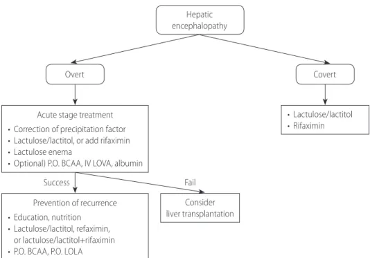
1) How should HE be diagnosed and classified? 2) How should overt HE be defined and diagnosed? 3) What are the precipitating factors of overt HE?
4) What differential diagnoses should be considered in diagnos-ing overt HE?
5) Is the measurement of serum ammonia helpful in diagnosing overt HE?
6) Is radiologic image evaluation of the central nervous system helpful in diagnosing overt HE?
7) What neurophysiological or neuropsychological tests are clin-ically necessary to diagnose overt HE?
8) How should the acute phase of overt HE be treated, and how should recurrence be prevented?
9) Are branched chain amino acids helpful in treating and pre-venting overt HE?
10) Is L-ornithine-L-aspartate (LOLA) helpful in treating and pre-venting overt HE?
11) Is proper education helpful in preventing the recurrence of and readmission for HE?
12) How should covert HE be defined and diagnosed? 13) What is the clinical significance of covert HE? 14) How should covert HE be treated?
15) How should the quality of life of HE patients be assessed?
Does treating HE improve patient quality of life?
Review of the manuscript and approval process
Each manuscript written by members was reviewed and ap-proved through meetings of the Committee. An updated manu-script was reviewed at a meeting of the advisory board and opened to a public hearing attended by KASL members, members of related organizations, and representatives from patient associa-tions. The final manuscript was approved by the KASL Board of Executives.
Release of the guidelines and plan for updates
The revised guideline (The KASL Clinical Practice Guidelines for Liver Cirrhosis: Varices, Hepatic Encephalopathy and Related Complications) was released at a KASL meeting on 22 June 2019. The Korean version of the guideline is available on the KASL web-site (http://www.kasl.org).
VARiCEs
Varices are a frequent complication of liver cirrhosis and a lead-ing cause of mortality in patients with liver cirrhosis. Varices were present in 52.2% of patients who received endoscopy for variceal screening,1 and the incidence of varices was significantly higher in patients with Child-Pugh class B/C than in those with Child-Pugh class A (35–43% vs. 48–72%).1,2 Portal hypertension, which is Table 1. Grading of Recommendations, Assessment, Development, and Evaluation (GRADE)
Criteria
Quality of evidence
High (A) Further research is very unlikely to change our confidence in the estimate of effect.
Moderate (B) Further research is likely to have an important impact on our confidence in the estimate of effect and could change the estimate.
Low (C) Further research is very likely to have an important impact on our confidence in the estimate of effect and is likely to change the estimate. Any change in estimate is uncertain.
Strength of recommendation
Strong (1) Factors influencing the strength of the recommendation include the quality of the evidence, presumed patient-important outcomes, and cost.
Weak (2) Variability in preference and values or relatively high uncertainty. Recommendation is made with less certainty or higher cost or resource consumption.
Of the quality levels of evidence, we excluded “very low quality (D),” which was originally included in the GRADE system, for convenience. (Guyatt GH, Oxman AD, Vist GE, Kunz R, Falck-Ytter Y, Alonso-Coello P, et al. GRADE: an emerging consensus on rating quality of evidence and strength of recommendations. BMJ 2008;336:924-926.)
the most common complication of liver cirrhosis, is the main de-terminant in the development of varices. Increased intrahepatic vascular resistance to portal flow leads to the development of portal hypertension, which is aggravated by splanchnic vasodila-tation and an increase in portal blood flow caused by hyperdy-namic circulation.3-5 When the portal pressure increases above a threshold, collaterals develop at the site of communication be-tween the portal and systemic circulation, of which varices are the most important. With the aggravation of portal hypertension, the collaterals grow and eventually rupture. Bleeding from varices is a major complication of portal hypertension and a leading cause of mortality in patients with liver cirrhosis. Therefore, preventing variceal development and progression, preventing bleeding from varices, appropriately managing acute bleeding from varices, and preventing variceal rebleeding are critical in patients with liver cir-rhosis.
The incidence of varices in cirrhotic patients without varices at baseline is 5–9% at 1 year and 14–17% at 2 years.6,7 The main risk factor for variceal development in these patients is a higher hepatic venous pressure gradient (HVPG).6 Small EVs often prog-ress to large varices; the incidence of progprog-ression from small to large EVs is 12% at 1 year and 25% at 2 years. The independent risk factors of EV progression are alcoholic cirrhosis, decompen-sated disease, and splenomegaly.7 The 1-year incidence of variceal bleeding in patients with cirrhosis and varices without a previous history of bleeding is approximately 12% (5% for small varices and 15% for large varices), and the main risk factors of bleeding are larger varices, the presence of redness over the varices, and decompensated disease.8 Although the mortality rate has de-creased significantly during the past several decades thanks to improvements in diagnostic and therapeutic modalities,9,10 it re-mains as high as 12–22%.11-14 In addition, rebleeding is frequent, up to 60% within 1 year, without appropriate treatment to pre-vent it.15
Surveillance of varices
Endoscopic surveillance of varices
Given the high prevalence of varices and poor prognosis with variceal bleeding, monitoring varices is important in patients with liver cirrhosis. Therefore, upon first diagnosis with liver cirrhosis, endoscopy should be performed to look for varices and assess the risk of bleeding. Diagnosis of liver cirrhosis is not difficult in pa-tients with decompensated liver cirrhosis accompanied by ascites or variceal bleeding, but a liver biopsy is needed to diagnose
pa-tients with compensated cirrhosis who have no clinical symptoms or signs. However, liver biopsy is an invasive procedure with a risk of serious complications.16 Furthermore, doubt has been cast on the accuracy of liver biopsy because of the risk of sampling er-rors17,18 and intra- and interobserver variability.18,19
Liver cirrhosis can disappear with appropriate treatment of the underlying liver disease,20,21 though portal hypertension can ac-company the severe stage of fibrosis (F3).22,23 Various practice guidelines recommend surveillance for hepatocellular carcinoma in patients with liver fibrosis, even before the development of cir-rhosis.24,25 Therefore, the alternative term compensated advanced chronic liver disease (cACLD) has been proposed for patients with severe fibrosis (F3) and compensated liver cirrhosis to better re-flect that the spectrum of severe fibrosis and cirrhosis is a contin-uum in asymptomatic patients and that distinguishing between these two conditions is often clinically impossible.26 A liver stiff-ness value, measured by transient elastography, of <10 kPa can rule out cACLD, and a value between 10 and 15 kPa is suggestive of cACLD but needs further tests for confirmation. A value >15 kPa is highly suggestive of cACLD.26 Endoscopic surveillance of all patients with cACLD can cause problems, such as an increase in medical costs due to an increase in unnecessary tests. Therefore, noninvasive screening tests have been proposed for patients with EVs, especially those whose EVs have a high risk of bleeding, to reduce unnecessary endoscopic surveillance. The Baveno VI crite-ria suggest that endoscopic surveillance can be avoided in cACLD patients with a liver stiffness <20 kPa and a platelet count >150×109/L because they are at very low risk for varices that need to be treated.26 Augustin et al.27 expanded the Baveno VI criteria to say that endoscopic surveillance can be avoided in cACLD pa-tients with liver stiffness <25 kPa and a platelet count >110×109/L. However, considering that noninvasive screening for varices that need to be treated is not particularly reliable28,29 and endoscopy is more easily accessed in Korea than in Western countries, we do not deem screening by noninvasive test to be useful in Korea. Surveillance of EVs
The incidence of EV development in cirrhotic patients without varices is 5–9% at 1 year and 14–17% at 2 years.6,7 Small EVs progress to large varices at the rate of 12% after 1 year and 25% after 2 years.7 Therefore, endoscopic surveillance should be per-formed more frequently in patients with small EVs than in those without EVs. In addition, because the type of underlying liver dis-ease (e.g., alcoholic cirrhosis) and liver function (e.g., decompen-sated cirrhosis) are risk factors for the progression of EVs, they
should be taken into account when determining the surveillance interval. Endoscopic surveillance should be performed at 2–3-year intervals in patients with compensated liver cirrhosis and at 1–2-year intervals in those with decompensated liver cirrhosis.30,31
EVs can be classified as large or small according to their size, with a breakpoint at 5 mm in diameter,32 or they can be classified as F1 (linearly dilated, small and straight varices), F2 (beady vari-ces, tortuous and occupying less than one third of the esophageal lumen), or F3 (nodular varices, large and occupying more than one third of the esophageal lumen).33 However, because the F2 and F3 classifications are fairly subjective and prophylactic treat-ment is recommended both for F2 and F3, F1 is usually classified as small, and F2 and F3 are classified together as large.
[Recommendations]
1. In patients diagnosed with liver cirrhosis, screening endoscopy is recommended to determine the presence of varices and assess the risk of bleeding. (A1)
2. In endoscopy, EVs are classified as small (F1) and large (F2 or F3), and the presence of redness should be evaluated. (B1) 3. To identify the development and progression of EVs,
endoscopic surveillance should be performed at 2–3-year intervals in patients with compensated liver cirrhosis and at 1–2-year intervals in those with decompensated liver cirrhosis. The frequency of endoscopic surveillance could be modified according to the type and severity of underlying liver disease. (B1)
Preventing the formation and progression of EVs
Appropriate treatment for the underlying liver disease can im-prove liver fibrosis, which could imim-prove portal hypertension and prevent the development of complications. In patients with hepa-titis B virus-related liver cirrhosis, the cirrhosis disappeared from the liver biopsy reports of 74% after 5 years of treatment with te-nofovir disoproxil fumarate,20 and in a meta-analysis, hepatic his-tologic improvement was observed in chronic hepatitis C patients treated with pegylated interferon±ribavirin.34 In an earlier study of patients with nonalcoholic fatty liver disease, the degree of weight loss correlated with the degree of histologic improve-ment.35 Furthermore, the incidence of EVs was significantly lower in patients with a sustained virologic response (SVR) to pegylated interferon+ribavirin treatment than in those without an SVR.36-38 In a recent study, portal pressure was significantly lower in
pa-tients with an SVR to direct-acting agents than in those without an SVR in patients with hepatitis C virus-related liver cirrhosis.39
Because the development of GEVs is a direct consequence of portal hypertension, reducing the portal pressure through the use of nonselective beta-blockers (NSBBs) from the early stage of liver cirrhosis could theoretically ameliorate the formation of GEVs. However, a placebo-controlled study to determine whether NSBBs could prevent the formation of varices in 213 patients with cirrho-sis and portal hypertension without GEVs, the incidence of varices or bleeding from varices did not differ between timolol group and the placebo group (39% vs. 40%, P=0.89), and serious adverse events developed more frequently in the timolol group than the placebo group (18% vs. 6%, P=0.006).6 Therefore, the use of NS-BBs to prevent the formation of varices is not recommended.
Several studies have evaluated whether NSBBs can prevent or delay the growth of small varices, and the results conflict. One study found a significant reduction in the rate of progression to large EVs in the nadolol group compared with the placebo group in patients with cirrhosis and small EVs (7% vs. 31% at 2 years, 20% vs. 51% at 5 years; P<0.001),40 but another study showed that propranolol offered no benefit for the prevention of progres-sion to large varices (23% in the propranolol group vs. 19% in the placebo group, P=0.786), even though the reduction in portal pressure was significantly greater in the propranolol group.41 A re-cent meta-analysis suggests that NSBBs are not effective in pre-venting the progression from small to large varices.42 Another study found that the incidence of progression to large varices across 24 months was significantly lower in the carvedilol group than the placebo group (20.6% vs. 38.6%, P=0.04), leading those researchers to suggest that carvedilol is a safe and effective way to delay the progression of small to large EVs in patients with cirrhosis.43
Carvedilol reduces portal pressure by means of an anti-a1-medi-ated decrease in intrahepatic resistance and splanchnic vasocon-striction. Because intrahepatic vasoconstriction is the main patho-logic mechanism in the development of portal hypertension during early-stage liver cirrhosis, it could be more effective than other medications in preventing the progression of varices in pa-tients with early-stage cirrhosis.44 However, further studies are needed to confirm the effects of carvedilol.
[Recommendations]
1. Appropriate treatment for the underlying liver disease is recommended to prevent the formation of EVs. (A1)
2. NSBBs (propranolol and nadolol) are not recommended to prevent the formation of EVs in cirrhotic patients without EVs. (A1)
3. In patients with small EVs that are not red, NSBBs (propranolol and nadolol) or carvedilol could be considered to prevent the progression of EVs. (B2)
Prevention of first variceal bleeding in patients with
EVs
In patients with liver cirrhosis and EVs, variceal bleeding occurs at a yearly rate of 5–15% of cases. Active prevention of the first variceal bleeding is indicated in patients at a high risk of bleeding, such as patients with large varices (F2, F3), decompensated cir-rhosis, or varices with red color signs on endoscopy.8,45
Prevention of first variceal bleeding in patients with small EVs
In cirrhotic patients with small EVs, the risk of bleeding is low (3% at 2 years and 8% at 4 years) and remains low in patients whose varices remain small at the follow-up endoscopy, though it increases significantly when the varices become large. An increase in Child-Pugh score during follow-up appears to be a significant predictor of enlarged varices and thus an increase in bleeding risk.46 The prevention of first bleeding in patients with small EVs depends on their risk of bleeding. Patients with small varices with red color signs on endoscopy or decompensated cirrhosis have an increased risk of bleeding and should consider using NSBBs.26,47 Prevention of first variceal bleeding in patients with large EVs
NSBBs and EVL
Meta-analyses of randomized controlled trials (RCTs) have shown that the use of NSBBs can prevent first variceal bleeding in cirrhotic patients with large EVs.48,49 A study comparing NSBBs and EVL as primary prophylaxis in patients with high-risk EVs found no significant difference between them in bleeding rates (relative risk [RR], 0.86; 95% confidence interval [CI], 0.55– 1.35).50 A meta-analysis of RCTs evaluating the efficacy of EVL and pharmacological therapy in preventing first EV bleeding in
patients with cirrhosis also found no significant difference in the rate of variceal bleeding between the two groups.51 Another me-ta-analysis found that EVL significantly reduced the rate of first variceal bleeding and severe adverse events than NSBBs in pa-tients with large EVs.52 Thus, in most studies, the efficacy of EVL in preventing first variceal bleeding was similar to that of NSBBs, and in some studies, the efficacy of EVL was superior to NSBBs. Therefore, either NSBBs or EVL is recommended for the preven-tion of first variceal bleeding in patients with large EVs. The choice of treatment should be based on clinician expertise and patient preference, characteristics, contraindications, and adverse events.26,47
Carvedilol
Carvedilol is known to be more effective in reducing portal pressure than propranolol.53-55 In a multicenter RCT comparing the efficacy of carvedilol and EVL in preventing first variceal bleeding in cirrhotic patients with large EVs, carvedilol had lower rates of first variceal bleeding (10% vs. 23%, P=0.04), but there was no significant difference in overall mortality or bleeding-related mor-tality during follow up.56 In another RCT comparing the efficacy of carvedilol and EVL for primary prophylaxis of EV bleeding, the carvedilol and EVL groups had comparable variceal bleeding rates (8.5% vs. 6.9%, P=0.61).57 In a study assessing the efficacy of carvedilol, propranolol, and EVL for the primary prevention of var-iceal bleeding in patients with large varices, no significant differ-ences among the groups were found in the risk of bleeding (15.4% vs. 10.8% vs. 10.2%, P=0.071), but the incidence of ad-verse events was the highest in the propranolol group.58 In studies comparing the efficacy of carvedilol, NSBBs, and EVL for the pri-mary prevention of EV bleeding, carvedilol was similar to NSBBs and EVL or superior to EVL. Therefore, carvedilol can also be used to prevent first variceal bleeding in patients with high-risk EVs.
Combination therapy of EVL and NSBBs
The combination of EVL and NSBBs for the primary prophylaxis of variceal bleeding could have a synergistic effect from the direct eradication of varices by EVL and the reduction of portal pressure by NSBBs. Several studies have compared the efficacy of combi-nation therapy with that of monotherapy based on that hypothe-sis. In RCTs comparing EVL plus propranolol with EVL alone for preventing first variceal bleeding in patients with high-risk EVs, the combination therapy did not show any difference from EVL alone in first bleed occurrence or mortality during follow up. How-ever, the recurrence of varices was lower in the combination
group than in the EVL alone group.59,60 No difference in the rate of first variceal bleeding was also found between EVL plus nado-lol combination therapy and nadonado-lol alone (14% vs. 13%, P=0.90).61 However, another study reported that EVL and pro-pranolol combination therapy lowered the rate of first variceal bleeding compared with propranolol alone (6% vs. 31%, P=0.03).62 Because most studies have shown that EVL and NSBBs combination therapy for the primary prophylaxis of EV bleeding do not differ in bleeding rate or mortality compared with mono-therapy, combination therapy is generally not recommended. However, some studies have reported that EVL and NSBBs combi-nation therapy reduced the rate of first variceal bleeding and vari-ceal recurrence compared with monotherapy. Therefore, combina-tion therapy can be considered in selected patients. A recent meta-analysis of RCTs showed that combination therapy with EVL and NSBBs reduced the rate of first variceal bleeding compared with placebo and isosorbide-5-mononitrate (ISMN).63
ISMN
In an RCT of cirrhotic patients with EVs, the ISMN group and propranolol group had no significant difference in bleeding rate, but the mortality rate during follow up was higher in the ISMN group (72.3% vs. 47.8% at 6 years, P=0.006).64 In a multicenter RCT comparing EVL, propranolol, and ISMN, the EVL and pro-pranolol groups did not differ significantly, but the EVL group had a significantly lower rate of first variceal bleeding than the ISMN group (7.5% vs. 33% at 2 years, P=0.03).65 A multicenter RCT compared propranolol plus placebo with propranolol plus ISMN for the prevention of EV bleeding. The rate of first variceal bleed-ing did not differ significantly between the groups (10.6% vs. 12.5% at 2 years, P>0.05).66 Therefore, ISMN alone or in combi-nation with NSBBs is not recommended for the prevention of first variceal bleeding.
Treatment practices for the prevention of first esophageal variceal bleeding
NSBBs
The advantages of NSBBs include low cost, ease of administra-tion, and not requiring follow-up endoscopies. Propranolol is started at 20–40 mg twice a day and adjusted every 2–3 days until the treatment goal (resting heart rate of 55–60 beats per minute) is achieved. The maximum dose is 320 mg daily in patients with-out ascites and 160 mg daily in patients with ascites. Nadolol is started at 20–40 mg once a day and adjusted every 2–3 days
un-til the treatment goal is achieved. The maximum dose is 160 mg daily in patients without ascites and 80 mg daily in patients with ascites. Systolic blood pressure should not decrease <90 mmHg.47
The disadvantages of NSBBs are that about 15% of patients have contraindications to therapy, and another 15% or so require dose reduction or discontinuation because of side effects.47 Con-traindications to NSBBs include sinus bradycardia, insulin-depen-dent diabetes mellitus, obstructive pulmonary disease, heart fail-ure, aortic valve disease, second- or third-degree atrioventricular heart block, and peripheral arterial insufficiency.67 Side effects of NSBBs include dizziness, fatigue, general weakness, dyspnea, headache, hypotension, bradycardia, and erectile dysfunc-tion.47,58,66,67 Discontinuing NSBBs can increase the risk of variceal bleeding and mortality. Thus, treatment with NSBBs should be continued indefinitely.68,69 In patients with contraindications or discontinuation due to severe side effects or poor compliance with NSBBs, EVL is recommended.68
In patients with end-stage liver disease, such as refractory asci-tes or spontaneous bacterial peritonitis, the administration of NS-BBs has not yet been established. In cirrhotic patients with refrac-tory ascites, the use of NSBBs can lower arterial pressure, decrease survival time,70 and increase the risk of paracentesis-in-duced circulatory dysfunction.71 In addition, among patients with cirrhosis and spontaneous bacterial peritonitis, NSBBs increase the risk of hepatorenal syndrome and acute kidney injury and re-duce survival time.72 However, other studies have reported that the use of NSBBs increased or did not affect survival time in cir-rhotic patients with refractory ascites.73,74 Another study found that treatment with low-dose propranolol (80 mg/day) increased survival time in patients with spontaneous bacterial peritonitis.75 The role of NSBBs in patients with refractory ascites or spontane-ous bacterial peritonitis thus remains uncertain, and clinicians must carefully consider the risks and benefits when deciding whether to administer them. If NSBBs are administered, thorough monitoring of blood pressure and renal function is necessary, and dose reduction or discontinuation should be considered in pa-tients who develop low blood pressure or impaired renal function. Discontinuation of NSBBs can increase the risk of EV bleeding; thus, if NSBBs are stopped, EVL should be considered.26
Carvedilol
Adjusting the dose of carvedilol is easier than adjusting the dose of NSBBs because it is not guided by heart rate. Carvedilol is started at 6.25 mg once a day (or 3.125 mg twice a day), and af-ter 3 days increased to 6.25 mg twice a day. The maximum dose
is 12.5 mg daily. Systolic blood pressure should not be decreased <90 mmHg.47
EVL
The advantages of EVL are that it can be performed in the same session as screening endoscopy, and it has few contraindications. The disadvantages of EVL are the side effects associated with se-dation and the risk of causing dysphagia, esophageal ulcerations, strictures, and bleeding. Although the incidence of side effects is higher with NSBBs, severe side effects, such as ulcer bleeding at the ligation site, are more likely to be associated with EVL.47 Some studies have reported that proton pump inhibitors (PPIs) signifi-cantly reduce the size of post-EVL ulcers or the rate of post-EVL ulcer bleeding.76-78 In cirrhotic patients, the long-term use of PPIs can increase the risk of spontaneous bacterial peritonitis and HE, so PPIs should be used with caution.79-81 Meanwhile, because EVL is a local therapy that does not act on the pathophysiology of portal hypertension, not only is it unable to prevent complications other than variceal bleeding, but it also requires follow-up endos-copies to assess variceal recurrence, even after variceal eradica-tion,47 defined as a case in which varices are not seen or become too small to be ligated. Repeat EVL can be performed at intervals of 2–8 weeks until variceal eradication is achieved. Follow-up en-doscopies should be performed 1–6 months after variceal eradi-cation and every 6–12 months thereafter.47,59,82
[Recommendations]
1. In cirrhotic patients with small EVs that have a high risk of bleeding (decompensated cirrhosis or red color signs on endoscopy), the use of a NSBBs (propranolol or nadolol) should be considered to prevent first variceal bleeding. (B1) NSBBs are adjusted every 2–3 days until the resting heart rate reaches 55–60 beats per minute.
2. In cirrhotic patients with large EVs, the use of a NSBBs (propranolol or nadolol), carvedilol, or EVL is recommended to prevent first variceal bleeding. (A1) A combination of NSBBs and EVL can also be considered. (B2)
Diagnosis and management of acute esophageal
variceal bleeding
Diagnosis of acute esophageal variceal bleeding
In patients with upper gastrointestinal bleeding, variceal bleed-ing caused by portal hypertension can be suspected if the patients
show jaundice, ascites, HE, splenomegaly, collateral circulation of the abdominal vessels, lower extremity edema, or spider angio-mas. A definite diagnosis can be established by endoscopic exam-ination. If blood clots or white nipples appear on the surface of the varices, or if blood is found in the stomach without a potential bleeding focus other than EVs, acute EV bleeding can be diag-nosed.45
General management of acute esophageal variceal bleed-ing
Acute EV bleeding is a medical emergency requiring intensive care. It is essential to protect the circulatory and respiratory status of the patient regardless of the cause of bleeding. Volume resus-citation via adequate fluid therapy and a packed red blood cell (PRBC) transfusion should be initiated to restore and maintain he-modynamic stability. A recent RCT showed that bleeding-related mortality (5% vs. 9%, P=0.02) and the incidence of serious ad-verse events (12% vs. 18%, P=0.01) were significantly decreased in the “restrictive” PRBC transfusion group (initiating PRBC trans-fusion at a hemoglobin threshold of 7 g/dL and maintaining it at 7–9 g/dL) compared with the “liberal” PRBC transfusion group.83 Improved survival in the restrictive transfusion group might be as-sociated with lower rates of hemostasis failure and serious ad-verse events. In patients with acute EV bleeding, adequate fluid therapy/PRBC transfusion should be performed while considering age, cardiovascular disease, presence or absence of ongoing bleeding, and hemodynamic status. Excessive fluid therapy/PRBC transfusion may increase the portal pressure and aggravate bleed-ing from the varices, so that should be taken into account.84 Re-garding correction of coagulopathy, clinical studies of recombi-nant factor VIIa have not shown a clear benefit, and therefore the routine use of fresh frozen plasma or recombinant factor VIIa is not recommended.85,86 Although the efficacy of platelet transfu-sion in patients with acute EV bleeding has not been proven be-cause of a lack of clinical studies, it can be considered in patients with severe thrombocytopenia.
Pharmacological treatment of acute esophageal variceal bleeding
Cirrhotic patients presenting with acute gastrointestinal bleed-ing have a high risk of developbleed-ing bacterial infections, therefore initiation of prophylactic antibiotic treatment at the time of admis-sion is necessary. Meta-analyses of RCTs have shown that the use of antibiotic prophylaxis reduces the risk of infections, recurrent bleeding, and bleeding-related death.87,88 A recent meta-analysis
demonstrated that prophylactic antibiotic treatment was associat-ed with a decrease in bleassociat-eding-relatassociat-ed mortality (RR, 0.79; 95% CI, 0.63–0.98), mortality from bacterial infections (RR, 0.43; 95% CI, 0.19–0.97), development of bacterial infections (RR, 0.35; 95% CI, 0.26–0.47), and rebleeding (RR, 0.53; 95% CI, 0.38– 0.74).88 However, another recent retrospective study questioned the usefulness of the routine antibiotic prophylaxis in cirrhotic pa-tients experiencing acute variceal bleeding because of a very low incidence of bacterial infections (2%) and mortality (0.4%) in Child-Pugh class A patients with acute variceal bleeding, even in the absence of prophylactic antibiotic treatment.89 No prospective study has evaluated the usefulness of antibiotic prophylaxis, and therefore the routine use of prophylactic antibiotics is recom-mended for all cirrhotic patients presenting with variceal bleeding, regardless of their Child-Pugh class. In a previous RCT comparing intravenous ceftriaxone (1 g every 24 hours) and oral norfloxacin (400 mg every 12 hours) for the prophylaxis of bacterial infection in cirrhotic patients with gastrointestinal bleeding, the incidence of proven or possible infections (11% vs. 33%, P=0.003), proven infections (11% vs. 26%, P=0.03), and spontaneous bacterial peritonitis or bacteremia (2% vs. 12%, P=0.03) was significantly lower in the ceftriaxone group.90 However, controversy remains about whether those results are applicable to general cirrhotic patients because that was study conducted in Spain among pa-tients with advanced cirrhosis, and most of the Gram-negative bacilli detected in the patients receiving oral norfloxacin were norfloxacin-resistant strains. Therefore, it is necessary to select appropriate antibiotics based on local antimicrobial susceptibility patterns. Generally, short-term (maximum 7 days) antibiotic pro-phylaxis with intravenous ceftriaxone (1 g every 24 hours) is rec-ommended in patients with acute variceal bleeding.
Vasoactive agents, such as vasopressin, terlipressin, somatosta-tin, and octreotide, are effective in supporting hemostasis in pa-tients with acute variceal bleeding by decreasing portal pressure. In a meta-analysis, the use of vasoactive agents in patients with acute variceal bleeding was significantly associated with a reduc-tion in 7-day mortality (RR, 0.74; 95% CI, 0.57–0.95) and an in-crease in the hemostasis rate (RR, 1.21; 95% CI, 1.13–1.30).91 In
patients with suspected variceal bleeding, vasoactive agents should be initiated as soon as possible, together with prophylactic antibiotics, before the diagnostic endoscopy. Vasopressin reduces portal pressure by inducing systemic and splanchnic vasoconstric-tion, but it is not now recommended for patients with acute vari-ceal bleeding because of the significant side effects, such as an increase in peripheral vascular resistance and reduction in cardiac output and coronary blood flow. Although terlipressin, a synthetic analogue of vasopressin, is the only drug proven to reduce bleed-ing-related mortality (RR, 0.66; 95% CI, 0.49–0.88),92 its side ef-fects, such as hyponatremia and myocardial ischemia due to coro-nary artery vasoconstriction, should be considered.93,94 A recent meta-analysis91 and a Korean multicenter RCT11 comparing three vasoactive agents (terlipressin, somatostatin, and octreotide) found no significant differences among them regarding the hemo-stasis rate and survival time. In patients with acute variceal bleed-ing, it is recommended that one of the vasoactive agents should be started as soon as possible (Table 2) and continued for 3–5 days.26,47
Endoscopic treatment of acute esophageal variceal bleed-ing
If acute variceal bleeding is suspected, endoscopy should be performed as soon as possible to confirm the hemorrhagic focus and hemostasis. Endoscopic hemostasis should be done when acute EV hemorrhage is confirmed by endoscopy. EVL is the endo-scopic treatment of choice for patients with acute bleeding from EVs. Endoscopic injection sclerotherapy (EIS) is no longer recom-mended as standard treatment for acute EV bleeding because of its higher incidence of treatment failure, bleeding-related mortali-ty, and adverse events compared with EVL.95-99 In a meta-analysis comparing EVL and EIS in patients with acute EV bleeding, bleed-ing-related mortality did not differ significantly (RR, 0.95; 95% CI, 0.77–1.17), but the risk of rebleeding was reduced (RR, 0.68; 95% CI, 0.57–0.81) and the rate of variceal eradication was in-creased (RR, 1.06; 95% CI, 1.01–1.12) in patients undergoing EVL compared with EIS.100 Most practice guidelines recommend en-doscopy within 12 hours after presentation with suspected varice-Table 2. Vasoactive agents used in the management of acute variceal bleeding
Type Initial dose Maintenance dose Side effects
Terlipressin 2 mg intravenously 1–2 mg intravenously every 4–6 hours Hyponatremia, myocardial ischemia, abdominal pain, diarrhea Somatostatin 250 μg intravenously 250 μg/hr intravenously Nausea/vomiting, abdominal pain, headache, hyperglycemia Octreotide 50 μg intravenously 50 μg/hr intravenously Nausea/vomiting, abdominal pain, headache, hyperglycemia
al bleeding, but that recommendation lacks evidence. A previous Taiwanese retrospective study reported that delayed endoscopy (>15 hours after admission) was an independent risk factor of in-hospital mortality (odds ratio [OR], 3.67; 95% CI, 1.27–10.39).101 In addition, a prospective observational study of 101 patients with acute EV bleeding showed that the 6-week rebleeding rate (18.9% vs. 38.9%, P=0.028) and mortality (27% vs. 52.8%, P=0.031) were significantly lowered in patients undergoing early endoscopy (≤12 hours) compared with those undergoing delayed endoscopy (>12 hours).102 However, because those studies were performed without randomization, several confounders that can delay the endoscopy, such as hemodynamic instability, might have influenced the results. Therefore, until the results of large RCTs are reported, endoscopy should be performed as soon as possible in patients with suspected acute EV bleeding. However, the spe-cific timing should be determined by the hemodynamic status of individual patients and the experience and medical resources of the institution.
Once endoscopy and EVL have been performed, early place-ment of a transjugular intrahepatic portosystemic shunt (TIPS) can be considered in carefully selected patients at high risk for re-bleeding. Early TIPS placement reduced the rates of treatment failure and bleeding-related mortality in an RCT103 of patients with a HVPG >20 mmHg and in an RCT104 of patients with Child-Pugh class C cirrhosis (score of 10–13) or Child-Pugh class B cirrhosis with active bleeding on endoscopy despite intravenous adminis-tration of a vasoactive agent. However, because these two trials excluded patients with Child-Pugh class A cirrhosis, Child-Pugh class B cirrhosis without active bleeding during endoscopy, Child-Pugh class C with a score of 14–15, patients >75 years, HCC be-yond the Milan criteria, or a creatinine level greater than 3 mg/dL, it should be considered that those study results apply to only a very small portion of patients with acute variceal bleeding. Nota-bly, a recent prospective observational study showed that the 1-year rebleeding risk was significantly decreased (3% vs. 49%, P<0.001), but 1-year survival did not differ between patients with and without a TIPS (66.8±9.4% vs. 74.2±7.8%, P=0.78).105 Fur-ther studies are needed to evaluate the beneficial effect of early TIPS placement.
Recently, the efficacy of applying hemostatic powder via endos-copy within 2 hours of admission was evaluated in 86 randomized patients with acute variceal bleeding.106 Cirrhotic patients with acute variceal bleeding received standard medical treatment and were randomized to receive either immediate endoscopy with he-mostatic powder application within 2 hours of admission followed
by early elective endoscopy the next day (that is, within 12–24 hours of admission) for definitive treatment (EVL for EV bleeding or endoscopic variceal obturation [EVO] for gastric variceal bleed-ing; study group) or early elective endoscopy only (control group). Improved rates of hemostasis and survival time in the study group suggested the therapeutic potential of endoscopic application of hemostatic powder, an easy procedure requiring minimal exper-tise.
Rescue treatment for patients with hemostasis failure Failure to control acute EV bleeding is defined as death or the need to change therapy (defined by one of the following criteria) within 5 days of an acute bleeding episode.107
- Fresh hematemesis of ≥100 mL of fresh blood ≥2 hours after the start of a specific pharmacological treatment or therapeu-tic endoscopy
- Development of hypovolemic shock
- 3 g drop in hemoglobin (9% drop in hematocrit) within 24 hours without transfusion
TIPS placement is considered the best rescue treatment for pa-tients with inadequate bleeding control despite combined phar-macological and endoscopic therapy.108 A prospective observa-tional study to evaluate the efficacy of TIPS in 58 patients who failed to achieve hemostasis after EIS and pharmacological treat-ment reported that the TIPS achieved control of the bleeding in 52 patients (90%), and 1-year and 3-year survival rates were 51.7% and 40.2%, respectively.108 Balloon tamponade is still used as a bridge therapy and provides hemostasis in 80–90% of pa-tients, but the rebleeding rate after deflation is as high as approx-imately 50%.109,110 Moreover, because it is associated with a high rate of serious complications, such as esophageal ulceration, esophageal rupture, and aspiration pneumonia, balloon tampon-ade should not exceed 24 hours.111 In a small RCT, a self-expand-able, esophageal covered metal stent was tested as an alternative to balloon tamponade in patients in whom pharmacological and endoscopic treatment failed to control bleeding.112 Although sur-vival in the esophageal stent group was not improved compared with the balloon tamponade group, bleeding control was higher (85% vs. 47%, P=0.037), and serious adverse events were lower (15% vs. 47%, P=0.077) in the esophageal stent group.112 This stent can be placed endoscopically without radiological guidance, and it can stay in place for up to 2 weeks. However, because only 28 patients were included in that study, further study is warrant-ed.
[Recommendations]
1. Endoscopy should be performed in patients with suspected esophageal variceal bleeding. (A1)
2. Endoscopic treatment should be performed in patients with acute esophageal variceal bleeding. (A1)
3. In patients with acute esophageal variceal bleeding, restrictive PRBC transfusion is recommended with the goal of maintaining a hemoglobin level of 7–9 g/dL. (A1)
4. Short-term antibiotic prophylaxis should be instituted in patients with acute esophageal variceal bleeding. (A1) 5. If esophageal variceal bleeding is suspected, vasoactive
agents should be initiated as soon as possible after admission. (A1)
6. Early TIPS placement can be considered in patients at high risk of rebleeding. (B2)
7. A TIPS is a possible rescue treatment for patients in whom bleeding control fails despite combined pharmacological and endoscopic therapy. (A2)
8. Balloon tamponade can be considered as a bridge therapy for patients who fail to achieve hemostasis after endoscopic treatment. (B2)
Prevention of esophageal variceal rebleeding
Definition of esophageal variceal rebleeding
EV rebleeding is defined as recurrent bleeding after an absence of bleeding for at least 5 days following recovery from acute EV bleeding.107 An average of 60% of patients with acute EV bleed-ing experience rebleedbleed-ing within 1–2 years, and the mortality rate from rebleeding is 33%. Therefore, appropriate treatment to pre-vent rebleeding is necessary.15,48
Diagnosis of esophageal variceal rebleeding
The diagnosis of EV rebleeding is the same as the diagnosis of acute EV bleeding. Clinically significant rebleeding can be sus-pected in a patient who has recurrent melena or hematemesis with 1) hospitalization or the need for a transfusion, 2) a decrease in hemoglobin of more than 3 g /dL, or 3) death within 6 weeks.107 Prevention of esophageal variceal rebleeding
NSBBs and EVL are the most common methods used to prevent EV rebleeding. NSBBs, which reduce portal pressure, have been reported to be more effective than placebo at preventing rebleed-ing in several RCTs.113-115 The combination of an NSBBs plus ISMN
could improve portal pressure reduction,116 but it could also in-crease the incidence of side effects such as headache and dizzi-ness.117 EVL is the endoscopic treatment of choice for the preven-tion of EV rebleeding. EVL should be repeated every 2–8 weeks until variceal eradication is achieved. Periodic endoscopic follow-up is needed to detect the recurrence of varices even after achievement of variceal eradication. Several systematic reviews and meta-analyses comparing EVL alone to NSBBs alone demon-strated no difference in the rebleeding rate,51,118,119 but the overall mortality rate during follow-up was significant higher with EVL alone (RR, 1.25; 95% CI, 1.01–1.55)51 or not different.119 In a long-term follow-up study, the rebleeding rate was higher (30% vs. 64%, P=0.001) but the survival time was longer (30% vs. 49%, P=0.013) in patients treated with the combination of an NSBBs plus ISMN.120
Several RCTs and meta-analyses comparing the combination of EVL plus NSBBs to EVL alone or NSBBs alone showed that the combination therapy had lower overall rebleeding and variceal re-bleeding.121-124 Therefore, the combination of EVL plus an NSBBs has been suggested as the primary treatment for preventing EV rebleeding. A recent meta-analysis demonstrated that the re-bleeding rate decreased (RR, 0.44; 95% CI, 0.28–0.69) and the mortality rate during follow-up tended to decrease with the bination of EVL plus a NSBBs (RR, 0.58; 95% CI, 0.33–1.03) com-pared with EVL alone. However, although the overall rebleeding rate tended to decrease (RR, 0.76; 95% CI, 0.58–1.00), the mor-tality rate during follow-up did not differ between the combina-tion of EVL plus NSBBs and NSBBs alone.125 These results suggest the importance of NSBBs in preventing EV bleeding.
RCTs comparing carvedilol to EVL (36.4% vs. 35.5%, P=0.857) and carvedilol to the combination of nadolol plus ISMN (51% vs. 43%, P=0.46) did not show any significant difference in rebleed-ing rate, and the side effects of carvedilol were less than those with the combination of nadolol plus ISMN (1.6% vs. 28.3%, P<0.0001).126,127 Therefore, the use of carvedilol to prevent EV re-bleeding can be considered, but no studies have compared the combination of EVL plus carvedilol with the combination of EVL plus an NSBBs, which is currently considered to be the primary treatment to prevent rebleeding. Further studies using carvedilol to prevent EV rebleeding are required.
In a meta-analysis of studies about preventing variceal rebleed-ing by usrebleed-ing NSBBs to reduce portal pressure, the risk of variceal rebleeding was significantly reduced (OR, 0.17; 95% CI, 0.09– 0.33; P=0.0001) when the HVPG was decreased to the target level (reduction in HVPG of ≥20% or to ≤12 mmHg) compared
with the non-responding group.128 A recent RCT comparing HVPG-based medical therapy with TIPS placement to reduce variceal re-bleeding showed lower incidence of rere-bleeding within 2 years (26% vs. 7%, P=0.002) in the TIPS group, but there was no sig-nificant difference in mortality during follow-up between the two groups, and the incidence of HE was lower (8% vs. 18%, P=0.05) in the HVPG-based medical therapy group.129 Considering that a TIPS is a limited treatment method, HVPG-based medical therapy is a useful way to prevent rebleeding if HVPG measurement is possible. However, because HVPG measurement is invasive, it is not widely practiced in many hospitals.
An RCT comparing TIPS placement with a combination of EVL plus an NSBBs to prevent variceal rebleeding found a lower vari-ceal rebleeding rate in the TIPS group (0% vs. 29%, P=0.001), but the incidence of HE within 1 year in that group was higher (35% vs. 14%, P=0.035). There was no difference in the follow-up mortality rate (32% vs. 26%, P=0.418) between the two groups.130 Therefore, the use of TIPS is not recommended as a pri-mary treatment for the prevention of variceal rebleeding, and it should instead be considered a rescue therapy for patients with primary treatment failure.131 In addition, liver transplantation is considered a rescue therapy for patients with recurrent variceal rebleeding because it exhibits good long-term results.132,133
[Recommendations]
1. In patients with acute esophageal variceal bleeding, treatment to prevent variceal rebleeding is recommended. (A1) 2. The combination of endoscopic variceal ligation (EVL)
plus NSBBs is recommended as the primary treatment for esophageal variceal bleeding. (A1) If the combination treatment is difficult to perform, use of a NSBBs or EVL alone is recommended. (A1)
3. If primary treatment for esophageal variceal rebleeding fails, TIPS placement should be considered as a rescue therapy. (B1) 4. Liver transplantation might be considered in patients with
recurrent variceal rebleeding. (B1)
Definition of gastric varices and prevention of
primary bleeding
Definition and classification of gastric varices
Gastric varices are enlarged submucosal veins of the stomach that cause critical upper gastrointestinal bleeding. GVs occur in approxi-mately 20% of patients with portal hypertension, and the bleeding rate in 2 years is known to be 25%.134 The incidence of gastric vari-ces is lower than that of EVs, but their rebleeding rate and mortality rate are higher because they cause severe bleeding.134-136
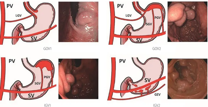
Figure 1. Classification of gastric varices. PV, portal vein; LGV, left gastric vein; SV, splenic vein; GOV, gastroesophageal varices; PGV, posterior gastric vein; SGV, short gastric vein; IGV, isolated gastric varices; GEV, gastric epiploic vein.
GOV1
IGV1
GOV2
Gastric varices are classified as gastroesophageal varices (GOV) or isolated gastric varices (IGV) depending on their location and relation to any EVs (Fig. 1). GOVs are classified by whether they extend along the lesser curvature (GOV1) or the gastric fundus (GOV2). IGV are classified as varices located in the fundus (IGV1) and those in any other region, i.e., stomach or duodenum (IGV2).134 The incidence of GOV1s is about 74%.
Prevention of primary bleeding of gastric varices
The risk factors for gastric variceal bleeding are location (IGV1>GOV2>GOV1), variceal size, redness, and severe liver dys-function.26,47,93,137-139
To prevent bleeding from GOV1s, follow the guidelines for the prevention of EV bleeding. In a Korean study of 85 patients with GOV1s, the GOV1s also disappeared when EVs were eliminated by EVL (64.7%).140 For GOV2s and IGV1s, EVO, balloon-occluded retrograde transvenous obliteration (BRTO), and vascular plug-as-sisted retrograde transvenous obliteration (PARTO) can be consid-ered to prevent bleeding.141,142 NSBBs are non-invasive and can be used because they can reduce other side effects in patients with cirrhosis.
One randomized study reported the prevention of first gastric variceal bleeding. It enrolled 89 patients with GOV2s or IGV1s larger than 10 mm.141 The effects of EVO (cyanoacrylate), an
NSBB, and simple observation were compared. For the prevention of gastric variceal bleeding, EVO (10%) was superior to an NSBB (38%) and simple observation (53%).141 The survival rate of the EVO group (93%) was higher than that of the simple observation group (73%), but it did not differ from that of the NSBB group (83%). In a meta-analysis of patients with a high risk of gastric variceal bleeding, BRTO was effective in preventing gastric varice-al bleeding (clinicvarice-al success rate, 97.3%).142 In a recent study of 73 patients, PARTO was found to be a safe procedure without seri-ous side effects that effectively prevented gastric variceal bleed-ing (Fig. 2).143,144
[Recommendations]
1. Primary prevention of bleeding for GOV1s follows the recommendations for EVs. (B1)
2. The group at high risk for bleeding (redness or severe liver dysfunction) from GOV2s or IGV1s can be treated with BRTO, PARTO, or EVO. (B2)
Management of bleeding from gastric varices
Bleeding from gastric varices is less common than from EVs; however, the risks of rebleeding or varix-related death are much
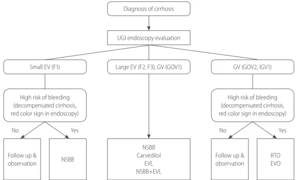
Figure 2. The prevention of initial variceal bleeding. UGI, upper gastrointestinal; EV, esophageal varix; GV, gastric varix; GOV, gastroesophageal varix; IGV, isolated gastric varix; NSBB, non-selective beta blocker; EVL, endoscopic variceal ligation; RTO, retrograde transvenous obliteration; EVO, endo-scopic variceal obturation.
Follow up & observation
High risk of bleeding (decompensated cirrhosis, red color sign in endoscopy)
Small EV (F1)
Yes
No No Yes
Large EV (F2, F3), GV (GOV1) UGI endoscopy evaluation
Diagnosis of cirrhosis
High risk of bleeding (decompensated cirrhosis, red color sign in endoscopy)
GV (GOV2, IGV1) Follow up & observation NSBB NSBB Carvedilol EVL NSBB+EVL RTO EVO
higher in patients bleeding from gastric varices. The gastric vari-ces that bleed are generally large and have high blood flow in the channel, which makes massive bleeding common in patients with large gastric varices.134,145,146 Gastric varices exhibit unique charac-teristics and have a greater variety of sizes, forms, locations, and collateral vessels than EVs. An individualized approach might be needed because few well-controlled clinical trials have tested treatments for gastric variceal bleeding. Until sufficient evidence accumulates, clinicians should seek the best option for each pa-tient based on the papa-tient’s general condition and bleeding pat-terns and the clinician’s medical resources and expertise.145 Management of bleeding from gastric varices
Endoscopic therapy
Urgent endoscopic examination, within 12 to 24 hours, is nec-essary when a patient is suspected to have active bleeding from gastric varices. Endoscopic examination can visualize the bleeding sites and directly enable proper hemostatic treatments.47,147
EVO
EVO achieves hemostasis and induces variceal eradication by an intravariceal injection of tissue adhesive agents (cyanoacrylates). Active or recent bleeding from fundic varices (GOV2s, IGV1s) or GOV1s can be managed with EVO. Special care is needed to pre-vent complications from the adhesive agents, such as ocular inju-ry, damage to endoscopic devices, or the impaction of an injection needle into a varix.148 Medical personnel are advised to wear gog-gles during the procedure. The working channel of a scope can be occluded by adhesive agent that spills during the procedure, so it can be helpful to flush the channel with olive oil in advance. To inject the sticky mixture quickly, a large needle is generally used (21 G or 22 G). The injection site is determined based on the di-rection of blood flow inside the varix. Because the intravariceal pressure is usually concentrated in the most protruding part of the varix, avoid that site if possible. The injection needle should be long enough to pass through the thick gastric wall (5 mm or lon-ger). 2-N-butyl cyanoacrylate, which is the most commonly used agent in Korea, is used as a 1:1 mixture with lipiodol to delay the polymerization reaction. About 1 mL of mixture is used in each session, and the injection can be repeated until hemostasis is achieved. The initial volume and ratio of the mixture can be ad-justed to accommodate the variceal size, intravariceal blood flow, and bleeding pattern (active or stabilized). If the bleeding is se-vere or the variceal size is large, the volume of the mixture can be
increased to 2 mL at a time. As soon as the injection is finished, 1 mL of distilled water or saline should be pushed into the catheter to ensure that the mixture remaining in the catheter is injected into the varix. Then, the needle should be retracted quickly to prevent intravariceal impaction of the needle. The success rate of EVO for hemostasis was 91–97%, and the rebleeding rate was 17–49% in patients with active gastric variceal bleeding.149-153 The common complications following EVO are systemic embolism, in-fection, fever, gastric perforation, gastric ulcer, and peritonitis.154
EVL
As with EVs, EVL is frequently performed for GOV1 bleeding. EVL for gastric varices showed an initial hemostasis rate of 80–90% and a rebleeding rate of 14–56% in patients with GOV1s.140,155-158 However, it should be noted that the depth and size of gastric varices differ from those of EVs. Ligation might not be adequate due to the thick gastric mucosa. Gastric ulcers, where the bands fall off, will expose submucosal varices directly to gastric acid and food materials. This situation could increase the risk of massive bleeding from the ulcers.154,155,158,159 In patients with fundal variceal bleeding, the effect or safety of EVL has not been fully explored. In a small randomized trial, EVL showed a significantly higher rebleeding rate than EVO in patients with IGV1 bleeding (83.3% vs. 7.7%, P=0.003).155
Radiologic intervention
Radiologic intervention is one useful hemostatic therapy for the management of bleeding from gastric varices. Sufficient consulta-tion with intervenconsulta-tional radiologists is needed in advance. Imag-ing tests, such as computed tomography (CT), should be per-formed before the procedure to confirm that the collateral veins are accessible and that no contraindications to the procedure are present.
TIPS
TIPS placement is a procedure that robustly decompresses por-tal hypertension by making a bypass between the hepatic vein and the portal vein. In small non-randomized trials, both TIPS and EVO achieved a hemostasis rate of more than 90%. Complica-tions, such as HE and stent occlusion, and medical costs were higher with the TIPS than with EVO.160,161 However, TIPS place-ment is a useful rescue therapy when initial hemostasis fails.162-164 The success rate of TIPS in controlling bleeding as a rescue thera-py is 90–100%, with a rebleeding rate of 16–40%.162-166 More-over, since non-covered stents have been replaced by covered
stents, the occlusion and stenosis rates have decreased to 8%.167,168 HE can be prevented by decreasing the stent diameter. In a randomized study, the incidence rates of HE within 2 years were 43% and 27% in patients with a conventional stent (10 mm) and those with a smaller one (8 mm), respectively (P=0.03).168 TIPS is contraindicated in patients with heart failure or severe pul-monary hypertension because it can abruptly increase preload to the heart. It is difficult to perform the procedure in patients with main portal vein thrombosis. When a cyst, abscess, or mass is blocking the accessible tract in the liver or the intrahepatic bile ducts are markedly dilated, it is difficult to perform TIPS.169
RTO
RTO obliterates gastric varices by infusing a sclerosant or em-bolic agent in a retrograde manner through a gastrorenal shunt. An accessible shunt should be confirmed by CT prior to the proce-dure. After occluding the shunt with a balloon catheter, a scle-rosant, such as ethanolamine oleate or sodium tetradecyl sulfate, is infused into the gastric varices.170,171 In a recent, large, retro-spective study, the technical success rate of BRTO was 95%.172 Another multicenter study, in which 23% of patients had GOV1s, had a technical success rate of 97%.173 However, the EVs recurred or became aggravated in 20–41% of patients after the proce-dure.172,173 A recent meta-analysis also showed favorable results. The technical success and major complication rates of BRTO were 96.4% and 2.6%, respectively. The clinical success rate, defined as no recurrence of gastric varices or complete obliteration of vari-ces on subsequent imaging, was 97.3%.142
If a shunt is too large for balloon catheter occlusion, BRTO is not possible. Moreover, BRTO requires that patients retain the balloon catheter for several hours, until the sclerosing agent has hardened in the varices. In rare cases, the balloon can rupture during the procedure, and a systemic embolism of the sclerosing agent can occur. Therefore, a novel intervention, PARTO, was re-cently developed. PARTO uses a vascular plug with or without coils instead of a balloon and uses a gelatin sponge as the embol-ic agent.143 A multicenter prospective study showed that complete thrombosis of gastric varices and shunts was achieved in 98.6% of patients. No recurrent variceal bleeding or development of HE occurred during follow-up. Moreover, 40% of patients showed improvement in their Child-Pugh scores.144 Thus, PARTO is a note-worthy treatment that can replace BRTO in patients with gastric varices and a gastrorenal shunt. However, more data on the long-term efficacy and safety of PARTO are needed.
Treatment of gastric variceal bleeding
General management of gastric variceal bleeding In patients with cirrhosis and acute upper gastrointestinal bleeding, a restrictive blood transfusion strategy (with a target range for the post-transfusion hemoglobin level of 7 to 9 g/dL) and antibiotic prophylaxis improved survival.83,174 Although pa-tients enrolled in the studies were small, the same transfusion strategy can be recommended for those with gastric variceal bleeding. The beneficial effects of vasoactive agents (terlipressin, octreotide, somatostatin) have not been fully proved in patients with gastric variceal bleeding, either. However, considering their ability to decrease portal hypertension, their use in patients with bleeding from gastric varices can be recommended.91,158,175,176
Treatment of GOV1 bleeding
GOV1s, which are an extended type of EV, develop along the lesser curvature and receive blood from the left gastric vein. When EVs are eradicated by endoscopic treatments, the gastric varices also concomitantly disappear in 60–65% of patients.134,140 Because of their close relationship in pathophysiology, the man-agement of bleeding from cardiac varices (GOV1s) is similar to that for EV bleeding.177 However, it should be noted that sufficient ligation can be difficult for gastric varices because of their large size and deeper location. Furthermore, subsequent post-ligation ulcers might be exposed to gastric acid or food material.154,155,158,159 According to small clinical trials and observational studies, EVO produces more favorable outcomes than EVL. The initial hemosta-sis rates with EVO and EVL in patients with GOV1 bleeding were 85–100% and 80–90%, respectively. The rebleeding rates fol-lowing EVO and EVL were 3–26% and 14–56%, respective-ly.140,155-158 However, most of those trials were small; the evidence needed to recommend one of these treatments over the other re-mains insufficient.140,155,157,158,178 Therefore, clinicians may choose either EVO or EVL based on their expertise, available medical re-sources, and the variceal condition (size or extent).
Treatment of GOV2 or IGV1 bleeding
GOV2s are a type of gastric varix that extends from EVs toward the fundus. IGV1s are varices localized in the fundus in the ab-sence of EVs.134 Both GOV2s and IGV1s are usually called gastric fundic varices. Unlike EVs, fundic varices are supplied with blood from the posterior gastric vein or short gastric vein.179,180 Bleeding from the fundus usually occurs in a stage of large varix. Manage-ment of fundic variceal bleeding can be difficult because massive