대한임상신경생리학회지 13(1):58~60, 2011 ISSN 1229-6414
Copyright 2011 by The Korean Society of Clinical Neurophysiology 58
Address for correspondence;
Jong Seok Bae, M.D.
Department of Neurology, Inje University College of Medicine, Busan Paik Hospital, 633-165 Gaegeum-dong,
Busanjin-gu, Busan 614-110, Korea
Tel: +82-51-890-6148 Fax: +82-51-895-6367 E-mail: [email protected]
Case Report
골수이식 이후의 다발근육염: 만성 이식편대숙주병의 드문 증상인가? 자가면역작용인가?
인제대학교 의과대학 부산백병원 신경과1, 병리과2, 인제대학교 임상연구소3
최원철
1
․정용한2
․양영일2,3
․배종석1
Polymyositis After Bone Marrow Transplantation: As an Uncommon Manifestation of Chronic Graft-Versus-Host Disease?
or Autoimmune Process?
Won-cheol Choi, M.D.
1
, Yong Han Jung, M.D.2
, Yeong Il Yang, M.D.2,3
, Jong Seok Bae, M.D.1
Departments of Neurology
1and Pathology
2, Inje University College of Medicine, Busan Paik Hospital, Institute for Clinical Research, Inje Univesity
3, Busan, Korea
Received 12 November 2010; received in revised form 15 December 2010; accepted 14 January 2011.
Chronic graft-versus-host disease (GVHD) is a well-known complication of allogeneic bone marrow transplantation (BMT) and has heterogeneous manifestations, with multi-organ involvement. Recently, polymyositis (PM) was reported to be a rare manifestation of chronic GVHD. Here, we report a 30-year-old woman who was diagnosed with PM after allogeneic BMT.
Key Words: Graft-versus-host disease, Polymyositis, Bone marrow transplantation
Graft-versus-host disease (GVHD) is a serious complication of allogeneic bone marrow transplantation (BMT). It involves an immunological reaction against antigens in the BMT recipient triggered by the immunocompetent donor graft.
1Chronic GVHD is a heterogeneous syndrome with multi-organ involvement.
2,3Thus, its major clinical manifestations are similar to those of autoimmune connective tissues diseases. Polymyositis (PM) is a very unusual manifestation of chronic GVHD; it can exist as the
sole manifestation of chronic GVHD or along with other various features of chronic GVHD.
1,4,5Case Report
A 30-year-old woman came to our department because of progressively increasing muscle weakness without myalgia. One year earlier, she noticed posterior neck pain and difficulty in maintaining neck extension. Six months earlier, she developed symmetrical proximal muscle weakness of all limbs. Subsequently, her proximal limbs showed obvious changes of muscle atrophy.
She was diagnosed with aplastic anemia 9 years earlier and
underwent allogeneic BMT 4 years ago. Two years ago, she
developed dry eyes and dry skin with a skin rash. GVHD after
BMT was diagnosed and a lower dosage of prednisolone was
골수이식 이후의 다발근육염: 만성 이식편대숙주병의 드문 증상인가? 자가면역작용인가?
Korean J Clin Neurophysiol / Volume 13 / June, 2011 59
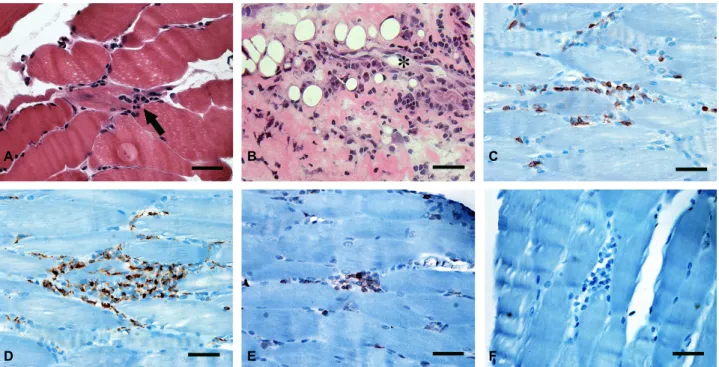
Figure 1. Microscopic images of muscle and fascia. Cryosections of (A) skeletal muscle and (B) fascia show a mononuclear cell infiltration in the interstitium and endomysium (arrow) of muscle and around a capillary (asterisk) in the fascia (hematoxylin and eosin staining). Immunohistochemical staining of skeletal muscle demonstrated that the infiltrating cells were (C) CD45RO and (D) CD3-positive T cells and (E) CD68+ monocytes, while no CD20-positive B cell was observed.
Scale bar=100 µm.
prescribed at another hospital. After she developed the muscle weakness and atrophy, the prednisolone was stopped under a working diagnosis of muscle weakness caused by steroid myopathy.
Despite discontinuing the steroid, her muscle weakness and atrophy progressed.
Her proximal muscle power was at most Medical Research Council (MRC) grade I in the upper limbs, grade II in the lower limbs, and her distal muscle power was grade V in all limbs. Her gait was a typical waddling gait. The muscle wasting was severe in all limbs. Deep tendon reflexes were absent bilaterally.
Needle electromyography detected mild spontaneous activity, consisting primarily of positive sharp waves, short amplitude, and short-duration myopathic motor unit action potentials, and early recruitment patterns. Histopathologically, a skeletal muscle biopsy showed typical features of polymyositis, such as scattered necrotic muscle fibers, regenerating muscle fibers, and interstitial and endomysial infiltration (arrow) of inflammatory cells (Figure 1A). The fascia contained an inflammatory cell infiltrate (arrow) around capillaries (asterisk), with mild deposition of collagen fibers (Figure 1B). The inflammatory infiltrates in the muscle and fascia were predominantly lymphocytes with some monocytes.
Immunohistochemical staining of muscle revealed that the majority of the cells in the endomysial infiltrate expressed T cell markers, such as CD45RO (Figure 1C) and CD3 (Figure 1D), and the monocyte/macrophage marker CD68 (Figure 1E). Conversely, no CD20
+B cell was observed in interstitium or endomysium (Figure 1F).
Initially, the serum creatine phosphokinase was 849 IU/L. A blood work-up for various connective tissue diseases was done.
Only antinuclear antibody was positive, with a nucleolar pattern.
The acetylcholine receptor antibody titer and thyroid function test was within the normal range.
We diagnosed her with PM, probably as a manifestation of chronic GVHD. She was started on a trial of high-dosage intravenous steroid and subsequently switched to maintenance high-dosage oral steroid therapy. Her muscle power improved slightly immediately after the initial intravenous steroid. During follow-up, however, her muscle weakness and wasting did not improve continuously or recover markedly. After therapy with oral steroid for about 6 months, her serum CPK had declined to 114 U/L.
A B C
D E F
최원철