∙ Received: October 15, 2012. Accepted: October 18, 2012.
∙ Corresponding author : Jo Jin U
Department of Nuclear Medicine, Inha University Hospital, 7-206 3rd ST, Jung-gu, Incheon, Korea
Tel: +82-32-890-3771, Fax: +82-32-890-3164 E-mail: [email protected]
Original Article
Brain SPECT 영상의 Attenuation Correction 방법들에 대한 비교인하대병원 핵의학과, 김천대학교 방사선학과1, 동남보건대학 방사선과2
조진우 ․ 김창호 ․ 나수경1 ․ 이귀원2Comparison of Attenuation Correction Methods for Brain SPECT Images
Jo Jin U, Kim Chang Ho, Na Soo Kyung1, Lee Gui Won2
Department of Nuclear Medicine, Inha University Hospital, Incheon, Korea
1
Department of Radiological Science, Gimcheon University, Gimcheon, Korea
2
Department of Radiological Technology, Dongnam Health College, Suwon, Korea
Purpose : The purpose of this study was to compare count between Chang's method and CT-based attenuation correction (AC-CT) among the attenuation correction (AC) methods for non-attenuation correction (AC-non) images of Brain SPECT (Single Photon Emission Computed Tomography). Materials and Methods : We injected
99m
Tc 37Mbq in a Hoffman 3D phantom filled with distilled water in the phantom study, and injected intravenously
99mTc-HMPAO 740Mbq in a normal volunteer in the patient study, and then obtained Brain SPECT images with Symbia T6 of Siemens and conducted quantitative brain analysis. Transverse images to which each method was applied were rebuilt at the same position, and 6 regions of interest (ROI) were drawn on each of Slice No. 10, 20 and 30 and then the counts of AC-non, AC-CT and Chang's method were compared.
Results : The mean counts of AC-non, AC-CT and Chang's method were 4606.8 ± 511.3, 16794.6 ± 2429.4, and 8752.6 ± 896.5, respectively, in the phantom study and 5460.8 ±519.6, 15320 ± 1171.6 and 12795 ± 1422.1, respectively, in the patient study. In the phantom study, the ratio of AC-CT to AC-non was 3.70 and the ratio of Chang's method to AC-non was 1.92, and in the patient study, they were 2.85 and 2.38, respectively. Conclusion : From this study, we found that AC-CT makes higher AC than Chang's method. In addition, when Chang's method was used, AC in the patient study was higher than that in the phantom study. These results need to be considered also in other examinations. (Korean J Nucl Med Technol 2012;16(2):120-125)
Key Words : Brain SPECT, Attenuation correction(AC), Chang's method
서 론
최근 고령화 사회에 접어 들면서 알츠하이머 병과 같은 뇌 신경계 질환이 사회 문제로 대두됨에 따라 Brain SPECT (Single Photon Emission Computed Tomography) 검사가 이 에 대응되는 검사로 필요성이 강조되고 있다. SPECT는 방사
성 의약품을 환자에게 투여한 후 즉시 또는 일정 시간이 지 난 후 감마카메라로 환자 주위를 회전 시켜 획득된 여러 방 향의 평면 영상을 단층영상으로 재구성 하여 방사성 추적자 의 깊이 분포에 대한 정보를 제공한다. 깊이에 따른 광자의 감쇠정도가 다르기 때문에 감쇠보정이 필요하고 기존의 SPECT에서는 Chang's method를 감쇠 보정에 이용하였
다.2.4) 하지만 최근 SPECT/CT가 이용되면서 CT의 X-ray 감
쇠정보를 이용한 감쇠보정이 사용되고 있고 이는 좀 더 정확 한 감쇠 보정이 가능하다고 Kazunzri I 등은 보고 하였다.4) 따라서 Brain SPECT/CT에서 CT의 역할은 perfusion image 와의 Fusion을 위한 형태학적인 영상뿐만 아니라 획득된 SPECT영상의 정확한 감쇠 정보를 제공하는데 있다고 보고
Fig. 1. Symbia T6 SPECT/CT (Siemens Medical Solution, USA).
Fig. 2. Hoffman 3D Brain Phantom (Data Spectrum Corporation,
USA).되고 있다. 그리고 Masuo H 등은 CT 영상을 이용한 감쇠보 정이 Chang's method를 이용한 감쇠보정보다 더 정확하다고 보고 하였다.3) 이에 우리는 Brain SPECT (Single Photon Emission Computed Tomography)의 Non-attenuation cor- rection (AC-non) 영상에 대한 감쇠보정 방법 중 Chang's method와 CT based attenuation correction (AC-CT) 사이의 count를 비교하기 위해 이 연구를 진행하였다.
실험재료 및 방법
1. 실험 재료
장비는 Symbia T6 SPECT/CT (Siemens Medical Solution, USA)를 사용하였으며(Fig. 1), collimator는 LEHR (Low en- ergy high resolution) collimator를 사용하여 실험 하였다.
phantom study에서는 hoffman 3D brain phantom (Data Spectrum Corporation, USA)를 사용하였다(Fig. 2).
2. Phantom Study
증류수로 채워진 hoffman 3D brain phantom에 99mTc 37 Mbq (1 mCi)을 투여하고 phantom을 scanner bed의 head holder위에 올려놓은 후 dual head 감마 카메라를 이용하여 SPECT를 시행하였다(step & shoot mode, 20sec/60view, zoom 1.45, 128×128 matrix).
3. Patient study
안정된 상태의 52세 남자 정상 지원자를 대상으로 scanner
bed위에 supine position 상태에서 눈을 감고 99mTc-HMPAO 740 Mbq를(20 mCi) 투여한 뒤 10분 뒤에 SPECT Phantom study와 같은 조건으로 SPECT를 시행 하였다.
4. CT scan
CT의 조건은 130kVp, 250 ref. mAs(CAREdose 4D), rota- tion cycle 1.5 sec, pitch 0.4로 설정 하였으며 reconstruction 조건은 kernel H31 medium smooth, window setting은 cerc-
A B
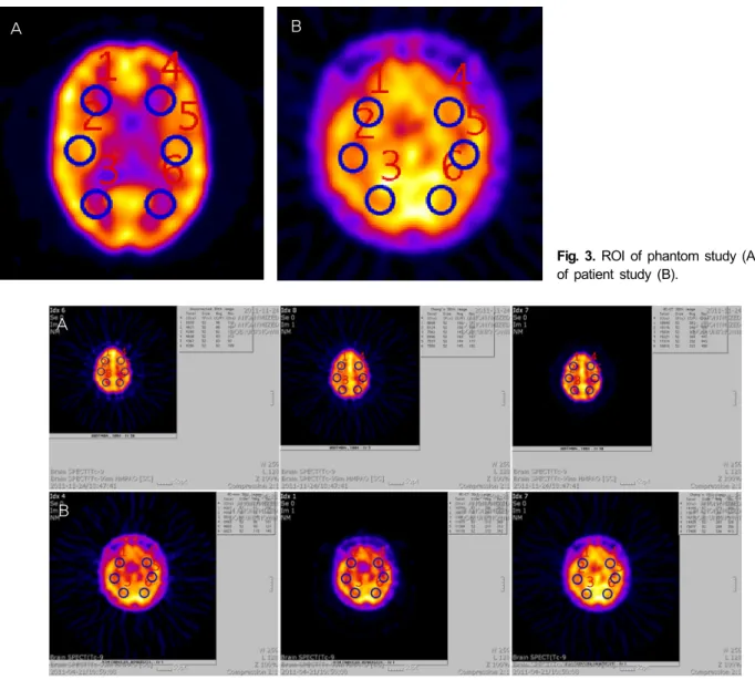
Fig. 3. ROI of phantom study (A), ROI
of patient study (B).A
B
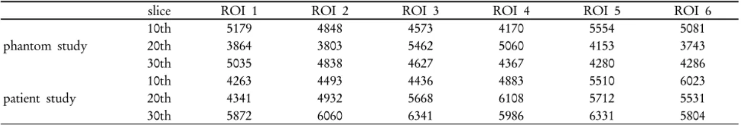
Fig. 4. ROI and count number of phantom study (A), ROI and count number of patient study (B).
brum으로 설정 하여 CT scan을 시행하였다.
5. 감쇠 보정
Chang's method를 이용한 감쇠 보정은 Attenuation co- efficient 값을 0.15cm-1을 적용하였다. Filtered backprojection 으로 재구성되었고 butterworth filter를 사용하여 cutoff 0.40, order 5를 적용하였다.
CT를 이용한 감쇠 보정은 μ-map으로 변환된 CT image를 이용하였고 SPECT 영상의 재구성은 3D OSEM방법을 이용 하여 subset 15, iteration 7, gaussian FWHM 8을 적용하였다.
phantom study와 patient study에서 획득된 data를 뇌 정량 분석을 하였으며 각각의 방법들을 적용한 transverse image는 같은 위치에서 재구성되었고 각각 10, 20, 30번째 slice에서
Fig. 3.에서 보이는 것처럼 6개의 region of interest (ROI)를 그려 AC-non과 AC-CT 그리고 Chang's method의 count를 비교하였다(Fig. 4).
결 과
획득된 영상의 10, 20, 30번째 영상에서 AC-NON, Chang's method, AC-CT ROI의 count값은 Table 1. 2. 3.과 같이 나 타났다.
1. Phantom study 결과
10번째 영상의 ROI count 평균은 AC-NON일 때 4900±
485, Chang's method를 이용하였을 때 9574 ± 840, AC-CT
Table 1. ROI count number of AC-NON
slice ROI 1 ROI 2 ROI 3 ROI 4 ROI 5 ROI 6
phantom study
10th 5179 4848 4573 4170 5554 5081
20th 3864 3803 5462 5060 4153 3743
30th 5035 4838 4627 4367 4280 4286
patient study
10th 4263 4493 4436 4883 5510 6023
20th 4341 4932 5668 6108 5712 5531
30th 5872 6060 6341 5986 6331 5804
Table 2. ROI count number of Chang's method
slice ROI 1 ROI 2 ROI 3 ROI 4 ROI 5 ROI 6
phantom study
10th 10474 9949 8548 8645 10379 9452
20th 7423 8080 10382 10398 8089 7790
30th 8668 8496 8124 7537 7562 7550
patient study
10th 14198 14929 13577 15077 16274 17480
20th 13302 15107 16062 17559 14755 14535
30th 15358 16180 15967 15596 15472 14334
Table 3. ROI count number of AC-CT
slice ROI 1 ROI 2 ROI 3 ROI 4 ROI 5 ROI 6
phantom study
10th 19056 16497 17654 14857 19938 17550
20th 12226 11707 21344 19493 13482 12355
30th 18840 19221 18118 17314 15834 16816
patient study
10th 10740 11037 10815 11304 13071 14176
20th 9647 11322 14069 14959 13420 13423
30th 13170 13403 15159 13551 14341 12717
0 5000 10000 15000 20000 25000
1 2 3 4 5 6
AC-CT Chang's method AC-non (count)
(ROI) AC-NON
4900 ±485 Chang’s method
9574 ±840 AC-CT
17592 ±1807
AC-CT 3.59 Chang’s method
1.95
AC-CT Chang's method AC-non
Fig. 5. ROI count number of 10th slice regarding Phantom study. Fig. 6. ROI count number of 20th slice regarding Phantom study.
일 때 17592 ± 1807이었다(Fig. 5).
20번째 영상의 ROI count 평균은 AC-NON일 때 4347±732, Chang's method를 이용하였을 때 8693 ± 1336, AC-CT일 때 15101 ± 4200이었다(Fig. 6).
30번째 영상의 ROI count 평균은 AC-NON일 때 4572 ± 315, Chang's method를 이용하였을 때 7989 ± 512, AC-CT 일 때 17690 ± 1280이었다(Fig. 7).
AC-CT의 경우 AC-NON보다 10번째 영상, 20번째 영상, 30번째 영상에서 각각 3.59, 3.47, 3.86배의 감쇠 보정을 하였
으며, Chang's method의 경우 AC-NON보다 1.95, 1.99, 1.74 배의 감쇠 보정을 해주었다.
2. Patient study 결과
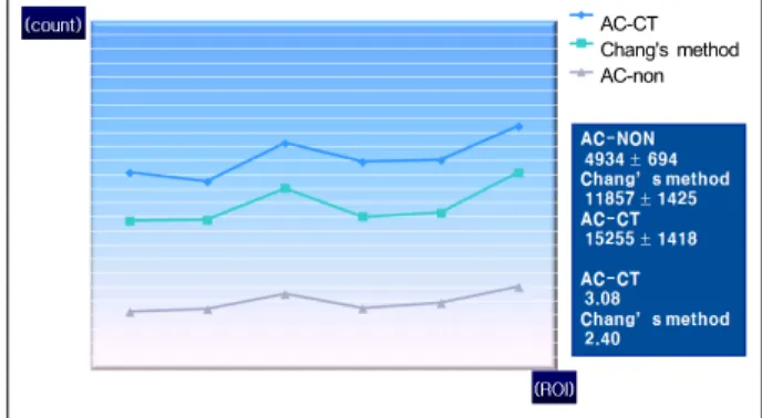
10번째 영상의 ROI count 평균은 AC-NON일 때 4934±
694, Chang's method를 이용하였을 때 11857 ± 1425, AC-CT일 때 15255 ± 1418이었다(Fig. 8).
20번째 영상의 ROI count 평균은 AC-NON일 때 5382 ±
0 5000 10000 15000 20000 25000
1 2 3 4 5 6
AC-CT Chang's method AC-non (count)
(ROI) AC-NON
4572 ± 315 Chang’s method
7989 ± 512 AC-CT
17690 ± 1280 AC-CT
3.86 Chang’s method
1.74
AC-CT Chang's method AC-non
Fig. 7. ROI count number of 30th slice regarding Phantom study. Fig. 8. ROI count number of 10th slice regarding Patient study.
AC-CT Chang's method AC-non
AC-CT Chang's method AC-non
Fig. 9. ROI count number of 20th slice regarding Patient study. Fig. 10. ROI count number of 30th slice regarding Patient study.
Table 4. The ratio of AC-CT to AC-NON and Chang's metod to AC-NON
Phantom study Patient study
AC-CT Chang's method AC-CT Chang's method
Count (mean) 16794.6 ± 2429.4 8752.6 ± 896.5 15320 ± 1171.6 12795 ± 1422.1
AC-NON (mean) 4606.8 ± 511.3 5460.8 ± 519.6
Ratio 3.70 1.92 2.85 2.38
636, Chang's method를 이용하였을 때 12806±1958, AC-CT 일 때 15220 ± 1453이었다(Fig. 9).
30번째 영상의 ROI count 평균은 AC-NON일 때 6065 ± 227, Chang's method를 이용하였을 때 13723 ± 882, AC-CT 일 때 15484 ± 643이었다(Fig. 10).
AC-CT의 경우 AC-NON보다 10번째 영상, 20번째 영상, 30번째 영상에서 각각 3.08, 2.82, 2.55배의 감쇠 보정을 하였 으며, Chang's method의 경우 AC-NON보다 2.40, 2.37, 2.26 배의 감쇠 보정을 해주었다.
3. 감쇠 보정 비교
Phantom study에서 AC-NON에 대한 AC-CT의 비는 3.70, Chang's method에 대한 비는 1.92로 나타났으며 patient study에서 AC-NON에 대한 AC-CT의 비는 2.85, Chang's method에 대한 비는 2.38로 나타났다(Table 4). AC-CT로 감
쇠보정 하였을 때 AC값이 Chang's methd를 이용한 AC값보 다 높게 나타났으며 이는 AC-CT를 이용하였을 때는 scatter correction을 같이 해주었기 때문에 나타난 결과이다.3)
결 론
우리는 이 연구를 통하여 AC-CT와 Chang's method 사이 에 감쇠 보정값이 다르다는 것을 알 수 있었고 AC-CT가 Chang's method보다 더 높은 감쇠보정을 해준다는 것을 알 수 있었다. 이는 scatter correction에 의한 결과라고 앞서 말 한 바 있다. 그리고 Chang's method는 patient study에서의 감쇠 보정 값이 phantom study에서의 감쇠 보정 값보다 더 높다는 것을 알 수 있었다. 이는 Skull bone에 의한 감쇠가 Chang method에서는 저평가된 것이다.4) 따라서 brain SPECT/CT를 시행하는 경우 scatter correction을 같이 시행 하고 bone에 의한 감쇠 정보를 반영할 수 있는 AC-CT가
chang's method보다 정확하다 할 수 있겠다. 추후 더 많은 정 상 지원자의 brain SPECT/CT 감쇠보정 data와 질병군과 비 교 연구가 필요할 것으로 사료된다.
요 약
이 연구의 목적은 Brain SPECT (Single Photon Emission Computed Tomography)의 Non-attenuation correction (AC- non) 영상에 대한 attenuation correction(AC) 방법 중 Chang's method와 CT based attenuation correction(AC-CT) 사이의 count를 비교하기 위함이다. phantom study는 증류 수로 채워진 hoffman 3D phantom에 99mTc 37Mbq을 투여하 였고, patient study는 normal volunteer에 99mTc-HMPAO 750Mbq를 정맥주입하고 Siemens사의 Symbia T6로 Brain SPECT 영상을 획득하였고 뇌 정량 분석을 하였다. 각각의 방법들을 적용한 transverse image는 같은 위치에서 재구성 되었으며 각각 10, 20, 30번째 slice에서 6개의 region of inter- est(ROI)를 그려 AC-non 과 AC-CT 그리고 Chang's meth- od의 count를 비교하였다. phantom study에서 AC-non, AC-CT, Chang's method의 각각 평균 count는 4606.8 ± 511.3, 16794.6 ± 2429.4, 8752.6 ± 896.5이었으며 patient study에서 5460.8 ± 519.6, 15320 ± 1171.6, 12795 ± 1422.1이 었다. phantom study에서 AC-CT와 AC-non 사이의 비는 3.70이고 Chang's method와 AC-non 사이의 비는 1.92였으
며 patient study에서는 각각 2.85, 2.38이었다. 우리는 이 연 구를 통하여 AC-CT가 Chang's method보다 더 높은 AC을 해준다는 걸 알 수 있었다. 그리고 Chang's method는 patient study에서의 AC 값이 phantom study에서의 AC값보다 더 높다는 것을 알 수 있었다. brain SPECT/CT를 시행하는 경 우 scatter correction을 같이 시행하고 bone에 의한 감쇠 정 보를 반영할 수 있는 AC-CT가 chang's method보다 정확하 다 할 수 있겠다.
REFERENCES
1. Hee-jeong Kim. Current status of imaging physics & in- strumentation in nuclear medicine. Nucl Med Mol Imaging 2008;42:83-87
2. Bohdan Bybel, Richard C. Brunken, Frank P. DF, Donald R.
Neumann, Guiyun Wu, Manuel D. Cerqueira. SPECT/CT Imaging : Clinical utility of an emerging technology. Radio Graphics 2008;28:1097-1113.
3. Masuo H Jun D, Keita U, Makoto Y, Tsuyoshi K, Masayasu T, Kenesi K, Isamu N. Comparison of methods of attenuation and scatter correction in brain perfusion SEPCT. J Nucl Med Technol 2005;33:224-229.
4. Kazunari I, Kohei H, Masahiro O, Seishi K, Yoshihiro K, Norio T, Makoto H, Takamichi M. Impact of CT attenuation correction by SPECT/CT in brain perfusion images. Ann Nucl Med 2012;26:241-247.