• 검색 결과가 없습니다.

Mammary gland tumors in three male dogs

N/A
N/A
Protected

Academic year: 2021

Share "Mammary gland tumors in three male dogs"

Copied!
4
0
0

로드 중.... (전체 텍스트 보기)

전체 글

(1)

pISSN 2466-1384 eISSN 2466-1392 大韓獸醫學會誌 (2016) 第 56 卷 第 4 號 Korean J Vet Res(2016) 56(4) : 229~232 https://doi.org/10.14405/kjvr.2016.56.4.229

229

<Original Article>

Mammary gland tumors in three male dogs

Jeong-Hee Han

1,†

, Kyeong-Soo Kim

2,†

, Jae-Hoon Kim

2,

*

1

College of Veterinary Medicine and Institute of Veterinary Science, Kangwon National University, Chuncheon 24341, Korea

2

College of Veterinary Medicine and Veterinary Medical Research Institute, Jeju National University, Jeju 63243, Korea (Received: September 13, 2016; Revised: November 7, 2016; Accepted: November 9, 2016)

Abstract: Mammary gland tumors are very rare in male dogs. In this study, four mammary gland tumors from 3 male dogs (2 intact, 1 neutered) were collected from local animal hospitals. The dogs included two purebred Shih Tzu (1 intact, 1 neutered) and one intact purebred Cocker Spaniel. The mean age of dogs with mammary gland tumors was 9 years (5-12 years). Two dogs had a solitary mass, whereas one dog had two mammary masses. Of the four tumor masses, three were observed in the fourth or fifth mammary glands, and one was observed in the third mammary gland. According to histopathologic examinations, all four mammary masses from three dogs were benign tumors including two benign mixed tumors in one case and two complex adenomas. There were no history of obesity, testicular tumors, diabetes, and sex hormonal therapy in any male dogs with mammary tumors. Surgical excision was the only reported treatment for these tumors. No recurrence or metastasis was recorded up to 25 months after surgery.

Keywords: dogs, male, mammary gland, neoplasms

Introduction

Mamamry gland tumors have been reported to comprise 50% of all neoplasms in the bitch [4]. They occur most often in bitches 10–11 years old and are very rare in dogs less than 5 years of age. Anatomically, about 70% of all mammary tumors deveolp in gland 4 or 5 [4].

In bitches and queens, early ovariectomy offers a great protective effect against mammary carcinoma [8]. Ovariohys- terectomy at or prior to first estrus dramatically decreases the risk of development of mammary tumors in female dogs, sug- gesting that sex hormone status is an important risk factor [4].

The incidence rate of mammary gland tumors that occur in male dogs ranges from 0 to 2.7% (average < 1%) [1, 10].

Some mammary tumors in male dogs are associated with hormonal abnormalities such as estrogen secreting Sertoli cell tumor of the testis [8]. Several cases of mammary gland tumors in male dogs with clinical and histopathologic fin- dings were demonstrated in many countries [1, 6, 11, 12, 14].

However, only two cases of mammary gland tumors in male dogs were previously described in Korea [2]. Two male dogs in that report were diagnosed as a mammary benign mixed tumor and a simple adenoma, but there were no available data such as clinical information and pathologic descriptions.

Here we describe the clinicopathologic findings in three male dogs with mammary gland tumors.

Materials and Methods

Case studies

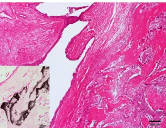
This study was performed using archival biopsy samples from three male dogs with mammary tumors that had been examined between 2013 and 2015 at the laboratory of Veter- inary Pathology in Jeju National University. Two histologic classification systems for canine mammary tumors and dys- plasia have been published: the first in 1974 and a modifica- tion in 1999. Since the publication of the second system, several new histologic subtypes of canine mammary neo- plasms have been described. Therefore, new histologic clas- sification and grading system of canine mammary tumors were proposed in 2011 [5]. According to recent classifica- tion, two masses in one case of mammary benign mixed tumor and two cases of complex adenomas were diagnosed by general histopathologic examinations.

Clinical information was taken from the biopsy sample request forms and from follow-up questions and telephone or e-mail communications with the clinicians. Taken informa- tion included age at presentation, precise site of tumor mass, duration of mammary mass before the surgery, recurrence of tumor, date of final examination, and associated medical his- tory (testicular diseases, therapy with hormone and obesity).

*Corresponding author

Tel: +82-64-754-3387, Fax: +82-64-702-9920 E-mail: kimjhoon@jejunu.ac.kr

The first two authors contributed equally to this work.

(2)

230 Jeong-Hee Han, Kyeong-Soo Kim, Jae-Hoon Kim

Histopathology

Surgically excised tissue samples were immediately fixed in 10% neutral buffered formalin. The samples were pro- cessed routinely for histopathologic examination and tissue sections were stained with hematoxylin and eosin. To con- firm the presence of bony tissues, von Kossa staining was also performed on paraffin embedded tissue sections.

Results

Clinical data

Three male dogs diagnosed with primary mammary gland tumors were identified. Clinical informations such as breed, age, sex, location and size of tumor mass, and periods of fol- low-up are summarized in Table 1. Two of the dogs were intact and the other one was neutered. All three dogs were purebred such as two Shih Tzu (1 intact, 1 neutered) and one intact Cocker Spaniel. The mean age of dogs with mammary gland tumors was 9 years (5–12 years). Grossly, the mam- mary tumors were firm to hard, tan to off-white, and round to oval pedunculated masses ranged from 1.5 to 3 cm in diame- ter (Fig. 1). The mammary masses were firm to hard, tan to off-white, round to oval and ranged from 2 to 3 cm in diam- eter in the cut surface of case 1 (Fig. 2). Of the four tumor masses, three were involved in the fourth or fifth mammary glands of 2 Shih Tzu, and one was involved in the third gland of Cocker Spaniel. One Shih Tzu dog had two mam- mary masses in the fourth and fifth glands. The duration of the mammary lesions was several months.

The follow-up period was ranged from 4 to 25 months.

Obesity, diabetes and sex hormone therapy were not recorded in all three male dogs based on the history taking. During four years before the surgical removal of mammary tumor, one dog (No. 3) had a history of cryptorchid testicles in abdominal cavity and castration. On four months after the surgery, this dog died from oral cancer unrelated to the mam- mary tumor. Two intact dogs were castrated at the time of mammary tumor removal. And these dogs were alive with- out any evidences of recurrence or metastasis at the end of the follow-up period. Surgical excision (simple mastectomy) was the only reported treatment for the mammary tumors in three cases. Surgical margins were considered to complete in all dogs through histopathologic examinations.

Histopathology

According to histopathologic examinations, all four mam- mary masses from three dogs were benign tumors including two benign mixed tumors in one case and two complex ade- nomas. Histopathologically, two tumor masses of case 1 were relatively well encapsulated with dense connective tissue.

The neoplastic foci were composed of very small area (only 5%) of acinar or tubular pattern structures, and large area (95%) of myoepithelial cell components. Most acinar type of neoplastic areas contained irregular sized mammary gland with single to double layer of luminal epithelial cells and intra-luminal fluids. Many neoplastic glands showed severe cystic dilatation with papillary growth of luminal epithelium.

Most area of mass was composed of cluster of spindle cells or stellate cells, and they produced mucinous or collagenous materials and formed multifocal big bonny tissues (Fig. 3).

These bones were stained black with von Kossa staining (Fig. 3, insert).

The tumor masses of cases 2 and 3 were well circum- Table 1. Clinical data of male dogs with mammary gland tumor

Number of dog Breed Age (yr) Sex Location Size (diameter) Diagnosis Follow-up (mo)

1 Shih Tzu 10 M 4th left

5th left

3 cm 2 cm

Benign mixed tumor

Benign mixed tumor 25

2 Cocker Spaniel 12 M 3rd right 1.5 cm Complex adenoma 17

3 Shih Tzu 5 NM 5th left 1.5 cm Complex adenoma 4

M, intact male; NM, neutered male.

Fig. 1. Gross findings of mammary masses. Note two masses (arrows) in the left 4th and 5th mammary glands (A, case 1) and single mass (arrow) in the left 5th mammary gland (B, case 3) of male dogs.

Fig. 2. The mammary masses are firm to hard, tan to off-white,

and round to oval and ranged from 2 to 3 cm in diameter in the

cut surface in case 1.

(3)

Mammary gland tumors in male dogs 231

scribed with dense connective tissue. The neoplastic foci were composed of large area of acinar or tubular pattern structures derived from mammary glands and adjacent small amounts of fibrous elements originated from myoepithelial cells (Fig.

4). Large areas of neoplastic foci contained irregular various sized mammary glands with single to triple layers of luminal epithelial cells. Some neoplastic glands showed cystic dilata- tion with papillary growth of luminal epithelium and intra- luminal fluids. Spindle or clear cell originated from myoepi- thelial cells produced basophilic homogeneous mucinous materials. The mammary masses of case 1 were diagnosed as a benign mixed tumor and cases 2 and 3 were as complex adenomas. Most tumor cells in three mammary cases did not show any invasive tendency and malignancy.

Discussion

Mammary cancer in men is very rare, accounting for less than 1% of all breast carcinomas [1, 13]. An increased risk of mammary cancer in men was most closely related with sev- eral factors such as undescended testes, orchiectomy, orchi- tis, testicular injury, late puberty, infertility, increased level of blood cholesterol, fast weight gain, obesity, amphetamine use, diabetes, and cigarette smoking [13]. And they sug- gested that mammary cancer in men developed in response to low amounts of androgen related with testicular malfunction and under conditions related with high level of estrogen. Sev- eral previous studies of mammary gland tumors in male dogs implicated hormonal abnormalities, particularly in associa- tion with testicular tumors including Sertoli cell tumor, as the cause of the tumors [14]. High serum estrogen and progester- one concentrations in male dogs with mammary gland tumors were also demonstrated in one study [7]. The dogs in this study had no history of testicular neoplasm or other abnormalities, and no signs of feminization. During four years before the surgical removal of mammary tumor, one Shih Tzu dog had a history of cryptorchid testicles in abdom- inal cavity and castration. Therefore, the occurrence of mam- mary tumor may not be closely associated with testicular deformity. In addition, there were no history of obesity, dia- betes, and sex hormonal therapy in any male dogs with mam- mary tumors.

According to a recent literature on 27 mammary tumors from 18 male dogs, breed and age appear to be significant factors in the development of mammary tumors in the male dogs [1]. Based on the previous reports [9, 12], mammary tumors were more prevalent in Cocker Spaniels and Terrier breeds. The average age at diagnosis in those studies was 9.2 years. In the present study, mammary gland tumors were observed in two Shih Tzu and one Cocker Spaniel, and the average age of onset for the tumors was 9 years.

Like mammary gland tumors in female dogs, most of the mammary gland tumors in male dogs were observed in the 4th (caudal abdominal) and 5th (inguinal) mammary glands.

The reason for this might be that the posterior glands have a greater mass of mammary glandular tissue to react to any injuries or carcinogenic stimulus [7]. The higher prevalence of mammary gland tumors in the fourth or fifth glands in male dogs is also attributed to the increased mass of mam- mary tissue in which a tumor might arise, although male mammary tissue is composed of only ductular components and is devoid of fully developed terminal duct lobular units [1].

According to literatures, the canine mammary tumors in male included the following diagnoses: malignant tumors such as adenocarcinoma perithelioma, papillary cystadeno- carcinoma, chondroadenocarcinoma, adenocarcinoma, cystic carcinoma, malignant mixed tumor, osteosarcoma, sarcocar- cinoma, cystadenochondroma, papillary cystadenoma with squamous cell carcinoma, and spindle cell sarcoma and benign tumors such as ductal papillary adenoma, simple ade- Fig. 3. Mammary benign mixed tumor had cystic dilated neo-

plastic gland and multifocal bony tissues (right side). Insert:

The bony tissue is shown black color by special stain. H&E stain or von Kossa stain (insert). Scale bars = 100 µm, 50 µm (insert).

Fig. 4. Neoplastic foci are composed of acinar or tubular struc- tures and islands of spindle cells (M) with mucinous materials in mammary gland complex adenoma (case 3). H&E stain.

Scale bar = 50 µm.

(4)

232 Jeong-Hee Han, Kyeong-Soo Kim, Jae-Hoon Kim

noma, complex adenoma, fibroadenoma, and benign mixed tumor [1, 6, 9, 10, 12, 14]. According to old literatures, 28 of 51 mammary gland tumors in male dogs were classified as malignant tumor [6, 9]. However, the overall incidence rate of malignant mammary gland tumors in male dogs is lower than that of female dogs [3, 12]. Recent two surveys demon- strated that all 27 mammary tumors in 18 male dogs in one survey and 7 (88%) of 8 male dogs in the other were con- firmed as benign tumors [1, 12]. These differences may be closely associated with the different histopathologic criteria for malignancy [1]. The most important histopathologic crite- ria for the diagnosis of malignant mammary tumors in the dog based on hematoxylin and eosin-stained sections were the following factors: tumor type, anaplasia characterized by nuclear and cellular pleomorphism, mitotic index, presence of necrosis within the neoploasm, destructive-invasive growth to adjacent blood vessels or lymphatics, and regional lymph node metastasis [5]. On the basis of recent classification of canine mammary tumors, all four mammary tumors in this study were well encapsulated or circumscribed with dense connective tissues and had benign histologic features. We could not find any evidences for the invasion to stroma and lymphatics around neoplastic foci of mammary tumors.

Surgery is the first choice for the treatment of dogs with most types of mammary gland tumors; the exceptions are inoperable diseases such as canine inflammatory carcinoma and distant metastasis [8]. Several surgical procedures including simple, regional, radical, and total mastectomy and lumpec- tomy have been applied in canine mammary tumors [2, 8].

Only surgical excision of mammary mass yields unsatisfac- tory results in dogs with malignant mammary tumors exhib- iting lymphatic or vascular invasion and metastasis. Therefore, the development of adjuvant treatments and the investigation of their antineoplastic efficacy have been studied for many years. In this study, only surgical excision of palpable tumors was performed in all male dogs. The complete surgical removal of localized tumors without metastatic involvement is the therapeutic procedure with the highest probability of cure [7].

Surgical excision of mammary gland tumors also allows his- tological examination to determine for malignancy, increases survival time, improves the patient’s quality of life, and can be curative [2, 7, 8].

The annual incidence rate of mammary gland tumors in female dogs has been estimated at 198/100,000 [8]. In con- trast, the incidence rate in the male dogs was 4/100,000 in previous literature [12]. Although rare, this study illustrated that mammary gland tumors can arise in male dogs in Korea.

Therefore, mammary gland tumor should take into consider-

ation in the differential diagnosis of subcutaneous mass around caudal abdominal and inguinal mammary glands and prepuce in male dogs.

Acknowledgments

This study has been worked with the support of a research grant of Kangwon National University in 2016.

References

1. Bearss JJ, Schulman FY, Carter D. Histologic, immunohis- tochemical, and clinical features of 27 mammary tumors in 18 male dogs. Vet Pathol 2012, 49, 602-607.

2. Chae HC, Byeon YE, Lee ST, Lim JH, Kweon OK, Kim WH. Mammary gland tumors treated by surgical excision: a retrospective study in 79 dogs. J Vet Clin 2007, 24, 331-336.

3. Gilbertson SR, Kurzman ID, Zachrau RE, Hurvitz AI, Black MM. Canine mammary epithelial neoplasms: biologic implications of morphologic characteristics assessed in 232 dogs. Vet Pathol 1983, 20, 127-142.

4. Ginn PE, Mansell JEKL, Rakich PM. Skin and appendages.

In: Maxie MG (ed.). Jubb, Kennedy, and Palmer’s Pathology of Domestic Animals. 5th ed. pp. 777-780, Elsevier Saunders, New York, 2007.

5. Goldschmidt M, Peña L, Rasotto R, Zappulli V. Classifi- cation and grading of canine mammary tumors. Vet Pathol 2011, 48, 117-131.

6. Jabara AG. Two cases of mammary neoplasms arising in male dogs. Aust Vet J 1969, 45, 476-480.

7. Maiti SK, Kumar DKM, Kumar S, Ravindran NA, Mathew D, Palakkara S, Muthalavi MA, Kumar V.

Mammary gland tumours in male dogs: a hormonal and tumour marker study. Vet Arh 2014, 84, 537-548.

8. Misdorp W. Tumors of the mammary gland. In: Meuten DJ (ed.). Tumors in Domestic Animals. 4th ed. pp. 575-606, Iowa State University Press, Ames, 2002.

9. Mitchell L, De la Iglesia FA, Wenkoff MS, Van Dreumel AA, Lumb G. Mammary tumors in dogs: survey of clinical and pathological characteristics. Can Vet J 1974, 15, 131-138.

10. Moulton JE, Taylor DO, Dorn CR, Andersen AC. Canine mammary tumors. Pathol Vet 1970, 7, 289-320.

11. Raflo CP, Diamond SS. Neoplasm of the mammary papilla in a male dog. Am J Vet Res 1980, 41, 953-954.

12. Saba CF, Rogers KS, Newman SJ, Mauldin GE, Vail DM.

Mammary gland tumors in male dogs. J Vet Intern Med 2007, 21, 1056-1059.

13. Thomas DB. Breast cancer in men. Epidemiol Rev 1993, 15, 220-231.

14. Walker D. Mammary adenomas in a male dog-probable

oestrogenic neoplasms. J Small Anim Pract 1968, 9, 15-20.

수치

Fig. 1. Gross findings of mammary masses. Note two masses (arrows) in the left 4th and 5th mammary glands (A, case 1) and single mass (arrow) in the left 5th mammary gland (B, case 3) of male dogs
Fig. 4. Neoplastic foci are composed of acinar or tubular struc- struc-tures and islands of spindle cells (M) with mucinous materials in mammary gland complex adenoma (case 3)

참조

관련 문서

다양한 번역 작품과 번역에 관한 책을 읽는 것은 단순히 다른 시대와 언어, 문화의 교류를 넘어 지구촌이 서로 이해하고 하나가

The index is calculated with the latest 5-year auction data of 400 selected Classic, Modern, and Contemporary Chinese painting artists from major auction houses..

1 John Owen, Justification by Faith Alone, in The Works of John Owen, ed. John Bolt, trans. Scott Clark, &#34;Do This and Live: Christ's Active Obedience as the

영문 : Histomorphometric analysis of bone formation in the bone defects around the implant in adult dogs according to grafts of low or high heat treated autogenous

Fourth, reality therapy group counseling program was effective in improving prisoners' social relationships which is one of the sub factors of

Fifth, in addition to the structural model (alternative model) in which sports confidence and athlete satisfaction mediate in parallel from the path

Purpose: Giant cell tumor of the tendon sheath are the most common tumors after ganglionic cysts in benign soft tissue tumors which could be recurred after surgical

All disputes, controversies, or differences which may arise between the parties out of or in relation to or in connection with this contract, or for the breach thereof,