Anti-inflammatory Effect of Achyranthoside E Dimethyl Ester in LPS-stimulated RAW 264.7 Cells
Soo Young Bang
1, Ji-Hee Kim
1, Hyung-In Moon
2and Young Hee Kim
1*
1
Department of Molecular Biology, College of Natural Sciences, Pusan National University, Busan 609-735, Korea
2
Department of Medicinal Biotechnology, College of Natural Resources and Life Science, Dong-A University, Busan 604-714, Korea
Received April 9, 2013 /Revised May 20, 2013 /Accepted May 23, 2013Achyranthoside E dimethyl ester (AEDE) is an oleanolic acid glycoside from Achyranthes japonica. In this study, we investigated the effects of AEDE on nitric oxide (NO) production and underlying mo- lecular mechanisms in lipopolysaccharide (LPS)-stimulated macrophages. AEDE inhibited LPS-induced NO secretion as well as inducible NO synthase (iNOS) expression, without affecting cell viability.
Further study demonstrated that AEDE induced heme oxygenase-1 (HO-1) gene expression. In addi- tion, the inhibitory effects of AEDE on iNOS expression were abrogated by small interfering RNA-mediated knock-down of HO-1. Moreover, AEDE induced nuclear translocation of nuclear factor E2-related factor 2 (Nrf2), a transcription factor that regulates HO-1 expression. AEDE-induced ex- pression of HO-1 was inhibited by inhibitors of phosphatidylinositol 3-kinase (PI-3K) and extracellular signal regulated kinase (ERK1/2). AEDE phosphorylated Akt and ERK1/2 as well. Therefore, these results suggest that AEDE suppresses the production of pro-inflammatory mediator such as NO by inducing HO-1 expression via PI-3K/Akt/ERK-Nrf2 signaling. These findings provide the scientific ra- tionale for anti-inflammatory therapeutic use of AEDE.
Key words : Achyranthoside E dimethyl ester, heme oxygenase-1, inducible nitric oxide synthase, NF-E2-related factor 2, nitric oxide
*Corresponding author
*Tel:+82-51-510-2526, Fax:+82-51-513-9258
*E-mail : [email protected]
This is an Open-Access article distributed under the terms of the Creative Commons Attribution Non-Commercial License (http://creativecommons.org/licenses/by-nc/3.0) which permits unrestricted non-commercial use, distribution, and reproduction in any medium, provided the original work is properly cited.
Journal of Life Science 2013 Vol. 23. No. 6. 736~742 DOI : http://dx.doi.org/10.5352/JLS.2013.23.6.736
Introduction
Achyranthoside E dimethyl ester (AEDE) is an oleanolic acid glycoside from Achyranthes japonica and Achyranthes bi- dentata [16]. A. japonica and A. bidentata have been used in traditional medicine for the treatment of edema, arthritis, and delayed menses and as a contraceptive and abortifacient [1]. A. japonica has been reported to inhibit platelet ag- gregation [36] and have anti-fungal [14] and anti-in- flammatory activities [9]. Our previous study demonstrated that A. japonica extract has anti-inflammatory activity [3]. A.
bidentata has been reported to have anti-inflammatory [10, 18, 33], osteoprotective [11, 35, 37] and neuroprotective [27, 35, 38] activities. AEDE from A. bidentata has inhibitory activ- ity on osteoclast formation [16]. However, little is known about the effects of AEDE on inflammation and underlying
mechanisms.
Nitric oxide (NO) is a free radical with multiple effects on various organ systems. The most prominent physiological actions of NO as a biological mediator include cGMP-de- pendent vasodilation, neural communication, host defense, inflammation, immune suppression and blood clotting [20].
NO is produced in physiological and pathophysiological conditions by NO synthase (NOS), and inducible NOS (iNOS) is induced by inflammatory cytokines and/or bacte- rial lipopolysaccharide (LPS) in various cell types including macrophages. A large amount of NO, particularly synthe- sized by iNOS, induces an inflammatory response to inhibit the growth of invading microorganisms and tumor cells.
This strong inflammatory response to foreign cells could also cause further damage for the neighboring cells and tissues of the host [19]. Therefore isozyme specific inhibitors of NOS are essential for therapeutic purposes and drugs that specifi- cally inhibit iNOS could be useful in treating diseases medi- ated by NO overproduction [28].
Heme oxygenase-1 (HO-1) is an inducible enzyme that
catalyzes the rate-limiting step in the oxidative degradation
of cellular heme into carbon monoxide (CO), biliverdin, and
free iron [25]. HO-1 and its enzymatic by-products provide
a host defense mechanism that can protect the body against oxidative injury and also contributes to the anti-in- flammatory activity of cells and tissues [22]. In activated macrophages, HO-1 expression or CO treatment inhibits the production of the pro-inflammatory cytokines and chemo- kines such as tumor necrosis factor-α (TNF-α), interleukin-1β (IL-1β), IL-6, monocyte chemoattractant protein-1, and mac- rophage inflammatory protein-1β [22]. Up-regulation of HO-1 expression or the administration of CO also sup- presses the production of pro-inflammatory mediators such as nitric oxide (NO) and prostaglandin E2 (PGE2) [30].
Moreover, an increasing number of therapeutic agents have been reported to induce HO-1 expression and exert their an- ti-inflammatory effects through HO-1 induction. These stud- ies support beneficial effects of HO-1 that may serve as a therapeutic target in inflammatory diseases.
HO-1 is primarily regulated on the transcriptional level via signaling pathways involved in survival and stress re- sponses in different cell types. Transcription factor NF-E2-related factor 2 (Nrf2) plays a central role for in- ducible expression of HO-1 [29]. In basal conditions, Nrf2 is sequestered in the cytoplasm by Kelch-like ECH-asso- ciated protein 1 (Keap1) and degraded by the ubiq- uitindependent 26S proteasome system [12]. Under activa- tion, Nrf2 released from Keap1 inhibition, translocates to the nucleus, heterodimerizes with Maf, and binds antioxidant response elements (AREs) located in the promoter regions of many detoxifying/antioxidant genes, including HO-1 [12, 21].
In the present study, we investigated the effects of AEDE from A. japonica on LPS-induced inflammatory response (NO release) in macrophages and further explored the possible mechanisms. Our results provide a molecular basis for un- derstanding the inhibitory effects of AEDE on endotox- in-mediated inflammation.
Materials and Methods Materials
LPS (phenol extracted Salmonella enteritidis), Tween-20, 3-(4,5-dimethylthiazol-2-yl)-2,5-diphenyltetrazolium bromide (MTT) and other reagents were purchased from Sigma Chemical Co. (St. Louis, MO, USA). Antibodies for iNOS, HO-1, Nrf-2, TATA-box binding protein (TBP), and α-tubulin were purchased from Santa Cruz Biotechnology (Santa Cruz, CA, USA). HO-1 siRNA was purchased from Bioneer
(Daejoen, Korea). INTERFERin siRNA Transfection Reagent was purchased from Polyplus transfection (France).
Dulbecco’s modified Eagle’s medium (DMEM) and fetal bo- vine serum (FBS) were purchased from Invitrogen Corporation (San Diego, CA, USA). AEDE (Fig. 1A) isolated from the methanol-soluble fraction of the dried roots of A.
japonica was prepared as described previously. The isolated compound was kept at 4°C in the dark until further experiments.
Cell culture
Murine macrophage RAW 264.7 cells were maintained in DMEM supplemented with glutamine (1 mM) and 10% FBS at 37
oC in an atmosphere of 5% CO
2.
Measurement of nitrite concentration
NO synthesis in cell cultures was measured by a micro- plate assay method. To measure nitrite, 100 μl aliquots were removed from conditioned medium and incubated with an equal volume of the Griess reagent (1% sulfanilamide/0.1%
N-(1-naphthyl)-ethylenediamine dihydrochloride/2.5%
H
3PO
4) at room temperature for 10 min. Nitrite concen- tration was determined by measuring the absorbance at 540 nm with a Vmax 96-well microplate spectrophotometer (Molecular Devices, Menlo Park, CA, USA). The sodium ni- trite was used as a standard.
Cell viability assay
The cytotoxicity of AEDE was assessed using the micro- culture tetrazolium (MTT)-based colorimetric assay. The re- maining cells after Griess reaction were used for MTT assay.
MTT was added to each well (final concentration is 62.5 μ g/ml). After incubation for 3 h at 37
oC and 5% CO
2, the supernatant was removed and the formed formazan crystals in viable cells were solubilized with 150 μl of DMSO. The absorbance of each well was then read at 570 nm using mi- croplate reader.
Western blot analysis
The cells were washed with phosphate buffered saline
(PBS) three times and scraped off and lysed with lysis buffer
(1% Triton X-100, 1% deoxycholate). Protein concentration
of lysates was determined using Bradford reagent (Bio-Rad,
Hercules, CA, USA) and equal amounts of protein were sep-
arated electrophoretically using 10% SDS-PAGE (sodium do-
decyl sulfate-polyacrylamide gel electrophoresis), and then
B A
C
D
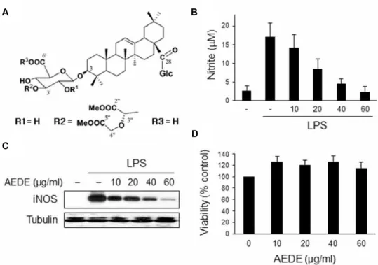
Fig. 1. Effect of AEDE on the NO secretion and iNOS expression in macrophages. (A) Chemical structure of AEDE isolated from the methanol-soluble fraction of the dried roots of
A. japonica
. (B) RAW 264.7 cells were incubated with various concentrations of AEDE for 1 h and then stimulated with LPS (0.1 μg/ml) for 20 h. The amount of nitrite released was measured by the method of Griess. (C) Cells were treated as mentioned above and equal cytosolic extracts were analyzed by Western blotting. (D) Cells were incubated with various concentrations of AEDE for 24 h and cell viability was measured by MTT assay according toMaterials and methods
. *p
<0.05 and **p
<0.01 vs. LPS-treated group.the gel was transferred to 0.45 μm nitrocellulose paper. The blot was incubated with specific antibody and then detected by the enhanced chemiluminescence detection system ac- cording to the recommended procedure (Amersham Biosciences, Piscataway, NJ, USA). TATA binding protein (TBP) was used as protein-loading controls for each lane.
Quantitative image analysis was performed using image analysis software, ImageJ (http://rsb.info.nih.gov/ij) and data were presented as fold of control.
Preparation of nuclear extract
Nuclear extracts were prepared as described previously [2]. In brief, cells were washed with ice-cold PBS, centri- fuged at 1,000× g for 5 min, resuspended in 400 μl of ice-cold hypotonic buffer (10 mM HEPES/KOH, 1.5 mM MgCl
2, 10 mM KCl, 0.5 mM DTT, 0.2 mM PMSF, pH 7.9), left on ice for 10 min, vortexed, and centrifuged at 15,000×
g for 30 sec. Pelleted nuclei were gently resuspended in 50 μ l of ice-cold buffer (20 mM HEPES/KOH, 1.5 mM MgCl
2, 420 mM NaCl, 0.2 mM EDTA, 25% glycerol, 0.5 mM DTT, 0.2 mM PMSF, pH 7.9), left on ice for 30 min, vortexed,
and centrifuged at 15,000× g for 5 min at 4
oC. Aliquots of the supernatant that contained nuclear proteins were stor- ed at -70
oC.
Interference of HO-1
The siRNAs for HO-1 (GenBank accession no. NM 010442.1) were synthesized by Bioneer (Daejoen, Korea). The siRNA 1 sequence is 5’-CACCAAGGAGGUACACAUC(d TdT)-3’ (sense) and siRNA 2 sequence is 5’-CCUGAAUC GAGCAGAACCA(dTdT)-3’ (sense). Cells were transfected with HO-1 siRNA (200 nM) or negative control siRNA using INTERFERin. Then cells were incubated for 48 h until the protein expression detection.
Statistical analysis
All results were expressed as means ± SE. Each experi-
ment was repeated at least three times. Statistical sig-
nificances were compared between each treated group and
analyzed by the Student’s t-test. Data with p<0.05 were con-
sidered statistically significant.
Results
Effect of AEDE on NO synthesis and iNOS expression in macrophages
To investigate the anti-inflammatory effect of AEDE, we examined the effect of AEDE on NO synthesis in macrophages. RAW 264.7 cells were incubated with AEDE for 1 h and stimulated with LPS for 20 h. The amount of NO released into culture medium was measured by the method of Griess. Whereas LPS-treated cells produced a large amount of NO, AEDE suppressed NO release into cul- ture supernatant in a dose-dependent manner (Fig. 1B). To determine whether the decreased nitric oxide synthesis is correlated with iNOS expression, we analyzed the amount of iNOS by Western blot analysis. Macrophages were treated with AEDE as mentioned above. The level of iNOS was dra- matically reduced by AEDE in a dose-dependent manner (Fig. 1C). On the other hand, cell viability was not affected by AEDE as measured by MTT assay (Fig. 1D). These results suggest that AEDE inhibits NO release by suppressing iNOS expression level without affecting cell viability.
AEDE exhibits anti-inflammatory effect through the induction of HO-1
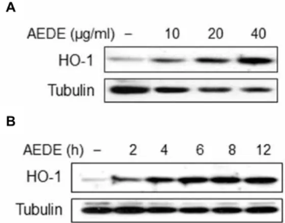
By-products of HO-1 are known to have anti-in- flammatory effect [25]. Accordingly, we investigated wheth- er AEDE exhibits its anti-inflammatory effect through the induction of HO-1 expression. We examined the HO-1-in- ducible activity of AEDE by Western blotting. HO-1 protein level was significantly increased by AEDE in a dose-depend- ent manner and reached peak at 6 h in RAW 264.7 cells (Fig.
2A, 2B). To confirm that AEDE suppresses iNOS expression through the induction of HO-1, we applied an HO-1 small interfering (si) RNA system to knock down HO-1 function.
Cells were transfected with si HO-1 or si control RNA, and the effects of AEDE on iNOS expression were examined by Western blotting. As shown in Fig. 3, decreased HO-1 ex- pression blocked AEDE-mediated suppression of LPS-stimu- lated iNOS expression, whereas transfection with control siRNA showed no effect. Taken together, these results in- dicate that HO-1 is involved in AEDE-induced inhibition of iNOS expression.
AEDE-induced HO-1 expression is mediated by Nrf2
Since the promoter region of HO-1 gene contains binding
A
B
Fig. 2. Induction of HO-1 by AEDE. Cells were incubated with various concentrations of AEDE for 8 h (A), or with AEDE (40 μg/ml) for indicated times (B). Cells were har- vested and equal cytosolic extracts were analyzed by Western blotting with anti-HO-1 antibody.
Fig. 3. Inhibitory effect of HO-1 on iNOS expression. Cells were transfected with HO-1 siRNA or control siRNA (Mock, M), after which they were pretreated with AEDE (40 μ g/ml) for 1 h and stimulated by LPS (0.1 μg/ml) for 6 h. Protein levels of iNOS and HO-1 were analyzed by Western blotting.
sites for transcription factor Nrf2 and the expression of HO-1 is known to be regulated by Nrf2 [29], we examined the effect of AEDE on nuclear accumulation of Nrf2 in RAW 264.7 cells. Cells were treated with AEDE, and accumulation of Nrf2 in nucleus was examined by Western blotting. Nrf2 nuclear accumulation was increased by AEDE in a dose-de- pendent manner and reached peak at 1 h (Fig. 4A, 4B). These results suggest that AEDE induces the expression of HO-1 through activating Nrf2.
AEDE-induced HO-1 expression is mediated by ERK1/2 pathway
We examined the signaling pathways associated with
AEDE-induced HO-1 expression. RAW264.7 macrophages
A
B
Fig. 4. Effect of AEDE on Nrf2 activation. Cells were incubated with indicated concentrations of AEDE for 1 h (A), or AEDE (40 μg/ml) for indicated times (B). Nuclear pro- teins were extracted and nuclear accumulation of Nrf2 was assayed by Western blotting.
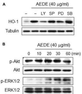
were exposed, separately, to one of pharmaceutical protein kinase inhibitors: LY294005 (a PI3K inhibitor), SP600125 (a JNK inhibitor), PD98059 (an ERK1/2 inhibitor) and SB203580 (a p38 MAPK inhibitor). As shown in Fig. 5A, AEDE-in- duced HO-1 expression was significantly inhibited by LY294005 and PD98059 but not by SP600125 and SB203580.
Further, AEDE transiently increased the phosphorylation of Akt at early time (20 min), whereas ERK1/2 was activated by AEDE at later time (30-60 min) (Fig. 5B). These results suggest that PI3K/Akt and ERK-related signaling could con- stitute the major signal pathways in the regulation of HO-1 expression by AEDE.
Discussion
In this study, we demonstrate that AEDE inhibited iNOS expression via the induction of HO-1. Inhibition of iNOS ac- tivity in macrophages may thus represent an interesting tar- get to treat various diseases including arthritis. In fact, ad- ministration of the selective inhibitors of iNOS has been re- ported to attenuate arthritis [7, 13]. Thus AEDE might reduce arthritis via inhibition of NO synthesis by affecting the iNOS expression level in macrophages.
Growing evidences have demonstrated that HO-1 exhibits anti-inflammatory activities by inhibiting production of pro-inflammatory mediators [31, 32], suggesting a potential therapeutic strategy for treating inflammatory diseases.
Although the contribution of HO-1 products (ie, CO, bili- verdin, and iron) has not been examined in this study, sev- eral studies point to HO-1-derived CO and biliverdin as the
A
B
Fig. 5. Intracellular signaling pathway involved in HO-1 ex- pression by AEDE. (A) RAW 264.7 cells were incubated with LY294002 (20 μM), SP600125 (20 μM), PD98059 (20 μM), and SB203580 (20 μM) for 1 h and then treated with AEDE (40 μg/ml) for 6 h. Equal amount of cyto- solic proteins were analyzed by Western blotting with HO-1 antibody. Alpha-tubulin was used as a loading control. (B) Cells were incubated with AEDE (40 μg/ml) for indicated times. Equal amount of cytosolic proteins were analyzed by Western blotting with anti-p-Akt or -p-ERK1/2 antibodies, respectively. Akt or ERK1/2 was used as a loading control.
potential metabolite to combat inflammation [4, 8, 23, 26].
In particular, a recent report demonstrated that CO and bili- verdin ameliorate murine collagen induced arthritis [4].
Moreover, HO-1 has been known not only to suppress in- flammation but also to work as anti-oxidant and protect cells from oxidative stresses [5, 17, 34]. AEDE induced HO-1 and protected cells from LPS-derived cytotoxicity (Fig. 2 and 1D, respectively). Therefore, it is possible to suppose that AEDE protect cells from various oxidative stresses as well as ex- hibit anti-inflammatory effects.
In mammalian cells, PI3K/Akt and three MAPKs repre-
sented by ERK1/2, JNK, and p38 MAPK has been reported
to be involved to some extent in HO-1 expression in re-
sponse to diverse stimuli [15, 24]. Recently, Cheng and Lee
described that MAPK and PI3K/Akt pathways are involved
in the phosphorylation of Nrf2 to facilitate disassociation
with Keap1 and nuclear translocation [6]. Thus AEDE-in-
duced activities of PI3K/Akt and ERK might promote the
disassociation from Keap1 and nuclear translocation of Nrf2,
although the effects of kinase inhibitors on Nrf2 were not examined in this study.
In conclusion, we demonstrated that AEDE inhibited NO release and iNOS expression in LPS-stimulated macro- phages, and these effects are mediated by Nrf2-induced HO-1 expression. Our finding could help us to understand the active principle included in the roots of A. japonica and the molecular mechanisms underlying anti-inflammatory ac- tion of AEDE.
Acknowledgement
This work was supported by a 2-Year Research Grant of Pusan National University.
References
1. Ahn, D. K. 2003. Illustrated book of Korean medicinal herbs, pp. 308, Kyohak Publishing Co.
2. Andrews, N. C. and Faller, D. V. 1991. A rapid micro- preparation technique for extraction of DNA-binding pro- teins from limiting numbers of mammalian cells.
Nucleic Acids Res
19, 2499.3. Bang, S. Y., Kim, J., Kim, H., Lee, Y. J., Park, S. Y., Lee, S. J. and Kim, Y. 2012.
Achyranthes japonica
exhibits anti-in- flammatory effect via NF-kB suppression and HO-1 in- duction in macrophages.J Ethnopharmacol
144, 109-117.4. Bonelli, M., Savitskaya, A., Steiner, C. W., Rath, E., Bilban, M., Wagner, O., Bach, F. H., Smolen, J. S. and Scheinecker, C. 2012. Heme oxygenase-1 end-products carbon monoxide and biliverdin ameliorate murine collagen induced arthritis.
Clin Exp Rheumatol
30, 73-78.5. Castilho, A., Aveleira, C. A., Leal, E. C., Simoes, N. F., Fernandes, C. R., Meirinhos, R. I., Baptista, F. I. and Ambrosio, A. F. 2012. Heme oxygenase-1 protects retinal en- dothelial cells against high glucose- and oxidative/nitro- sative stress-induced toxicity.
PLoS One
7, e42428.6. Cheng, S. E., Lee, I. T., Lin, C. C., Kou, Y. R. and Yang, C. M. 2010. Cigarette smoke particle-phase extract induces HO-1 expression in human tracheal smooth muscle cells:
role of the c-Src/NADPH oxidase/MAPK/Nrf2 signaling pathway.
Free Radic Biol Med
48, 1410-1422.7. Cuzzocrea, S., Chatterjee, P. K., Mazzon, E., McDonald, M.
C., Dugo, L., Di Paola, R., Serraino, I., Britti, D., Caputi, A. P. and Thiemermann, C. 2002. Beneficial effects of GW274150, a novel, potent and selective inhibitor of iNOS activity, in a rodent model of collagen-induced arthritis.
Eur J Pharmacol
453, 119-129.8. Fagone, P., Mangano, K., Coco, M., Perciavalle, V., Garotta, G., Romao, C. C. and Nicoletti, F. 2012. Therapeutic poten- tial of carbon monoxide in multiple sclerosis.
Clin Exp Immunol
167, 179-187.9. Han, B. H., Chi, H. J., Han, Y. N. and Ryu, K. S. 1972.
Screening on the anti-inflammatory activity of crude drugs.
Korean J Pharmacognosy
3, 205-208.10. Han, S. B., Lee, C. W., Yoon, Y. D., Lee, J. H., Kang, J. S., Lee, K. H., Yoon, W. K., Lee, K., Park, S. K. and Kim, H.
M. 2005. Prevention of arthritic inflammation using an ori- ental herbal combination BDX-1 isolated from
Achyranthes bidentata
andAtractylodes japonica
.Arch Pharm Res
28, 902-908.11. He, C. C., Hui, R. R., Tezuka, Y., Kadota, S. and Li, J. X.
2010. Osteoprotective effect of extract from
Achyranthes bi- dentata
in ovariectomized rats.J Ethnopharmacol
127, 229-234.12. Itoh, K., Mimura, J. and Yamamoto, M. 2010. Discovery of the negative regulator of Nrf2, Keap1: a historical overview.
Antioxid Redox Signal
13, 1665-1678.13. Jarvinen, K., Vuolteenaho, K., Nieminen, R., Moilanen, T., Knowles, R. G. and Moilanen, E. 2008. Selective iNOS in- hibitor 1400W enhances anti-catabolic IL-10 and reduces de- structive MMP-10 in OA cartilage. Survey of the effects of 1400W on inflammatory mediators produced by OA carti- lage as detected by protein antibody array.
Clin Exp Rheumatol
26, 275-282.14. Kim, J. C., Choi, G. J., Lee, S. W., Kim, J. S., Chung, K.
Y. and Cho, K. Y. 2004. Screening extracts of
Achyranthes japonica
andRumex crispus
for activity against various plant pathogenic fungi and control of powdery mildew.Pest Manag Sci
60, 803-808.15. Kim, K. C., Kang, K. A., Zhang, R., Piao, M. J., Kim, G.
Y., Kang, M. Y., Lee, S. J., Lee, N. H., Surh, Y. J. and Hyun, J. W. 2010. Up-regulation of Nrf2-mediated heme oxygen- ase-1 expression by eckol, a phlorotannin compound, through activation of Erk and PI3K/Akt.
Int J Biochem Cell Biol
42, 297-305.16. Li, J. X., Hareyama, T., Tezuka, Y., Zhang, Y., Miyahara, T. and Kadota, S. 2005. Five new oleanolic acid glycosides from
Achyranthes bidentata
with inhibitory activity on osteo- clast formation.Planta Med
71, 673-679.17. Liu, S., Hou, W., Yao, P., Li, N., Zhang, B., Hao, L., Nussler, A. K. and Liu, L. 2012. Heme oxygenase-1 mediates the pro- tective role of quercetin against ethanol-induced rat hep- atocytes oxidative damage.
Toxicol In Vitro
26, 74-80.18. Lu, T., Mao, C., Zhang, L. and Xu, W. 1997. The research on analgestic and anti-inflammatory action of different proc- essed products of
Achyranthes bidentata
.Zhong Yao Cai
20, 507-509.19. MacMicking, J., Xie, Q. W. and Nathan, C. 1997. Nitric oxide and macrophage function.
Ann Rev Immunol
15, 323-350.20. Moncada, S., Palmer, R. M. and Higgs, E. A. 1991. Nitric oxide: physiology, pathophysiology, and pharmacology.
Pharmacol Rev
43, 109-142.21. Motohashi, H., Katsuoka, F., Engel, J. D. and Yamamoto, M. 2004. Small Maf proteins serve as transcriptional co- factors for keratinocyte differentiation in the Keap1-Nrf2 regulatory pathway.
Proc Natl Acad Sci USA
101, 6379-6384.22. Otterbein, L. E., Bach, F. H., Alam, J., Soares, M., Tao Lu, H., Wysk, M., Davis, R. J., Flavell, R. A. and Choi, A. M.
2000. Carbon monoxide has anti-inflammatory effects in- volving the mitogen-activated protein kinase pathway.
Nat
초록:LPS로 인한 RAW 264.7 세포의 염증반응에 미치는 achyranthoside E dimethyl ester의 효과
방수영
1․김지희
1․문형인
2․김영희
1*
(
1부산대학교 자연과학대학 분자생물학과,
2동아대학교 생명자원과학대학 의약생명공학과)
Achyranthoside E dimethyl ester (AEDE)는 Achyranthes japonica에서 분리한 oleanolic acid glycoside이다. 본 연구에서는 대식세포에서 lipopolysaccharide (LPS)로 인한 nitric oxide (NO)의 생성에 미치는 AEDE의 효과를 관찰하고 그 작용 기전을 연구하였다. AEDE는 NO 생성과 inducible NO synthase (iNOS) 발현을 억제하였으며 세포에 독성을 유도하지 않았다. 또한 AEDE는 heme oxygenase-1 (HO-1)의 발현을 유도하였으며, HO-1 siRNA 를 처리했을 때 AEDE가 iNOS의 발현을 억제하지 못하였다. AEDE는 HO-1의 발현에 관여하는 전사인자인 nu- clear factor E2-related factor 2 (Nrf2)를 핵으로 이동시켰다. 한편 AEDE에 의한 HO-1의 발현은 phosphatidyli- nositol 3-kinase (PI-3K) 및 extracellular signal regulated kinase (ERK1/2) 억제제에 의해 감소되었으며, AEDE가 Akt와ERK1/2의 인산화를 유도하였다. 이상의 결과를 종합해보면, AEDE는 대식세포에서 PI-3K/Akt/ERK-Nrf2 신호전달과정을 통해 HO-1의 발현을 유도함으로써 NO와 같은 염증매개물질의 생성을 억제한다는 것을 알 수 있다. 이러한 연구결과는 AEDE가 항염증제로 사용될 수 있음을 시사한다.
Med
6, 422-428.23. Overhaus, M., Moore, B. A., Barbato, J. E., Behrendt, F. F., Doering, J. G. and Bauer, A. J. 2006. Biliverdin protects against polymicrobial sepsis by modulating inflammatory mediators.
Am J Physiol Gastrointest Liver Physiol
290, G695-703.24. Paine, A., Eiz-Vesper, B., Blasczyk, R. and Immenschuh, S.
2010. Signaling to heme oxygenase-1 and its anti-in- flammatory therapeutic potential.
Biochem Pharmacol
80, 1895-1903.25. Ryter, S. W., Alam, J. and Choi, A. M. 2006. Heme oxygen- ase-1/carbon monoxide: from basic science to therapeutic applications.
Physiol Rev
86, 583-650.26. Sarady-Andrews, J. K., Liu, F., Gallo, D., Nakao, A., Overhaus, M., Ollinger, R., Choi, A. M. and Otterbein, L.
E. 2005. Biliverdin administration protects against endotox- in-induced acute lung injury in rats.
Am J Physiol Lung Cell Mol Physiol
289, L1131-1137.27. Shen, H., Yuan, Y., Ding, F., Liu, J. and Gu, X. 2008. The protective effects of Achyranthes bidentata polypeptides against NMDA-induced cell apoptosis in cultured hippo- campal neurons through differential modulation of NR2A- and NR2B-containing NMDA receptors.
Brain Res Bull
77, 274-281.28. Southan, G. J. and Szabo, C. 1996. Selective pharmacological inhibition of distinct nitric oxide synthase isoforms.
Biochem Pharmacol
51, 383-394.29. Srisook, K., Kim, C. and Cha, Y. N. 2005. Molecular mecha- nisms involved in enhancing HO-1 expression: de-re- pression by heme and activation by Nrf2, the "one-two"
punch.
Antioxid Redox Signal
7, 1674-1687.30. Suh, G. Y., Jin, Y., Yi, A. K., Wang, X. M. and Choi, A.
M. 2006. CCAAT/enhancer-binding protein mediates
carbon monoxide-induced suppression of cyclooxygenase-2.
Am J Respir Cell Mol Biol
35, 220-226.31. Takagi, T., Naito, Y., Uchiyama, K. and Yoshikawa, T. 2010.
The role of heme oxygenase and carbon monoxide in in- flammatory bowel disease.
Redox Rep
15, 193-201.32. Wu, M. L., Ho, Y. C., Lin, C. Y. and Yet, S. F. 2011. Heme oxygenase-1 in inflammation and cardiovascular disease.
Am J Cardiovasc Dis
1, 150-158.33. Xiang, D. B. and Li, X. Y. 1993. Effects of Achyranthes bi- dentata polysaccharides on interleukin-1 and tumor necrosis factor-alpha production from mouse peritoneal macro- phages.
Zhongguo Yao Li Xue Bao
14, 332-336.34. Yu, J., Zhu, X., Qi, X., Che, J. and Cao, B. 2013. Paeoniflorin protects human EA.hy926 endothelial cells against gam- ma-radiation induced oxidative injury by activating the NF-E2-related factor 2/heme oxygenase-1 pathway.
Toxicol Lett
218, 224-234.35. Yuan, Y., Shen, H., Yao, J., Hu, N., Ding, F. and Gu, X.
2010. The protective effects of
Achyranthes bidentata
poly- peptides in an experimental model of mouse sciatic nerve crush injury.Brain Res Bull
81, 25-32.36. Yun-Choi, H. S., Kim, S. O., Kim, J. H., Lee, J. R. and Cho, H. I. 1985. Modified smear method for screening potential inhibitors of platelet aggregation from plant sources.
J Nat Prod
48, 363-370.37. Zhang, R., Hu, S. J., Li, C., Zhang, F., Gan, H. Q. and Mei, Q. B. 2012.
Achyranthes bidentata
root extract prevent OVX-induced osteoporosis in rats.J Ethnopharmacol
139, 12-18.38. Zhou, S., Chen, X., Gu, X. and Ding, F. 2009.