Aspectual Comparison of the Skin Changes in Hairless Mice According to the Aging Type
Kyoung-Hwa Choi, Young-Chul Kim* and Bae-Hwan Kim*
Department of Public Health, Keimyung University, Daegu, Korea
The purpose of this study was to compare the skin changes in female SKH-1 hairless mice between UVB irradiated photo-aged group and endogenous aged group. The UVB irradiation and endogenous aged groups showed poor skin conditions when compared with normal (N) group in terms of the skin erythema, water content and TEWL (transepidermal water loss). For the changes in gross observation and replica image analysis on wrinkle of the skin tissue, UVB irradiation group showed thicker, wider and deeper wrinkles than the changes seen in N group, whereas endogenous aged group showed thinner, narrower and shallower wrinkles than that of UVB irradiation group. In histopathological findings, UVB irradiation group and endogenous aged group showed thickened epidermis, increased dermal inflammatory cells, decreased collagen and elastic fiber content, increased number of degranulated dermal/subcutaneous mast cells, and lower expression quantity of TGF-
βin dermal layer when compared with N group, but to a lesser extent in aged group than the changes in UVB irradiation group. UVB irradiation group and endogenous aged group showed significantly higher xanthine oxidase activity, lower superoxide dismutase and catalase activities, and higher expression of MMP-3 mRNA in skin than N group. Therefore, aspectual comparison of the skin change in hairless mice between photo-aged and endogenous aged groups showed different each other, and these results will be useful for skin aging research.
Key words: Hairless mice, skin aging, photo-aging, endogenous aging
Received 25 March 2010; Revised version received 10 May 2010; Accepted 10 June 2010
인간 수명이 길어지면서 노인인구의 비율이 늘어나고 있고
,
노화에 대한 관심 또한 점점 증가하고 있다.
노화 에 의한 많은 현상 중 가장 뚜렷한 변화를 보여주는 피 부는 내적,
외적인 요소에 의한 노화 현상을 설명하기에최적의 장기이다
.
그러나 자외선 노출에 의한 인체 진피 결합조직의손상 유발은 오래 시간이 소요될 뿐 아니라,
인간의피부에 인위적인 자외선 노출은 부적합하기 때문 에 오늘날 노화연구에 있어서 인간의 광노화와 유사한 변
화양상을 보이는
hairless
마우스의 광노화 모델이 많이사용되어지고 있다
(Kligman, 1996).
피부의 경우 노화가 진행되면 점진적으로 정상적인 구 조와기능적인 특징이변화하는데 이는 내인성노화와 외 인성 노화로 구분된다
.
내인성 노화는 나이가 들어감에 따라 일어나는 자연노화로서 다른 장기의 노화와 유사한방식으로 피부에 영향을 주며
,
외인성 노화는UVB
등의환경적인 요소들이 원인으로 작용하는 노화를 말한다
(El- Domyati et al., 2002).
내인성 노화는 비교적 경미한 양상으로 잔주름
,
피부 건조증,
탄력감소등이 발생하며(Seo
et al., 2001),
조직학적으로 표피와 진피의 경계부가 편평해져 분리가 쉽게 일어나고
(Waller and Maibach,
2006),
멜라닌 세포와 랑게르한스 세포 수 감소로 인해노인에서의 피부 면역기능 저하를 유발한다
(Sunderkötter et al., 1997).
자외선 손상 때문에 발생하는 광노화는 내인성 노화에 비하여 굵고 깊은 주름이 발생하며
,
피부에 불규칙한 색소침착 등의 질환도증가시킨다
.
피부는매우 거칠고,
건조해지며
,
탄력성이 감소하여 심한 경우 피부를 처지게한다
(Seo et al., 2001).
이는 자외선에의한 진피 결합 조직의 심각한 손상과 관련되어 있는데
,
교원섬유 및 탄력*Corresponding authors: Young-Chul Kim, Department of Public Health, Keimyung University, 1000 shindang-dong, Dalseogu, Daegu 704-701, Korea
Tel: +82-53-580-5931 Fax: +82-53-588-5233 E-mail: [email protected]
Bae-Hwan Kim, Department of Public Health, Keimyung University, 1000 shindang-dong, Dalseogu, Daegu 704-701, Korea
Tel: +82-53-580-5933
Fax: +82-53-588-5233
E-mail: [email protected]
섬유 등의 기질단백질이 손상되어 피부 내 교원섬유 양 이 부족해지고 탄력섬유가 변성되어 주름살이 유발된다 고 볼 수 있다
(Rittie and Fisher, 2002).
피부가 자외선에 노출되면 염증반응
,
활성산소(reactive oxygen species, ROS)
의 생성및 세포자멸사(apoptosis)
등의 반응을 통해 손상을 일으키고 자극이 지속되면 세포
DNA
돌연변이,
과증식,
종양 등이 발생한다(Matsumura and Ananthaswamy, 2004).
활성산소는 교원섬유를 직접분해하기도 하고
, matrix metalloproteinase (MMP)
억제물질을 불활성화 시키거나
MMP
를 합성하여 교원섬유 분해에 작용한다
.
결체 조직을 분해시키는 다양한MMP
는in vivo 및 in vitro 실험에서
UVA
및UVB
조사에 의해선량 의존적으로 유도되는 것이 확인 되었으며
,
이중MMP-3
는 제4
형 콜라젠,
프로테오글라이칸,
파이브로넥틴
,
라미닌을 분해에 관여한다(Inomata
et al., 2003).
Transforming growth factor-beta(TGF-
β)
는 대식세포와섬유아세포의 동원
,
기질의 합성 자극,
혈관생성 자극 등다양한 생물학적 활성을 갖는 사이토카인으로서 혈소판 을 비롯하여 대식세포
,
섬유아세포,
평활근 세포 등에서생성 되며
,
섬유아세포의 교원질 합성을 증가시키는것으로 알려져 있다
(Peltonen
et al., 1990).
섬유아세포에 의한 교원섬유 합성은
insulin-like growth factor (IGF), epidermal growth factor (EGF), interleukin-1
및tumor necrotic factor (TNF)-
α를 포함한 다양한 성장 인자에 의해 자극되지만 in vivo에서는
TGF-
β가 가장 강력한 자극인자라 할 수 있다
(Fitzpatrick and Rostan, 2003). TGF-
β는 상처치유 과정에서도 필수적인 사이토카인으로 알려 져 있지만
(Martin
et al., 1992),
노화피부에서정상피부에비해 발현이감소됨이 알려져있다
(Schmid
et al., 1993).
광노화는 과거에는 내인성노화와 혼동되어 단순한 노 화
,
미성숙 노화(premature aging)
또는 촉진된 노화(accelerated aging)
라고불리기도 하였다(Yaar and Gilchrist,
1999).
초기의 연구자들은 피부의 광노화는 피부의 내인성 노화와 질적으로 동일하며 다만 양적인 차이가 있는 것이라고 주장하였지만 점차 질적인 차이가 인정되면서 오늘날에는광노화와 내인성 노화를 구분하고 있다
.
외국에서는 오래 전부터 내인성 노화와 광노화에 따른 피부 의 조직학적 변화에 대하여연구가 이루어지고 있으나 국 내에서는 거의 연구가 이루어지지 않는 실정이다
.
이에 본 연구는
hairless
마우스를 대상으로6
주간반복적으로
UV
를 조사한 광노화군과 자연적으로 노화된 군을 비교하여 피부노화에 관련된 인자들의 변화양상을 알 아봄으로써 피부노화동물평가시험을 위한 객관적이고 과 학적인 측정지표를 제시하고자하였다
.
재료 및 방법
실험동물 및 처치
실험동물은
9
주령(12
마리)
과41
주령(6
마리)
의SKH-1 hairless mouse (Charles-River, Japan)
를 분양받아1
주일간사육실에서 적응시킨 후
, 9
주령 마우스는 무처치 정상군(Normal, N)
및자외선 처치 광노화유발군(Experiment 1, E1)
으로6
마리씩 할당하고,
자연노화군(Experimental 2, E2) 6
마리를 포함하여 총3
군, 18
마리를 실험에 사용하였다.
실험 전 기간 동안 사료와 물은 자유롭게 공급하였으며
,
사육실은 온도
22
±3
oC,
상대습도50
±10%,
조명주기12
시간을 유지하였다
.
동물실험은 실험윤리위원회 원칙을준수하고 동물보호법의
3R
및 윤리적인 측면을 고려하면서 수행하였다
.
피부 광노화 유발
자외선 조사장치의 광원은
302 nm
의UVB
를 방출하는sunlamp (UVM-225D, Mineralight Lamp UVP, USA)
를사용하였다
.
자외선 조사량은UV-radiometer (HD 9021, Delta OHM, Italy)
로 측정하였으며,
마우스를 자외선 조사용
cage
에 가둔 후 등 부위에 격일 간격으로1
주일에3
회, 6
주간[1
주: 60 mJ/cm
2(1 MED), 2
주: 2 MED, 3
주
: 3 MED, 4-6
주: 4 MED]
조사하였다.
피부 홍반도, 수분함량 및 TEWL 측정
홍반도
,
수분함량,
경피수분손실량(transepidermal water loss, TEWL)
은CK electronic
사(Germany)
의Mexameter (MX18), Corneometer (CM825)
및Tewameter (TM300)
를 사용하여 각각비침습적 방법으로 실험
6
주에1
회 측정하였다
.
피부의 형태학적 관찰
6
주째 되는 날에 염산케타민(100 mg/kg BW)
으로 가볍게 마취를 하고 디지털 카메라로 사진 촬영을 한 후
replica
를 떠서Visioline (VL650, CK electronic GmbH, Germany)
을 사용하여 주름의 총넓이(total winkle area),
주름의 수
(no. of wrinkles),
총길이(total length),
평균길이(mean length),
평균깊이(mean depth)
등 주름지표5
개항목을 분석하였다
.
피부의 조직학적 관찰
절취한 피부조직을 실온에서
10%
중성 포르말린용액에 고정한 후 통상적인 방법으로 수세
,
탈수,
투명,
침투과정을 거친 다음
paraffin
에 포매하고4
µm
두께로 절편만들었다
.
절편은hematoxylin and eosin (H&E)
염색 후피부조직의 변화양상을 관찰하였으며
, Masson’s trichrome
염색 후 진피층 내 교원섬유
(collagen fiber)
의 양과 형태를 관찰하였다
. Verhoeff
염색을 통해 피부 진피층 내의탄력섬유
(elastic fiber)
의 변화,
소실 및 퇴행성 변화 등을관찰하고
, Toluidine blue
염색 후 진피층 및 피하층 내mast cell
의 분포양상 및 탈 과립 정도도 관찰하였다.
또한 조직내
TGF-
β 발현양을 관찰하기 위해 면역조직화학 염색도 실시하였다
.
면역염색은 자동염색기(Ventana Medical Systems Inc., USA, NEXES IHC)
로 실시하였으며, 1
차 항체는TGF-
β(Dako, Japan, dilution 1:100), 2
차항체는
iView DAB Detection kit (Ventana Medical Systems Inc., USA, 760-091)
를 사용하였고, Hematoxylin II (Ventana Medical Systems Inc., USA, 790-2208)
와Bluing Reagent (Ventana Medical Systems Inc., USA,
760-2037)
로 대조 염색하여 현미경으로 관찰하였다.
피부조직내 항산화능 측정
피부조직 중 일정량을 칭량한 후
4
배량의0.25 M
sucrose
용액을넣고glass teflon homogenizer
를 이용하여20%(w/v)
마쇄균질액을 만들었다.
이 균질액을600
×g
에서
10
분간 원심 분리하여 핵 및 미마쇄 부분을 제거한상층액을
10,000
×g
에서20
분간 원심 분리한 후 그 상층액을 효소활성도 측정에 사용하였다
.
피부조직 중 단백질함량은
Lowry
등(1951)
의 방법에 따라 측정하였다.
피부조직 중
Xanthine oxidase (XO)
의 활성도는xanthine
을 기질로 사용하여
30
oC
에서20
분간 반응시켜 생성된요산을
292 nm
에서 흡광도를 측정하는Stripe
와Della
(1969)
의 방법에 준하여 측정하였다.
활성도단위는 효소액 중에 함유된 단백질
1 mg
이1
분간 반응하여 기질인xanthine
으로부터 생성된uric acid
의 양을nmole
로 표시하였다
.
피부조직 중
Superoxide dismutase (SOD)
의 활성도는hematoxylin
자동산화의억제 정도를 관찰하는Martin
등(1987)
의 방법에 따라0.1 mM EDTA
가 함유된50 mM
인산 완충액
(pH 7.5)
에10
µM hematoxylin
및 효소액을가해
25
oC
에서 반응시켜 생성된hematein
을560 nm
에서측정하여 효소의 활성도를 산정하였다
.
활성도 단위는 효소액을 넣지 않은 반응액 중의
hematoxylin
자동산화를50%
억제하는 정도를1 unit
로 단백질1 mg
이1
분 동안반응한
unit
로 표시하였다.
피부조직중
Catalase (CAT)
활성도는hydrogen peroxide
를 기질로 하여 환원되는 정도를 파장
240 nm
에서 그흡광도를 읽고 분자흡광계수
(E=0.04 mM
−1cm
−1)
를 이용하여 활성을 산출하는
Aebi (1974)
의 방법에 준하였다.
활성도 단위는 피부조직 중에 함유된 단백질
1 mg
이1
분 동안 반응하여 감소되는
hydrogen peroxide
양을nmole
로 표시하였다피부조직의 MMP-3 유전자 발현량 측정
Deep freezer
에 냉동보관 하였던 피부조직에trizol (Invitrogen, USA)
을 첨가하여 마쇄한 후RNA
를 정량하였다
. BioNEER
사의cycleScript RT PreMix kit
를 이용하여cDNA
를 합성한 후PCR (Accupower PCR PreMix kit, BioNEER)
과 전기영동을 수행하였다. Template 2
µL, forward primer
와reverse primer (10 pmole/L, BioNEER, Korea)
각각1.4
µL,
멸균된 증류수15.2
µL
를 섞고PCR
반응
(Bio-RAD, Mycycler
TMthermal cycler, USA)
을 실시하였다
. Primer
는 대조군으로GAPDH (57
oC, 35 cycle),
실험군으로
MMP-3 (60
oC, 35 cycle)
을 사용하였으며 사용된
primer
들의 염기서열은Table 1
과 같다. Tris-acetate ethylenediaminetetra acetic acid (TAE) buffer
를 이용하여1.5% agarose gel
에 전기영동시켜ethidium bromide
에 염색한 후 수세하고
UV
를 조사하여DNA band
를 확인하였고
Gel Logic 100 Imaging System (Kodak, USA)
을이용하여 발현량을 수치화하여 통계처리 하였다 자료분석
통계적 분석은
SPSS 17.0 for windows (SPSS Inc., USA)
를 이용하여one-way ANOVA
를 이용 하였고,
정상군
,
실험군(E1, E2)
간의 차이를비교하기위하여Duncan
’s multiple range test
를 이용하여사후분석을 실시하였다.
통계학적인 유의성 검증은 α
=0.05
이하에서 실시하였다.
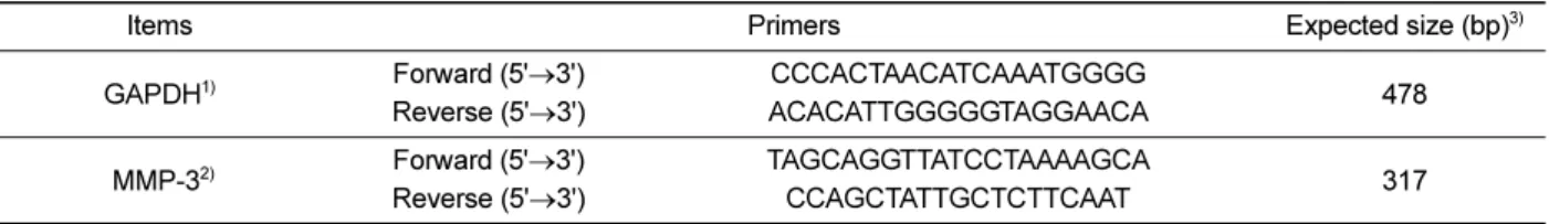
Table 1.
Nucleotide sequence of the primers and expected size of PCR products
Items Primers Expected size (bp)
3)GAPDH
1)Forward (5' Reverse (5'
→→3') 3') ACACATTGGGGGTAGGAACA CCCACTAACATCAAATGGGG 478 MMP-3
2)Forward (5' Reverse (5'
→→3') 3') TAGCAGGTTATCCTAAAAGCA CCAGCTATTGCTCTTCAAT 317
1)
GAPDH: Glyceraldehyde-3-phosphate dehydrogenase
2)
MMP-3: Matrix metalloproteinase-3
3)
bp: basepair
결 과
피부 홍반도, 수분함량, TEWL 비교
홍반도는 정상군에 비해 광노화군과 자연노화군 모두 유의한 증가를 보였으며 자연노화군과 광노화군 간의 유 의한 차이는 보이지 않았다
.
수분함량은 정상군에 비해 광노화군이 유의한 감소를 보였으며 자연노화군은 수치 가 낮았으나 유의한 차이를 보이지 않았다.
자연노화군과광노화군간에는 유의한 차이를 보이지 않았다
.
경표피수분손실량은 정상군에 비해 광노화군은 유의한 증가를 보 였으며 자연노화군은 정상군과 비슷한 수치를 보였다
.
자연노화군에 비해 광노화군은 유의한 증가를 보였다
(Table 2).
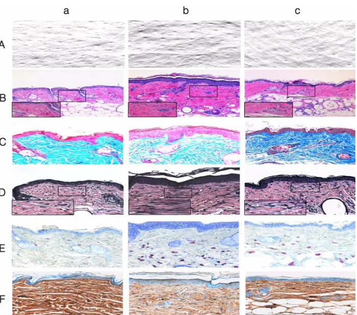
피부 조직의 형태학적 비교
정상군과 비교할 때 광노화군은 주름능선의 두께가 굵 고 간격이 넓으며 주름이 깊게 형성된 반면
,
자연노화군은 광노화군에 비해 상대적으로 주름 능선의 두께가 얇
고 간격이 좁으며 주름이 얕게 형성되어 있었다
(Figure
1A).
피부주름을Visioline
을 이용하여 측정한 결과,
주름의 총넓이
(total winkle area),
주름의 수(No. of wrinkles),
총길이
(total length),
평균길이(mean length),
평균깊이(mean depth)
등5
개 지표모두에서 정상군에 비해광노화군과 자연노화군은 유의하게 증가하였으며자연노화군 에 비해 광노화군에서도 유의하게 증가함이 관찰되었다
(Table 3).
피부의 조직학적 비교
H&E
염색을통해 피부조직 형태및 염증관련 세포를 관찰한 결과
,
정상군은 표피와 진피층 배열이 규칙적이며염증관련 세포들이 거의 보이지 않은 반면
,
광노화군은표피와 진피가 두꺼워졌고진피층 내 림프구
,
대식구 및호중구가 상당수 관찰되었으며
,
표피층의 비후와 피지선의 증식이 관찰되었다
.
자연노화군은 표피층이 얇아졌고표피능의 소실이 관찰되었다
(Figure 1B).
Masson
’s trichrome
염색을 통해진피층 내 교원섬유의 양과 형태를 관찰한 결과,
정상군은 교원섬유의 밀도가조밀하고 배열이 규칙적인 반면 광노화군은 교원섬유가 파괴되어 밀도가엉성하고 배열이 불규칙적이고 양도 많 이 줄어 있었다
.
자연노화군은 광노화군에 비해교원섬유의 밀도와 양의 변화가 적게 나타났다
(Figure 1C).
Verhoeff
’s
염색을 통해 피부 진피층 내 탄력섬유의 변화
,
소실 및 양과 형태를 관찰한 결과,
정상군은 탄력섬유가 규칙적인 반면
,
광노화군은 변성된 탄력섬유가 많이관찰되었다
.
자연노화군은 광노화군에 비해 탄력섬유의변성이 적었으며 탄력 섬유의 직경이 감소하고 잘게 끊 어진 것 같이 길이가 짧았다
(Figure 1D).
Toluidine blue
염색을 통해 진피층 및 피하층 내 비만세포가 분포양상과 탈과립 정도를 관찰한 결과
,
정상군은비만세포가 적게 관찰되었고 탈과립이 거의 없는 반면
,
광노화군은 비만세포가 현저히 많이 관찰되었고 탈과립 정도가 심하게 나타났다
.
자연노화군은 광노화군에 비해비만세포의 수가 적었다
(Figure 1E).
면역조직화학염색결과 진피층 내
TGF-
β 발현 양을관찰한 결과
,
정상군에 비해 광노화군과자연노화군은 발현양이 줄어들었으며 자연노화군에 비해 광노화군이 더 많 이 줄어듬이 관찰되었다
(Figure 1F).
항산화능 비교
적출한 피부조직에서 유해산소를 생성하는 효소인
XO
활성을 측정한 결과
,
정상군에 비해 광노화군과자연노화군에서 유의하게 높았으며
,
자연노화군에 비해 광노화군이 유의하게 높았다
.
유해산소를 제거하는 효소인SOD
Table 2.
Comparison of erythema index, water capacity, TEWL in SKH-1 hairless mice skin at 6 weeks after the beginning of experiment
Items Normal Experimental
N E1 E2
Erythema
index
1)98.07±20.83 261.67±68.85*** 208.81±31.01**
Water
capacity
1)61.83±6.90 43.40±10.87* 49.42±6.19 TEWL
2)6.67±1.42 47.84±20.95
***6.43±1.73
###Values are mean±SD of 6 mice.
1)Unit: AU (Arbitrary Unit)
2)Unit:
g//h TEWL: Transepidermal water loss, N: non-treatment group, E1:
UVB irradiation group, E2: aged group
*
P<0.05, **
P<0.01, ***
P<0.001 compared to the N group, and
###
P<0.001 compared to the E1 group by ANOVA and Duncan's multiple range test.
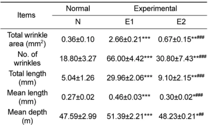
Table 3.
Comparison of wrinkle parameters in SKH-1 hairless mice skin at 6 weeks after the beginning of experiment
Items Normal Experimental
N E1 E2
Total wrinkle
area (mm
2) 0.36±0.10 2.66±0.21*** 0.67±0.15**
###No. of
wrinkles 18.80±3.27 66.00±4.42*** 30.80±7.43**
###Total length
(mm) 5.04±1.26 29.96±2.06*** 9.10±2.15**
###Mean length
(mm) 0.27±0.02 0.46±0.03*** 0.30±0.02*
###Mean depth
(m) 47.59±2.99 51.39±2.21*** 48.23±0.21*
##Values are mean±SD of 6 mice.
N: non-treatment group, E1: UVB irradiation group, E2: aged group,
*
P<0.05, **
P<0.01, ***
P<0.001 compared to the N group, and
##P
<0.01,
###P<0.001 compared to the E1 group by ANOVA and
Duncan's multiple range test.
활성은 정상군에 비해 광노화군과 자연노화군에서 유의 하게 낮았으며 자연노화군과 광노화군 간에는 유의한 차
이는 없었다
. CAT
활성은 정상군에 비해 광노화군과 자연노화군은 유의하게낮았으며 자연노화군과 광노화군 간 에는 유의한 차이는 없었다
(Table 4).
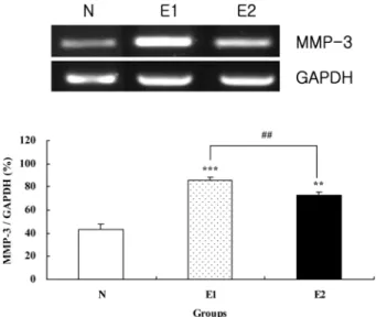
피부조직의 MMP-3 유전자 발현량 비교
피부조직의
MMP-3 mRNA
발현량을 측정한 결과,
정상군에 비해 광노화군과 자연노화군은 유의하게 높았으 며 자연노화군에 비해광노화군은 유의하게 높았다
(Figure 2).
고 찰
많은 연구자들이 피부노화에 지속적인 관심을 보이는 것은 주름살등의 노화현상을막고자하는 이유도 크지만
,
피부는 내적
,
외적인 요소에 의한 노화 현상을 설명하기에 최적의 장기이기 때문이다
(Seo
et al., 2001). Hairless
마우스의 피부에
UVB
를 조사하면 표피가 두꺼워지고 정상적인 교원섬유의구조가 파괴되면서 배열이 무질서해지 며 탄력섬유증
(fibroelstosis)
이 발생하고glycosaminoglycan
(GAG)
가 증가하는 등 자외선에 손상 받은 인간의 피부와거의유사한 피부변화를 보인다
(Fisher
et al., 2002).
이에Figure 1.
Comparison of replica images in SKH-1 hairless mice at 6 weeks after the beginning of experiment a: normal group, b:
UVB irradiation group, c: aged group (A). Histological observation on SKH-1 hairless mouse skin at 6 weeks after the beginning of experiment. H&E stain,
×100 &
×400 (enlarged box) (B). Masson’s trichrome stain,
×200 (C). Verhoeff’s stain,
×200 &
×
400(enlarged box) (D). Toluidine blue stain,
×200 (E). Immunohistochemical staining for TGF-
βin SKH-1 hairless mouse skin at 6
weeks after the beginning of experiment,
×200 (F).
본 연구는
hairless
마우스를 대상으로 광노화군과 자연노 화군을 비교하여 피부노화에관련된 육안적 관찰,
조직학 적 관찰,
분자생물학적 관찰인자들의 변화양상을 알아봄으로써 피부노화 동물평가시험을 위한 객관적인 측정지 표를 제시하고자 하였다
.
홍반은 각질 세포 및 진피 세포들이 관여하여 진피 혈 관을 확장시키는 반응으로
UV
의 파장,
광량,
피부의 조건
,
환경조건 등에 따라 달라지며, UVB
는 진피 상부층까지 도달하여 급속한 화상이나 홍반을 일으킨다
(Trevithick
et al
., 1992).
본 연구에서 홍반도를 측정한결과 정상군에 비해 광노화군과 자연노화군 모두 유의한 증가를 보 였다
.
각질층의 지질은 각질층내 수분 유지기능과 외부 수분 투과에 대한 장벽대로서 중요한 역할을 하는데
(Blanken
et al
., 1989),
자외선에 의한 피부 장벽 손상은 피부표면수분함량 감소와 각질층의 유연성 상실을유발하여 건성 피부나 거친 피부를생성하게한다
(Haratake
et al., 1997;
Jiang
et al., 2007).
피부의 보습과 주름의 중요 인자인hyaluronic acid (HA)
는 세포 외 기질의 주요성분으로 수분 보유
,
세포간 간격 유지,
세포 성장인자 및 영양성분의 저장 및 확산에 관여하며
(Kim
et al., 2005),
피부의노화와 함께 감소되어 피부 탄력저하 및 수분함유량 감 소의 직접적인 원인이라 여겨지고 있다
(Ghersetich
et al.,
1994).
이에 본 연구에서는 수분함량,
경표피수분손실량측정과 조직학적 관찰을 통해 피부손상 정도를 확인하였 다
.
수분함량은 정상군에 비해 광노화군이유의한 감소를보였으며 자연노화군은 수치가 낮았으나 유의한 차이는 보이지 않았다
.
경피수분손실량은 정상군에 비해 광노화군은 유의한 증가를보였지만 자연노화군은 정상군과 비 슷한 수치를 나타내피부장벽의 손상 정도가 상대적으로 적음을 유추할 수 있었다
.
피부 노화 중 주름 생성은 여러 가지원인이 있겠으나
나이의 증가와 환경적인 요인 외에 유전적 요인과 에스 트로겐 등의 호르몬에 의해서도 영향을 받는다
.
반복적인자외선 노출로인한 피부
MMP
증가는교원섬유를 분해하여 진피내의 기질 단백질을 감소시키며 진피층을 얇아
지게 하여 피부주름 형성을 가속화 시킨다
(Wlaschek
etal
., 1995). Oba
와Edwards (2006)
는UVB
를 조사한hairless
마우스에서 조사 기간별로 진피 내 교원섬유 변성 정도를 비교해서 진피내 결합조직의
3
차원적인 구조변화로 주름 형성을 야기한다고 보고했다
.
본 연구에서광노화군은 정상군에 비해 육안적인 관찰에서주름의 깊 이가 깊고 선이 굵은 주름이 넓게 형성되었지만 자연노 화군은 광노화군에 비해 주름의 깊이가 얕고 선이 얇은 주름이 좁게 형성되었다
.
주름양상을 보다 객관적으로분석하기 위해
Visioline
을 사용하여5
개 주름지표(
총 넓이,
주름수
,
총길이,
평균길이,
평균깊이)
를 측정한 결과 광노화군과 자연노화군 모두 정상군보다 유의하게높게 나타 나 피부노화가 상당히 진행되었음을 확인할 수 있었다
.
또한 자연노화군에 비해 광노화군이
5
개 주름지표 모두유의하게 높아
,
광노화군은 자연노화군보다 피부 노화의대표적인 징후인 주름이 현저하게 생성됨을 확인하였다
.
자외선은피부에 활성산소의생성
,
세포자멸사(apoptosis)
등의 반응을 통해
(Matsumura and Ananthaswamy, 2004)
진피에 염증성세포의 침윤을 일으킨다
(Cole
et al., 2010).
본 연구에서 피부 조직의 염증정도를 관찰한 결과 정상 군에비해 광노화 및 자연노화군은 비만세포가현저히 증 가하고 탈과립 정도도 심하였으며
,
염증세포 침윤도 심하게 나타났다
.
자연노화군에 비해 광노화군은 그 정도가더 심하게 관찰되었다
.
Table 4.
Comparison of xanthine oxidase (XO), superoxide dismutase (SOD) and catalase activities (CAT) in SKH-1 hairless mice skin at 6 weeks after the beginning of experiment
ltems Normal Experimental
N E1 E2
XO
1)3.69±0.40 9.24±01.55*** 5.77±1.23*
##SOD
2)20.49±2.02 14.71±0.98* 12.89±1.01**
CAT
3)8.10±1.45 3.47±1.47** 4.54±1.06**
Values are mean±SD of 6 mice.
1)
Unit: nmole uric acid formed/mg protein/min
2)
Unit: U (50% inhibition of autoxidation of hematoxylin)/mg protein/min
3)
Unit: nmole H
2O
2reduced/mg protein/min
N: non-treatment group, E1: UVB irradiation group, E2: aged group
*
P<0.05, **
P<0.01, ***
P<0.001 compared to the N group, and
##P
<0.01 compared to the E1 group by ANOVA and Duncan's multiple range test.
Figure 2.
Comparison of MMP-3 expression in hairless mice
skin at 6 weeks after the beginning of experiment. Values are
mean±SD of 6 mice. **
P<0.01, ***
P<0.001 compared to the N
group by ANOVA and Duncan's multiple range test.
피부의 진피 조직 속에는 피부탄력과 관련된 교원섬유 와 탄력섬유가 그물망 구조를 하고 있는데
(Lee
et al.,
1999),
자연노화가 진행되면 표피가 얇아지고 기저세포층의 유사분열이 감소하며 진피
-
표피 경계부가 편평해지고멜라닌 세포와 랑거한스 세포의 수가 감소한다
.
또한 진피의 섬유모세포 감소
,
세포외기질의 위축 및 교원섬유와탄력섬유의 감소도 동반된다
(Makrantonaki and Zouboulis,
2007).
광 손상 받은 피부는 표피층이 두꺼워지며 진피층도 섬유모세포와 염증세포가 증가하고교원섬유의 무 질서한 배열과 탄력섬유증으로 인해 두꺼워진다
(Seite
etal
., 2006).
본 연구에서 광노화군은 표피와 진피가 비후되고 진피내 교원섬유가많이 파괴되어 관찰되었으며
,
자연노화군도 표피와 진피의 두께가 얇고교원섬유 밀도 감 소 등이 관찰되었나 광노화군에비하면 정도가 심하지 않 았다
.
Chung
등(2001)
은 광노화 피부는 자연노화 피부에 비해 교원섬유 합성은 증가하지만 기질단백 분해 효소인
MMP
의 활성이 증가되어 교원섬유 분해가 더욱 증가하기 때문에 상대적인양은 크게감소한다고 하였다
.
본 시험결과
,
피부조직의MMP-3 mRNA
발현량은 정상군에 비해 광노화군과 자연노화군은 유의하게 높았으며
,
자연노화군에 비해 광노화군도유의하게 높아이론을 뒷받침 하 고 있다고 판단되었다
.
교원섬유는 전체 피부 단백질의
90%
를 차지하는 성분으로
TGF-
β 의해 생산이 촉진되며, activator-1 (AP-1)
이활성화시킨
MMP
에 의해 분해가 촉진된다(Massague, 1998).
자외선에 노출되면TGF-
β 생산은 감소되나AP-1
에 의한
MMP
의 기능은 향상되어 교원섬유의 분해 및변성이 촉진된다
(Fisher
et al., 1996; Quan
et al., 2002).
본 연구에서 진피층 내TGF-
β 발현 양을 관찰한결과 광노화군과 자연노화군에서 발현 양이 줄어들었으 며 광노화군의 감소가 더 두드러졌다
.
세포에 자외선을 조사하여 산화적 스트레스가 유발되 면
ROS
인free radical
이 생성되어 조직의 손상을 초래하며
(Sander et el., 2002),
항산화 효소 등은감소한다(Rittie and Fisher, 2002).
이에 본 연구에서는 피부의 노화유형에 따른 효소적 항산화방어체계의 관련성을 알아보았다
. XO
활성은 정상군보다 광노화군과 자연노화군이 유의하게 높았으며
,
광노화군의 정도가 더 심했다. SOD
활성은 정상군에 비해 광노화군과 자연노화군이 유의하게 낮 았으며 자연노화군과 광노화군은 유의한 차이가 없었다
. CAT
활성은 정상군에 비해 광노화군과 자연노화군이 유의하게 낮았으며 자연노화군과 광노화군은 유의한 차이 는 없었다
.
이는 광노화군이 자연노화군에 비해 생체 피부조직의항산화 방어체계에더 부정적인 작용을하는 것 으로 판단된다
.
이상의 실험결과를 종합해 볼 때
hairless
마우스에서노화유형에 따른 피부변화 양상은 뚜렷한 차이를 보였으 며
,
이들자료는 목적에 맞게 광노화 또는자연노화에 대 한 피부노화 효능을 동물모델을 이용하여평가할 경우기 초 자료로 유용하게 사용될 것으로 기대되었다.
감사의 글
본 연구는
2008
년도 계명대학교 비사(
신진)
연구기금에의하여 수행되었습니다