I Spontaneously Migrated Tip of anImplantable Port Catheter into theAxillary Vein in a Patient with SevereCough and the Subsequent Interventionto Reposition It
4
0
0
전체 글
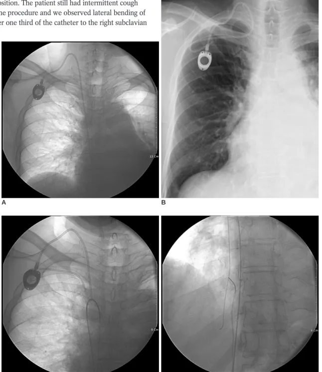
(2) Ahn et al.. Murray Hill, NJ) with a cobra catheter (Cook, Bloomington, IN) was inserted via the second puncture site and it was used to grasp the end of the guide wire in the suprarenal IVC (Fig. 1D). By simultaneous pulling of the wire loop, repositioning of the tip of the port catheter was successfully achieved and the tip was relocated at the initial position. The patient still had intermittent cough during the procedure and we observed lateral bending of the upper one third of the catheter to the right subclavian. A. vein when the patient was coughing. After the reposition procedure, when we induced the patient to cough, we were able to again demonstrate this movement on fluoroscopy (Figs. 1E, F). This was very suggestive to have. B. C D Fig. 1. Migration of implantable port catheter in 64-year-old man. A. Implantable port catheter is inserted via jugular approach. Tip of catheter is well-placed in right atrium. B. Four days after implantation procedure, chest PA radiograph shows coiled catheter and migration of tip from right atrium to right axillary vein. Associated pulmonary edema, cardiomegaly, air space consolidation with volume loss in left lung and pleural thickening with effusion in left hemithorax are also noted. C. 5-Fr pigtail catheter is advanced to right subclavian vein to hook migrated port catheter. D. Gooseneck snare wire and cobra catheter were used to capture wire.. S82. Korean J Radiol 9(Suppl), July 2008.
(3) Implantable Port Catheter Migration into Axillary Vein and Interventional Reposition. E. F Fig. 1. Migration of implantable port catheter in 64-year-old man. E, F. After repositioning (E), catheter shows normal position and curve. However, when we induced patient to cough (F), bending of catheter toward subclavian vein (arrow) was found on fluoroscopy. G. Two days later, recurrent catheter migration was found on chest radiograph. Coiling is noted in middle of catheter, but catheter tip is still located in superior vena cava.. G. caused the catheter migration. Two days later, migration and coiling of the catheter were again demonstrated on the chest posteroanterior radiograph (Fig. 1G) in spite that there was no active physical movement by the patient. Because the tip of the catheter was still located in the superior vena cava and there was a considerable risk of remigration due to the patient’s sustained cough, additional repositioning was not attempted thereafter. There was no additional positional change of the catheter seen on the serial follow up chest radiographs. Chemotherapy was Korean J Radiol 9(Suppl), July 2008. delayed because the patient developed pneumonia. The port was used for administering central venous fluid and the function of the port catheter was preserved well. Sadly, about four weeks later, the patient expired due to aggravated pneumonia and respiratory failure.. DISCUSSION An implantable port device provides an easily accessible central route for long-term chemotherapy patients. Many S83.
(4) Ahn et al.. immediate and late complications of implantable port catheters have been reported on. The early complications include pneumothorax, hematoma, malposition, embolism or arrhythmia, and these are often related to the placement technique. The delayed complications include skin necrosis, infection, catheter fracture, occlusion or thrombosis (2). Migration is one of the well-known complications of implantable port catheters. The incidence of spontaneous migration of a port catheter is reported to be about 0.91.8%, yet the mechanism of migration is not clear. The intravascular and extravascular portions of the catheter are not fixed and both sides are movable. The extravascular component of the port device can be moved by changing the body position or by physical movement, and especially in obese persons or woman with big breasts. Initial positioning of the port is important to prevent this kind of migration. The intravascular portion of the catheter is also movable and this is related with the inherent flexibility of the catheter. The intravascular component of the port catheter can be influenced by high intrathoracic pressure that’s induced by coughing, sneezing, straining or weight lifting. A high infusion flow rate can also make the tip migrate. A case of tip migration after the catheter flushing has been reported (3). Some authors have reported spontaneous migration of the catheter from the subclavian vein into the ipsilateral jugular vein (1, 3 5). Wu et al. (1) reported two cases of implantable port catheter tip migration in patients with severe cough. Our case also presented recurrent catheter migration and this was probably related to the patient’s sustained cough. We identified the bending of the catheter that was induced by cough on fluoroscopy, and this strongly suggested that the migration of the catheter was the result of coughing. If the predisposing factors for this kind of the migration are not corrected, then remigration can occur. Periodic check ups of the catheter location by performing chest radiograph are crucial to detect catheter tip migration. Some authors have recommended monitoring the catheter’s position at least bimonthly when it is not in use and more frequently when it is in use (3). Once migration is detected, prompt correction is important because catheter tip migration can result in further compli-. S84. cations such as thrombosis, venous phlebitis or occlusion. Revision or replacement is usually performed, but if the initial location of the catheter is ideal, then radiologic intervention by transfemoral snaring is useful to correct the tip position (6). The transfemoral approach has several advantages such as avoiding surgery and the associated risk of infection, and it decreases the patient’s discomfort. This repositioning technique showed a high initial success rate of over 80% (7). The transfemoral snaring technique was a quick and easy method to reposition the catheter tip in our patient, and it was convenient for both the operator and the patient. We present here a case of migration of the tip of a port catheter from the right atrium to the right axillary vein in a patient with severe cough. We are sure the coughing was the cause of the catheter tip migration, and we corrected the position of the catheter tip by transfemoral snaring. Radiologists should be familiar with catheter-related complications and their management, and they should pay attention to the catheter position not only during the procedure, but also on the follow up chest radiographs, and especially for the patients who suffer with severe cough.. References 1. Wu PY, Yeh YC, Huang CH, Lau HP, Yeh HM. Spontaneous migration of a Port-a-Cath catheter into ipsilateral jugular vein in two patients with severe cough. Ann Vasc Surg 2005;19:734736 2. Ballarini C, Intra M, Pisani Ceretti A, Cordovana A, Pagani M, Farina G, et al. Complications of subcutaneous infusion port in the general oncology population. Oncology 1999;56:97-102 3. Rasuli P, Hammond DI, Peterkin IR. Spontaneous intrajugular migration of long-term central venous access catheters. Radiology 1992;182:822-824 4. Roblin D, Porter JC, Knight RK. Spontaneous migration of totally implanted venous catheter systems from subclavian into jugular veins. Thorax 1994;49:281-282 5. DiGiacomo JC, Tarlian HS. Spontaneous migration of long-term indwelling venous catheters. JPEN J Parenter Enteral Nutr 1991;15:574-577 6. Lois JF, Gomes AS, Pusey E. Nonsurgical repositioning of central venous catheters. Radiology 1987;165:329-333 7. Hartnell GG, Gates J, Suojanen JN, Clouse ME. Transfemoral repositioning of malpositioned central venous catheters. Cardiovasc Intervent Radiol 1996;19:329-331. Korean J Radiol 9(Suppl), July 2008.
(5)
수치

관련 문서
Since every classical or virtual knot is equivalent to the unknot via a sequence of the extended Reidmeister moves together with the forbidden moves, illustrated in Section 2,
• I owe a great debt of gratitude to the President of the Congress for asking me to speak here today... • I’m happy to have this opportunity to speak with you today and
1 John Owen, Justification by Faith Alone, in The Works of John Owen, ed. John Bolt, trans. Scott Clark, "Do This and Live: Christ's Active Obedience as the
At Wolmi Observatory on the top of Wolmisan Mountain, you can enjoy the view of Incheon Port, the West Sea, and Incheon International Airport. Take a break with a cup of
A crack in a thin sheet a steeper R curve than a crack in a thick plate because of a low degree of stress triaxiality (3축) at the crack tip in the thin sheet, while
Bent posture, and as a fast check of the measurement major pectoral muscle case, the patient lying immediately behind the head comes along,
At the end of the study, a reevaluation of each study case was performed with the same questionnaire. The result shows there has been a meaningful result in the group
[r]
