A Novel Decorin Gene Mutation in Congenital Hereditary Stromal Dystrophy: A Korean Family
5
0
0
전체 글
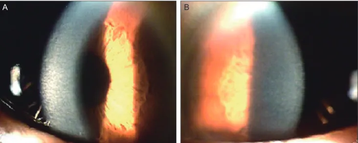
(2) Korean J Ophthalmol Vol.26, No.4, 2012. A. B. C. D. Fig. 1. Slit lamp photography of the patient. (A,B) Right eye. No gross abnormalities of the corneal endothelium, iris and lens were observed. Clouding of the cornea is noticeable under the arcuate slit beam. With magnification, ground-glass corneal opacities are more clearly seen in the anterior stroma, and identifiable small flakes and spots are present throughout the entire stoma. (C,D) Left eye. Density of corneal clouding is less than that of the right cornea.. a flaky pattern of stroma was noted throughout the entire cornea. The right eye had decreased vision and exhibited relatively denser homogenous opacities than the left (Fig. 1). The family members stated that corneal changes had been detected only in the patient’s mother at 69 years of age, and no specific issues had arisen in any other family member or relative. The patient’s father had reported no ophthalmic abnormalities before his death, and his mother had been diagnosed with diffuse corneal opacities of unknown etiology in both eyes three years previously (Fig. 2). She explained that she had experienced decreased vision since childhood, but these deficiencies produced no difficulties in her daily life. The patient’s brother and sister had no symptoms at all and no ophthalmic or systemic abnormalities. As far as the family knew, no one in the paternal 302. or maternal lineage or offspring of the patient had experienced any eye problems except for the patient’s mother (Fig. 3). The endothelium and Descemet’s membrane of the right eye were identified as normal following slit lamp examination. No gross abnormalities, such as Haab’s striae or features of posterior polymorphous corneal dystrophy, were detected in the right eye. The patient’s past medical records from another hospital demonstrated that his endothelial cells of both eyes presented with a normal shape and numbers under a specular microscope about six years ago. However, endothelial cells were found as indeterminate forms using specular microscopy due to the barrier of stromal opacity at the time of our study. The endothelial cells of the left eye were counted using a Konan Noncon.
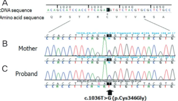
(3) JH Lee, et al. Novel Decorin Gene Mutation in CHSD. A. B. Fig. 2. Slit lamp photography of the patient’s mother. (A) Right eye. Corneal stroma with arcuate slit beam shows diffuse clouding in the right eye. (B) Left eye. Ground-glass corneal opacities and small flakes are similar to that of the right eye.. Light and electron microscopic findings. Fig. 3. Pedigree of the family with stromal dystrophy. ■ and ● represent affected persons.. Robo-8400 noncontact specular microscope (Konan Medical Inc., Hyogo, Japan) as 2564 cells/m2. We assumed that the right eye would have a similar amount of endothelial cells and a relatively uniform morphologic pattern as those of the left. Ultrasound corneal pachymetry (Humphrey Instruments Inc., San Leandro, CA, USA) revealed a central corneal thickness of 658 μm in the right eye and 632 μm in the left. The patient was suspicious for CHSD based upon clinical evidence, and he was scheduled for penetrating keratoplasty of the right eye. A corneal button was sent for light and electron microscopic analysis. There was no problem with corneal wound healing after keratoplasty, and the grafted cornea restored its transparency within two weeks. After 12 months, the corneal graft remained clear, and the patient’s best-corrected visual acuity was 20 / 50 in the right eye.. Light microscopy with hemotoxylin and eosin staining revealed a normal epithelium and uninterrupted Bowman’s membrane. The stromal lamellae were separated slightly from one another, forming a relatively compact space in between the anterior and the posterior stroma (Fig. 4). No Descemet’s membrane or endothelium was detected in the original corneal button, apparently as the result of inappropriate specimen handling. Any infiltration, vessels, inflammatory, or storage material could not be detected. Electron microscopy revealed a criss-crossing pattern of corneal collagen fibers with a relatively electron dense and lucent structure and collagen fibers irregular in shape and size (Fig. 5A). Keratocytes extended widely through the zone of low filaments (Fig. 5B). Genetic analysis Blood was sampled from the patient and family members for DNA collection and analysis [3]. DNA sequencing analysis of the decorin gene in chromosome 12q22 was positive in both the patient and his mother. The novel mutation of a heterozygous, nucleoside substitution (c.1036T>G) point mutation in the decorin gene was detected in both patients (Fig. 6). Lumican and keratocan sequence variants, which are closely located within the decorin gene, did not reveal any mutations. The c.1036T>G mutation resulted in a change of amino acid sequence (p.Cys346Gly). However, no genetic mutations were detected in other family members.. 303.
(4) Korean J Ophthalmol Vol.26, No.4, 2012. A. B. ×400. C. ×100. ×400. Fig. 4. Patient’s light microscope findings. (A) Irregularity of corneal collagen fibril formation. Relatively loose arrangement of collagen fibers in the anterior stroma (B) and a relatively dense arrangement in the posterior stroma (C) are observed.. A. B. ×2,500. ×2,000. Discussion CHSD is an autosomal dominant inherited disease characterized by flaky or feathery stromal clouding. Its disease penetration is variable, and the condition is usually treated with keratoplasty in early childhood. It can be differentiated from congenital hereditary endothelial dystrophy by the relatively normal corneal thickness and absence of corneal 304. Fig. 5. Patient’s transmission electron microscope findings. (A,B) A criss-crossing pattern of corneal fibers are shown with a relatively electron-dense (D) and lucent (L) structure. Irregularity in the collagen fibrils’ shape and thickness is also seen. A keratocyte (K) is apparent in the electron lucent area (L).. edema. Since the first report by Witschel et al. in 1978 [4], CHSD has been considered a rare corneal dystrophy; only four families have been reported: two French, one Belgian and one Norwegian [1,2,4,5]. To the best of our knowledge, our report includes the fifth family of CHSD worldwide and the first to be documented in Asia. The pathogenesis of CHSD is not yet well known. However, previous studies have reported characteristic histologic electron microscop-.
(5) JH Lee, et al. Novel Decorin Gene Mutation in CHSD. A. B C. Fig. 6. Mutation analysis of the decorin gene. Partial sequence chromatograms displaying the wild-type DNA sequence of an unaffected person (A) and the DNA sequence of the mother (B) and patient (C), who were heterozygous for the decorin c.1036T point mutation.. ic findings, which are characterized by multiple abnormal zones with an abnormal lucent ground substance separating the normal corneal stromal lamellae [1]. This pathologic characteristic of CHSD is principally attributable to abnormal fibrillogenesis in the cornea, which ultimately results in apparent stromal opacities and loss of corneal transparency. Experimental evidence indicates that decorin proteins (which consist of dermatan sulfate proteoglycans) contribute to both the lamellar adhesive properties of collagen and the control of regular fibril-fibril spacing observed in the cornea [6-8]. Because corneal transparency requires the regular spacing of collagen fibrils of a uniform diameter and regular interfibrillar space, reduction in vision and associated symptoms can occur as the result of this abnormal fibrillogenesis caused by the truncated decorin protein. The decorin gene mutation was initially reported as a single base pair deletion (c.967delT) detected in a Norwegian family by Bredrup et al. in 2005 [1]. In 2006, Rodahl et al. [2] described another deletion mutation of the decorin gene (c.941delC) in a Belgian family. In this case report, we detected a novel nucleotide substitution (c.1036T>G) in two members of an affected CHSD patient pedigree. This novel decorin gene mutation is different from previous mutations in that it is a nucleotide substitution rather than deletion. Interestingly, our electron microscopic findings were somewhat different from those of previous CHSD cases. The criss-crossing pattern of corneal collagen fibers was relatively intact. Moreover, the degree of visual impairment from corneal opacity seemed to be relatively minimal, since the patient maintained good vision until his late thirties. Usually, patients with CHSD lose their vision in early childhood. Considering the above. distinctive features, we concluded our case as a relatively mild form of CHSD, since the nucleotide substitution may result in less structural or functional changes of the decorin protein than a nucleotide deletion. The authors also assert that the location of the mutation on the decorin gene can modulate the penetration of disease symptoms as well as the onset, duration, severity, and other associated ocular signs. Further investigations of the decorin gene and its diagnostic values are clearly warranted. In summary, the patient in this report evidenced a late manifestation and mild symptoms with normal vision in the unaffected eye. The slight histopathologic changes in the cornea, mild deterioration of vision and novel decorin gene point mutation are dispositive for a mild form of CHSD. However, the unique process of disease and the aforementioned inconsistent tissue arrangements also raise salient questions as to the appropriateness of the diagnosis of CHSD. We categorized this case as an inherited disorder associated with a decorin gene mutation, as was reported by Bredrup et al. in 2005 [1]. Furthermore, future study will be required to evaluate the correlations between this disease and previously published cases of CHSD.. Conflict of Interest No potential conflict of interest relevant to this article was reported.. References 1. Bredrup C, Knappskog PM, Majewski J, et al. Congenital stromal dystrophy of the cornea caused by a mutation in the decorin gene. Invest Ophthalmol Vis Sci 2005;46:420-6. 2. Rodahl E, Van Ginderdeuren R, Knappskog PM, et al. A second decorin frame shift mutation in a family with congenital stromal corneal dystrophy. Am J Ophthalmol 2006;142:520-1. 3. O’Connell JR, Weeks DE. PedCheck: a program for identification of genotype incompatibilities in linkage analysis. Am J Hum Genet 1998;63:259-66. 4. Witschel H, Fine BS, Grutzner P, McTigue JW. Congenital hereditary stromal dystrophy of the cornea. Arch Ophthalmol 1978;96:1043-51. 5. Van Ginderdeuren R, De Vos R, Casteels I, Foets B. Report of a new family with dominant congenital heredity stromal dystrophy of the cornea. Cornea 2002;21:118-20. 6. Bonfield JK, Rada C, Staden R. Automated detection of point mutations using fluorescent sequence trace subtraction. Nucleic Acids Res 1998;26:3404-9. 7. Michelacci YM. Collagens and proteoglycans of the corneal extracellular matrix. Braz J Med Biol Res 2003;36:103746. 8. Danielson KG, Baribault H, Holmes DF, et al. Targeted disruption of decorin leads to abnormal collagen fibril morphology and skin fragility. J Cell Biol 1997;136:729-43.. 305.
(6)
수치


관련 문서
Neuroendocrine Tumors in the Stomach, Duodenum, and Pancreas Accompanied by Novel MEN1 Gene Mutation
Thus, we report a novel MEN1 gene mutation associated with specific pheno- typic features, including brain meningioma and three NETs in the stomach, pancreas and duodenum..
Mutation analysis of the SALL1 gene in the patient shows a single base pair deletion at nucleotide position 1470 (c.1470delG) in exon 2. The same type of mutation was found
A novel de novo mutation in the serine-threonine kinase STK11 gene in a Korean patient with Peutz-Jeghers syndrome.. Jong-Ha Yoo 1 , Jee-Hyoung Yoo 2 , Yoon-Jung Choi 3 , Jung-Gu
A Novel Mutation (A148V) in the Glucose 6-phosphate Translocase ( SLC37A4 ) Gene in a Korean Patient with Glycogen Storage Disease Type 1b.. We report a Korean patient with
Herein, we report a patient with Barth syndrome who had a novel mutation in TAZ gene and experienced unexpected acute exacerbation after contrast dye injection for computed
We report a patient with X-linked CGD who carried a novel mutation, a c.1133A>G (paAsp378Gly) missense mutation, in the CYBB gene.. Key Words: Chronic granulomatous
A novel p.Leu699Pro mutation in MFN2 gene causes Charcot-Marie-Tooth disease type 2A.. Sa-Yoon Kang, Keun Hyuk Ko, and
In three patients with lattice CD type 1 (LCD1), one known mutation (p.R124C) and two novel variants (p.L569Q and p.T621P) in the TGFBI gene were identified.. Conclusions: This
