ISSN 2234-3806 • eISSN 2234-3814
http://dx.doi.org/10.3343/alm.2015.35.3.336
Mutation Analysis of the TGFBI Gene in Consecutive Korean Patients With Corneal Dystrophies
Ju Sun Song, M.D.1,*, Dong Hui Lim, M.D.2,3,*, Eui-Sang Chung, M.D.2, Tae-Young Chung, M.D.2, and Chang-Seok Ki, M.D.1
Department of Laboratory Medicine and Genetics1, Samsung Medical Center, Sungkyunkwan University School of Medicine; Department of Ophthalmology2, Samsung Medical Center, Sungkyunkwan University School of Medicine; Department of Preventive Medicine3, Catholic University School of Medicine, Seoul, Korea
Background: Mutations in the transforming growth factor β-induced gene (TGFBI) are major causes of genetic corneal dystrophies (CDs), which can be grouped into TGFBI CDs. Although a few studies have reported the clinical and genetic features of Korean pa- tients with TGFBI CD, no data are available regarding the frequency and spectrum of TGFBI mutations in a consecutive series of Korean patients with clinically diagnosed CDs.
Methods: Patients with any type of CD, who underwent both ophthalmologic examination and TGFBI gene analysis by Sanger sequencing at a tertiary care hospital in Seoul, Korea from 2006 to 2013, were enrolled in this study.
Results: Among a total of 89 patients, 77 (86.5%) were diagnosed as having clinical TGFBI CD. Seventy-three out of 74 patients (98.6%) with granular CD type 2 (GCD2), had the p.R124H mutation. Of particular note, one patient with rapidly progressive CD had the p.R124H mutation as well as a novel nonsense variant with unknown clinical significance (p.A179*). In three patients with lattice CD type 1 (LCD1), one known mutation (p.R124C) and two novel variants (p.L569Q and p.T621P) in the TGFBI gene were identified.
Conclusions: This study provides epidemiological insight into CDs in a Korean population and reaffirms that GCD2 is the most common TGFBI CD phenotype and that p.R124H is the only mutation identified in patients with GCD2. In addition, we broaden the spectrum of TGFBI mutations by identifying two novel missense variants in patients with LCD1.
Key Words: Granular corneal dystrophy type 2, Korean, Lattice corneal dystrophy type 1, Mutation, TGFBI
Received: September 12, 2014 Revision received: December 8, 2014 Accepted: March 10, 2015
Corresponding author: Tae-Young Chung Department of Ophthalmology, Samsung Medical Center, Sungkyunkwan University School of Medicine, 81 Irwon-ro, Gangnam-gu, Seoul 135-710, Korea Tel: +82-2-3410-3563
Fax: +82-2-3410-0074 E-mail: [email protected]
Co-corresponding author: Chang-Seok Ki Department of Laboratory Medicine and Genetics, Samsung Medical Center, Sung- kyunkwan University School of Medicine, 81 Irwon-ro, Gangnam-gu, Seoul 135-710, Korea
Tel: +82-2-3410-2709 Fax: +82-2-3410-2719 E-mail: [email protected]
*These authors contributed equally to this work.
© The Korean Society for Laboratory Medicine This is an Open Access article distributed under the terms of the Creative Commons Attribution Non-Commercial License (http://creativecom- mons.org/licenses/by-nc/3.0) which permits unrestricted non-commercial use, distribution, and reproduction in any medium, provided the original work is properly cited.
INTRODUCTION
Corneal dystrophies (CDs) comprise a group of hereditary and non-inflammatory corneal diseases with progressive accumula- tion of deposits in different layers of the cornea, resulting in a loss of corneal transparency and visual impairment [1]. The genes that are currently known to be responsible for CD include TGFBI, GSN, K12, K3, M1S1, CHST6, COL8A2, and SLC4A11 [2]. Among these, mutations in the transforming growth factor
beta-induced gene (TGFBI; OMIM 601692) have been associ- ated with several CDs. These CDs include granular CD type 1 (GCD1 or classic CD); granular CD type 2 (GCD2 or Avellino CD);
granular CD type 3 (GCD3 or Reis-Bücklers CD); and lattice CD type 1 or TGFBI type (LCD1). According to the International Committee for Classification of Corneal Dystrophies (IC3D) clas- sification, these CDs belong to category 1, meaning that they are well-defined CDs for which the gene has been mapped and identified and specific mutations are known [3].
An apparent genotype-phenotype correlation has emerged from TGFBI molecular studies. Several recurrent mutations have been shown to be associated with four specific pheno- types: p.R124H in GCD2, p.R555W in GCD1, and p.R124C in LCD1 [4]. Moreover, a number of cases with phenotypic varia- tion for the same mutation, including atypical or variant pheno- types [5-8], have been reported, and novel mutations have also been described. Cumulatively, these data suggest phenotypic and allelic heterogeneity with respect to the TGFBI gene.
A few studies reported the clinical and genetic features of Ko- rean patients with CDs associated with TGFBI mutations; how- ever, these studies selected only patients with genetically con- firmed mutations [9-12]. Owing to the phenotypic and allelic heterogeneity in the TGFBI gene, we analyzed the TGFBI se- quences in Korean patients with multiple types of clinically diag- nosed CD. The purpose of this study was thus to investigate the frequency of TGFBI mutations in patients with multiple types of clinically diagnosed CD and to examine genotype-phenotype correlations and the spectrum of TGFBI mutations.
METHODS
We retrospectively enrolled a consecutive series of unrelated patients who had been clinically diagnosed as having CD and underwent TGFBI gene analysis by Sanger sequencing. Patients were enrolled at a tertiary care hospital in Seoul, Korea, from 2006 to 2013. Slit-lamp examination was performed by profes- sional ophthalmologists for all patients, and environmental and systemic factors were excluded. In addition, none of the patients
showed any response to steroid treatment. The clinical pheno- type of CD was categorized according to the IC3D classification into TGFBI CDs and other CDs [3]. This study was approved by the Institutional Review Board of the Samsung Medical Center.
Genomic DNA was extracted from peripheral blood leuko- cytes by using the Wizard Genomic DNA Purification Kit (Pro- mega, Madison, WI, USA) according to the manufacturer’s in- structions. Each TGFBI exon was amplified by PCR, and the re- sultant amplicons were sequenced on an ABI 3730xl Genetic Analyzer (Applied Biosystems, Foster City, CA, USA). For all pa- tients, TGFBI exon 4 (which can harbor the p.R124H mutation) was analyzed. If negative, other exons (1, 5, 6, 11, 12, 13, and 14) that are also known to harbor frequently detected mutations were analyzed. The remaining exons were also sequenced in patients without any mutations in these exons using primers de- signed by the authors (available on request). TGFBI NM_
000358.2 and TGFBI NP_000349.1 were used as cDNA and protein reference sequences, respectively.
For variants of unknown significance (VUS) identified in the TGFBI gene, in silico analysis was performed with Polymor- phism Phenotyping v2 (PolyPhen-2; http://genetics.bwh.har- vard.edu/pph2) and Sorting Intolerant From Tolerant (SIFT;
http://sift.jcvi.org) to predict pathogenicity. In addition, the loci with VUS were queried in dbSNP142 (http://www.ncbi.nlm.nih.
gov/projects/SNP/), 1000 Genome Project data (http://
www.1000genomes.org/), the National Heart Lung Brain Insti- tute Exome Sequencing Project (ESP) database (http://evs.
gs.washington.edu/EVS/), and an in-house collection of 150 exomes to determine the VUS allele frequencies.
Table 1. Clinical phenotype and spectrum of mutations in the TGFBI gene for 89 patients with corneal dystrophies
Clinical phenotype TGFBI mutation
TGFBI corneal dystrophies Granular corneal dystrophies type 2
(Avellino corneal dystrophies) N=74 p.R124H
p.R124H (;) p.A179*
Not detected
N=72 N=1 N=1 Lattice corneal dystrophies, TGFBI type N=3 p.R124C
p.L569Q p.T621P
N=1 N=1 N=1
Other corneal dystrophies Macular corneal dystrophies N=3 Not detected* N=3
Fuchs endothelial corneal dystrophies N=3 Not detected† N=2
Fleck corneal dystrophies N=2 Not detected N=2
Posterior polymorphous corneal dystrophies N=1 Not detected N=1
Epithelial basement membrane dystrophy N=1 Not detected N=1
Lattice corneal dystrophies, gelsolin type N=1 Not detected N=1
Other‡ N=1 Not detected N=1
*Two patients were analyzed for only exon 4; †One patient was analyzed for only exon 4; ‡Clinical phenotype is not in accordance with a specific category.
RESULTS
A total of 89 patients with CD were included in this study. The median patient age at diagnosis was 48 yr (range, 16-74 yr), and 38.2% of the patients were male. The proportion of patients with TGFBI CD was 86.5% (77/89). Seventy-four patients (83.1%, 74/89) were diagnosed as having as having GCD2 and three (3.3%, 3/89) were diagnosed as having as having LCD1.
No patients with GCD1 or GCD3 were identified in this study (Table 1).
Among the 74 patients clinically diagnosed as having GCD2, 73 (82.0%, 73/89) had the p.R124H mutation, while one pa- tient had no mutations in the TGFBI gene (Fig. 1). One patient with the p.R124H mutation had an additional nonsense variant, p.A179* (c.535C>T) that is predicted to produce a truncated TGFBI protein (Fig. 2A). The patient’s father was also diagnosed as having GCD2 and had the p.R124H mutation but not the p.A179* variant, while her mother did not have an eye problem but refused TGFBI gene testing.
Of the three patients with LCD1, one patient had the p.R124C mutation, while two other patients had novel variants, p.L569Q (c.1706T>A) in exon 13 and p.T621P (c.1861A>C) in exon 14 (Fig. 2B and C). These novel variants were predicted to be pathogenic by in silico analyses and were not identified in db- SNP142, 1000 Genome Project data, the ESP database of more than 13,000 control alleles, or an in-house collection of 150
exomes. A TGFBI mutation was not detected in any of the 12 patients with other types of CD (13.4%, 12/89).
DISCUSSION
This study reaffirms that GCD2 is the most common type of CD in the TGFBI CDs as well as in the multiple types of CD. This high proportion of GCD2 is consistent with the fact that, world- wide, exacerbation of GCD2 after refractive corneal surgery is frequently reported [13]. Thus, our results highlight the impor- tance of early diagnosis of GCD2 through genetic screening in Korea.
The patient with GCD2 who lacked any mutations in the TGFBI gene was a young 26-yr-old woman who showed a few discrete granular deposits in the anterior stroma with unilateral manifestation (Fig. 3A). Although fine, discrete round opacities in the anterior stroma are indicative of early GCD2 symptoms, unilateral manifestation is somewhat atypical [14]. Because the patient refused to provide information regarding her family his- tory, we could not determine whether this condition was inher- ited or not.
A previous study on 42 Chinese patients with particular CD subtypes did not detect TGFBI mutations in three patients with clinically diagnosed GCD2; interestingly, whole gene sequencing revealed that GCD2 was caused by non-TGFBI mutations in these patients [15]. Since TGFBI CD appears to represent a dis-
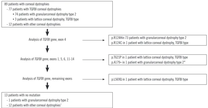
89 patients with corneal dystrophies - 77 patients with TGFBI corneal dystrophies • 74 patients with granularcorneal dystrophy type 2 • 3 patients with lattice corneal dystrophy, TGFBI type - 12 patients with other corneal dystrophies
13 patients with no mutation
- 1 patients with granularcorneal dystrophy type 2 - 12 patients with other corneal dystrophies†
p.R124Hin 73 patients with granularcorneal dystrophy type 2 p.R124C in 1 patient with lattice corneal dystrophy, TGFBI type Analysis of TGFBI gene, exon 4
Analysis of TGFBI gene, exons 1, 5, 6, 11-14
Analysis of TGFBI gene, remaining exons
p.T621P in 1 patient with lattice corneal dystrophy, TGFBI type p.A179* in 1 patient with granularcorneal dystrophy type 2*
p.L569Q in 1 patient with lattice corneal dystrophy, TGFBI type
Fig. 1. Flow chart of the TGFBI mutation analysis protocol and the results of the 89 patients included in the present study.
*The p.A179* mutation was additionally found in one patient with the p.R124H mutation; †Three patients underwent sequence analysis of only exon 4.
ease spectrum, rather than discrete phenotypes, with increas- ingly atypical or variant phenotypes, genetic diagnosis some- times challenges the apparent clinical diagnosis when few TGFBI mutations are found in the atypical group [16].
Both a novel p.A179* variant and the p.R124H mutation were identified in a 35-yr-old woman who experienced sudden deteri- oration of visual acuity in both eyes. In addition, both corneas exhibited progressed granular opacities with a dense confluent pattern that covered most of the corneal surface (Fig. 3B). We confirmed that the p.R124H mutation was inherited from her fa- ther, who also had GCD2. However, we could not test whether the patient’s mother had the p.A179* variant or not. Assuming the p.A179* variant did not occur de novo, the patient was likely compound heterozygous for the two variants. The clinical signifi- cance of p.A179* is not clear because this variant might be in- herited from the patient’s mother who had no apparent eye problems. Previously, a Japanese patient with compound hetero- zygous variants (p.R124H and p.G470*) has been reported and Fig. 2. Sequence electrophoerograms of novel TGFBI variants. (A) The c.535C>T (p.A179*) variant identified in a patient with granu- lar corneal dystrophy type 2 (arrow). (B) The c.1706T >A (p.
L569Q) variant identified in a patient with lattice corneal dystrophy, TGFBI type (arrow). (C) The c.1861A>C (p.T621P) variant identi- fied in a patient with lattice corneal dystrophy, TGFBI type (arrow).
Control
Control
Control Patient
Patient
Patient A
B
C
the pathogenicity of the nonsense variant (p.G470*) was unclear because the patient’s daughter with the p.G470* variant was un- affected [17].
Two novel variants, p.L569Q and p.T621P, were detected in each patient affected by LCD1. These two patients showed a phenotype of larger lattice lines and recurrent corneal erosions with typical lattice lines, retrospectively (Fig. 3C and D). Lattice CD variants (types IIIA, I/IIIA, IV, and polymorphic amyloidosis), which belong to LCD1, are caused by more than two dozen dis- tinct heterozygous mutations and are known to exhibit muta- tional and phenotypic variability [7]. In particular, lattice CD, type IIIA may exhibit larger lattice lines, although the lattice pattern depends on age; moreover, corneal erosions are a typical sign of Fig. 3. Slit-lamp photographs. (A) Patient with GCD2 without any TGFBI mutations and a few discrete granular deposits in the anteri- or stroma with unilateral manifestation. (B) Patient with GCD2 car- rying a novel p.A179* variant, as well as the p.R124H mutation, with progressed granular opacities in a dense confluent pattern covering most of the corneal surface. (C) Patient with LCD1, carry- ing the p.L569Q variant and large lattice lines. (D) Patient with LCD1, carrying the p.T621P variant with recurrent corneal erosions with typical lattice lines.
A
B
C
D
lattice CD, types IIIA and I/IIIA [3]. Nearly all of these mutations are located in the fourth FAS1 domain of TGFBI; two novel vari- ants in these two patients were also located in the fourth FAS1 domain of TGFBI [3].
Other non-TGFBI CD phenotypes were not uncommon. None of the 12 patients with these types of CD had a mutation in the TGFBI gene. However, among these patients, two with macular CD and one with Fuchs endothelial CD only underwent se- quence analysis of exon 4. To exclude TGFBI mutations, analy- sis of all exons is needed for these two patients.
This is the first study to investigate the clinical phenotypes and spectrum of mutations in the TGFBI gene in consecutive Korean patients with CD. Previous reports studying TGFBI fo- cused only on patients with TGFBI mutations confirmed by ge- netic testing. The population of the present study is thus a wider representation of the range of clinical and genetic characteris- tics of patients with CD. GCD2 was the most common CD phe- notype observed, and the p.R124H mutation was the only causal mutation of GCD2 while two novel missense variants in lattice CD and one novel nonsense variant in GCD2 were also identified. Moreover, sequence analysis of the entire TGFBI gene in patients with clinical phenotypes suggestive of homozy- gous mutations is needed to rule out the possibility of com- pound heterozygosity.
Authors’ Disclosures of Potential Conflicts of Interest
No potential conflicts of interest relevant to this article were re- ported.
Acknowledgments
This study was supported in part by a grant of the SMC-KIST Translational Research Program in 2014.
REFERENCES
1. Zhang T, Yan N, Yu W, Liu Y, Liu G, Wu X, et al. Molecular genetics of Chinese families with TGFBI corneal dystrophies. Mol Vis 2011;17:380- 7.
2. Klintworth GK. Corneal dystrophies. Orphanet J Rare Dis 2009;4:7.
3. Weiss JS, Moller HU, Lisch W, Kinoshita S, Aldave AJ, Belin MW, et al.
The IC3D classification of the corneal dystrophies. Cornea 2008;27 Suppl 2:S1-83.
4. Zhu Y, Shentu X, Wang W. The TGFBI R555W mutation induces a new granular corneal dystrophy type I phenotype. Mol Vis 2011;17:225-30.
5. Abazi Z, Magarasevic L, Grubisa I, Risovic D. Individual phenotypic variances in a family with Avellino corneal dystrophy. BMC Ophthalmol 2013;13:30.
6. Aldave AJ and Sonmez B. Elucidating the molecular genetic basis of the corneal dystrophies: are we there yet? Arch Ophthalmol 2007;125:
177-86.
7. Nowinska AK, Wylegala E, Janiszewska DA, Dobrowolski D, Aragona P, Roszkowska AM, et al. Genotype-phenotype correlation of TGFBI cor- neal dystrophies in Polish patients. Mol Vis 2011;17:2333-42.
8. Yoshida S, Yoshida A, Nakao S, Emori A, Nakamura T, Fujisawa K, et al.
Lattice corneal dystrophy type I without typical lattice lines: role of mu- tational analysis. Am J Ophthalmol 2004;137:586-8.
9. Kim HS, Yoon SK, Cho BJ, Kim EK, Joo CK. BIGH3 gene mutations and rapid detection in Korean patients with corneal dystrophy. Cornea 2001;20:844-9.
10. Jun RM, Tchah H, Kim TI, Stulting RD, Jung SE, Seo KY, et al. Avellino corneal dystrophy after LASIK. Ophthalmology 2004;111:463-8.
11. Lee JH, Cristol SM, Kim WC, Chung ES, Tchah H, Kim MS, et al. Preva- lence of granular corneal dystrophy type 2 (Avellino corneal dystrophy) in the Korean population. Ophthalmic Epidemiol 2010;17:160-5.
12. Cho KJ, Mok JW, Na KS, Rho CR, Byun YS, Hwang HS, et al. TGFBI gene mutations in a Korean population with corneal dystrophy. Mol Vis 2012;18:2012-21.
13. Han KE, Kim TI, Chung WS, Choi SI, Kim BY, Kim EK. Clinical findings and treatments of granular corneal dystrophy type 2 (avellino corneal dystrophy): a review of the literature. Eye Contact Lens 2010;36:296-9.
14. Holland EJ, Daya SM, Stone EM, Folberg R, Dobler AA, Cameron JD, et al. Avellino corneal dystrophy. Clinical manifestations and natural histo- ry. Ophthalmology 1992;99:1564-8.
15. Yam GH, Wang K, Jhanji V, Choy KW, Baum L, Pang CP. In vitro amy- loid aggregate forming ability of TGFBI mutants that cause corneal dys- trophies. Invest Ophthalmol Vis Sci 2012;53:5890-8.
16. Vincent AL, de Karolyi B, Patel DV, Wheeldon CE, McGhee CN. TGFBI mutational analysis in a New Zealand population of inherited corneal dystrophy patients. Br J Ophthalmol 2010;94:836-42.
17. Sakimoto T, Kanno H, Shoji J, Kashima Y, Nakagawa S, Miwa S, et al. A novel nonsense mutation with a compound heterozygous mutation in TGFBI gene in lattice corneal dystrophy type I. Jpn J Ophthalmol 2003;
47:13-7.