INTRODUCTION
Complement component 7 (C7) is one of the five terminal complement components that generate a multi-protein com- plex known as the membrane attack complex (MAC) upon activation of either the classical or alternative complement system. MAC is comprised of the complement components C5b, C6, C7, C8, and C9. On cell membranes, this complex becomes MAC, which is capable of forming trans-membrane channels through which ions migrate, leading to cell lysis and cell death (1). The killing function of MAC is best demon- strated in individuals with a homozygous deficiency of MAC protein: since MAC is essential to maintain localization and to prevent dissemination of infection caused by Gram-nega- tive organisms such as Neisseria gonorrhoeae and N. meningitides, these individuals have increased susceptibility to develop a disseminated disease of such infections (2).
The deficiency of C7 leads to recurrent meningococcal infec- tions, and it is often discovered by complement assays in such conditions (3). The C7 deficiency is an autosomal recessive disorder and has mostly been reported in Caucasians (4). The gene encoding the C7 protein has been assigned to the chro- mosome band 5p13 (5). The molecular bases of C7 deficiencies have been reported, as well as those of subtotal deficiencies
of C7 (6-8). In this report, we describe a Korean patient with meningococcal meningitis who was diagnosed as having C7 deficiency based upon complement assays and C7 gene muta- tion analysis. This is the first confirmed case of C7 deficien- cy in Korea and the two mutations detected in the patient have not been reported previously.
MATERIALS AND METHODS Subjects
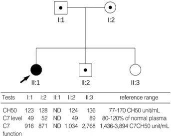
The proband, an 11-yr-old Korean girl, was admitted be- cause of meningococcal meningitis. Laboratory screening tests for underlying immunodeficiency revealed that the total he- molytic activity (CH50) was undetectable in the serum of the patient. Under the suspicion of an inherited complement deficiency, her family members, both parents and two siblings, were recruited for further studies after obtaining informed consent. The total serum hemolytic activities in these family members were within normal range (Fig. 1). Subsequent anal- ysis by radial immunodiffusion assay with antibodies against the main principal complement components revealed that there was no detectable C7 in the serum of the patient, whereas
Chang-Seok Ki, Jong-Won Kim, Hee-Jin Kim, Sung-Min Choi*, Gyoung-Yim Ha�, Hee Jung Kang�, Won-Duck Kim*
Department of Laboratory Medicine, Samsung Medical Center, Sungkyunkwan University School of Medicine, Seoul; Departments of Pediatrics*and Laboratory Medicine�, College of Medicine, Dongguk University, Gyeongju; Department of Laboratory Medicine�, Hallym University College of Medicine, Anyang, Korea
Address for correspondence Won-Duck Kim, M.D.
Department of Pediatrics, Dongguk University, College of Medicine, 707 Sukjang-dong, Gyeongju 780-714, Korea
Tel : +82.54-770-8253, Fax : +82.54-741-2093 E-mail : [email protected]
*The first two authors contributed equally to this work.
220
Two Novel Mutations in the C7 gene in a Korean Patient with Complement C7 Deficiency
Complement C7 deficiency is an autosomal recessive disorder well known to be associated with increased susceptibility to meningococcal infection and has mostly been reported in Caucasians. In the Korean population, no case of C7 deficiency has been reported to date. Recently we experienced an 11-yr-old girl with meningo- coccal meningitis who was diagnosed as having C7 deficiency based upon the unde- tectable serum C7 protein on radial immunodiffusion and the undetectable serum total and C7 hemolytic activities. To identify the genetic basis of the C7 deficiency of the patient, we performed a mutation analysis for the C7 gene and found two novel mutations; a point mutation at the 3′splice acceptor site of intron 4 (c.281-1G>T) and a large deletion mutation encompassing almost the whole C7 gene from exon 1 to exon 17 (c.1-?_2350+?del). A haplotype analysis showed that the large deletion mutation was inherited from the patient’s father. To the best of our knowledge, this is the first confirmed case of C7 deficiency in Korea.
Key Words : Complement 7; Deficiency; C7 Gene; Mutation, Missense; Meningitis, Meningococcal; Koreans
Received : 6 August 2004 Accepted : 6 November 2004
the C7 concentrations in the sera of both parents and younger brother were 49-52% of the normal plasma level (reference range, 80-120%). The functional deficiency of C7 was con- firmed by a hemolytic assay that showed the patient’s serum was unable to recover the hemolytic activity of C7-deficient serum. The C6 levels of the patient and the family members were all within normal range.
Direct sequencing of the C7 gene
Genomic DNA was isolated from peripheral blood leuko- cytes using the Wizard Genomic DNA Purification kit ac- cording to the manufacturer’s instructions (Promega, Madison, WI, U.S.A.). All coding exons and flanking intron regions of the C7 gene were amplified by using primer sets designed by the authors (available upon request). The polymerase chain reaction (PCR) was performed with a thermal cycler (model 9600, Applied Biosystems, Foster City, CA, U.S.A.) as fol- lows: 32 cycles of denaturation at 94℃for 30 sec, annealing at 60℃for 30 sec, and extension at 72℃for 30 sec. The amplicon (5 L) was treated with 10 U shrimp alkaline phos- phatase and 2 U exonuclease I (USB Corp., Cleveland, OH, U.S.A.) at 37℃for 15 min and was incubated at 80℃for 15 min for enzyme inactivation. Cycle sequencing was per- formed on the ABI Prism 3100 Genetic Analyzer with the BigDye Terminator Cycle Sequencing Ready Reaction kit (Applied Biosystems, Foster City, CA, U.S.A.).
Paternity testing and haplotype analysis
Paternity testing was performed using 5 short tandem re- peat (STR) markers from 5 different chromosomes (D7S820, D8S1179, D18S51, D21S11, and TH01 on chromosome
11p15.5). Haplotype analysis for the C7 gene region was per- formed using 7 STR markers flanking the C7 gene (D5S630, D5S416, D5S2031, D5S419, D5S1993, D5S674, and D5- S426) and 3 intragenic single nucleotide polymorphisms (SNP; c.665G>A, c.1166G>A, and c.1182+10G>A). All STR markers were obtained from the ABI PRISM Linkage Mapping Set v2.5 (Applied Biosystems) and were analyzed on the ABI Prism 3100 Genetic Analyzer and the Genescan software (Applied Biosystems).
Gene dosage analysis
To confirm the deletion mutation of the C7 gene, a multi- plex PCR system was designed. The fourth exon of the low- density lipoprotein receptor (LDLR) gene was arbitrarily se- lected and was simultaneously amplified with each exon of the C7 gene. After electrophoresis of the PCR products on a 2% agarose gel, the density (D) of each band was measured by using the Gel-Doc 1000 Documentation System and Qu- antity One Software (Bio-Rad Laboratories, Hercules, CA, U.S.A.). Any exon with a density ratio (DR) less than 0.75, an arbitrary cutoff, was considered as being deleted: DR={[patientDC7 exon/ (patientDC7 exon+patientDLDLR exon 4)]/[controlDC7 exon/(controlDC7 exon+control DLDLR exon 4)], where D is the band density of any exon of the C7 gene or exon 4 of the LDLR gene.
RESULTS
The sequencing analysis indicated that the patient was a
I:1 I:2
II:1 II:2 II:3
Tests I:1 I:2 II:1 II:2 II:3 reference range CH50 123 128 ND 124 136 77-170 CH50 unit/mL C7 level 49 52 ND 49 89 80-120% of normal plasma C7 916 871 ND 1,034 2,768 1,436-3,894 C7CH50 unit/mL function
Fig. 1.Pedigree of the family with C7 deficiency. The solid circle represents the proband (arrow) and squares/circle with a central dot represent carriers. The table at the bottom shows the relevant complement profile in each individual. ND, not detectable.
Fig. 2.Direct sequencing analysis of the family demonstrated a novel G-to-T transversion (c.281-1G>T) at the 3′splice acceptor site of intron 4 (arrow). The patient’s mother and brother were het- erozygous for the same mutation. However, the patient’s father did not carry the mutation.
Father (I:1)
Mother (I:2)
Proband (II:1)
Brother (II:2)
Sister (II:3)
G/G
T G G T G T T C A GG T C A G T G C A Intron 4 Exon 5
G/T
T/T
G/T
G/G
homozygous carrier of a G-to-T transversion mutation in the consensus splice-acceptor site of the intron 4 (c.281-1G>T), presumably resulting in skipping of exon 5 of the C7 gene during the RNA processing and splicing step. This led us to suspect that the patient’s parents would be heterozygous carriers of this splice-site mutation; however, the sequencing analysis revealed that the patient’s father was homozygous for the wild type allele for this locus, while her mother was a het- erozygous carrier for the mutation (Fig. 2). In addition, the patient was homozygous for three intragenic SNPs (c.665G>
A in exon 7, c.1166G>A in exon 10, and c.1182+10G>A in intron 14), while her father was homozygous for the oppo- site alleles for the three SNPs.
From this observation, we first performed paternity testing with five STR markers (D7S820, D8S1179, D18S51, D21- S11, and TH01), which indicated a paternity probability of 0.999 between the patient and his father (data not shown).
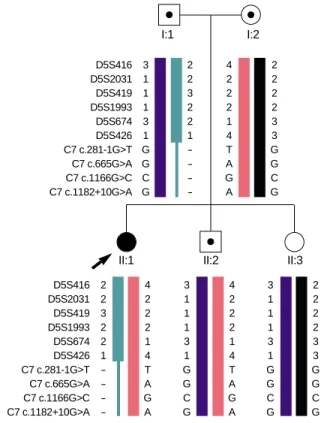
Haplotype analysis using 7 STR markers flanking the C7 gene and 3 intragenic SNPs showed that the patient inherited bi- parental alleles for all STR markers but had only mother’s alleles for the c.281-1G>T mutation and all 3 intragenic SNPs (Fig. 3). Therefore, the patient and her father were suspected to have a large deletion mutation encompassing at least from the splice-site mutation to intron 14 of the C7 gene. Further analysis by a semi-quantitative gene dosage analysis using the multiplex PCR method showed that all exons of the C7
gene except for exon 18 were deleted (Fig. 4). To delineate the telomeric breakpoint of the deletion, three exons of the CARD6 gene, located immediately telomeric to C7, were tested and were shown to have a normal gene dosage, indicat- ing that the breakpoint should lie between the CARD6 gene and the C7 gene, whereas the centromeric breakpoint occurred between exon 17 and exon 18 of the C7 gene (standard nomen- clature: c.1-?_2350+?del).
The younger brother of the patient inherited the c.281-1G>
T mutation from his mother. All heterozygous carriers (the patient’s father, mother, and younger brother) showed decreased C7 levels and functions but had normal levels of CH50.
DISCUSSION
C7 is a single-chain polypeptide composed of 821 amino acid residues and is structurally similar to the other compo- nents of MAC; C6, C8 , C8 , and C9 (9, 10). The C7 gene spans about 80 kb of DNA and is encoded by 18 exons (10).
To date, more than 15 different molecular defects leading to total or subtotal C7 deficiency have been reported (11, 12).
Here we report two novel mutations of the gene in a Korean family with C7 deficiency. In order to identify mutations in the C7 gene, we performed a sequencing analysis for all exons
Fig. 3.Haplotype analysis in the family. The solid circle represents the proband (arrow) and squares/circle with a central dot repre- sent carriers. The patient inherited a large deletion in the C7 gene from her father.
I:1
D5S416 3 D5S2031 1 D5S419 1 D5S1993 1 D5S674 3 D5S426 1 C7 c.281-1G>T G C7 c.665G>A G C7 c.1166G>C C C7 c.1182+10G>A G
2 2 3 2 2 1 - - - -
4 2 2 2 1 4 T A G A
2 2 2 2 3 3 G G C G
D5S416 2 D5S2031 2 D5S419 3 D5S1993 2 D5S674 2 D5S426 1 C7 c.281-1G>T - C7 c.665G>A - C7 c.1166G>C - C7 c.1182+10G>A -
4 2 2 2 1 4 T A G A
3 1 1 1 3 1 G G C G
4 2 2 2 1 4 T A G A
3 1 1 1 3 1 G G C G
2 2 2 2 3 3 G G C G I:2
Proband
Exon 8
IC T Dagj
=0.6
Dagj
=3.4
Dagj
=1.3
Dagj
=3.0 Exon 18
IC T LDLR exon4 (IC) C7 exons (T)
Exon 8
IC T Exon 18
IC T Control
M Exon 2 3 4 8 15 16 17 18
Exon DR
2 0.154
3 0.364
4 0.364
8 0.455
15 0.529
16 0.583
17 0.667
18 1.125
A
B
Fig. 4.Gene dosage analysis by multi- plex PCR method. (A) The fourth exon of the LDLR gene [internal control (IC);
expected band size, 543 bp] was simul- taneously amplified with each exon of the C7 gene (T). The adjusted band density of each C7 exon [Dadj=DC7 exon/ (DC7 exon+DLDLR exon 4)] in the proband was calculated and was compared with that in a healthy control, giv- ing a density ratio (DR) of each exon [DR=(Dadjof proband)/(Dadj of control)], where D is the band density of any C7 exon or exon 4 of the LDLR gene.
II:1 II:2 II:3
and their flanking introns of the gene. The patient was homo- zygous carriers of a splicing mutation (c.281-1G>T) and three polymorphisms of the C7 gene (c.665G>A, c.1166G>A, and c.1182+10G>A). As expected, the patient’s mother was a heterozygous carrier of the mutation and all three polymor- phisms, but the patient’s father had wild type alleles for all these variations. Extended haplotype analysis with 7 STR markers flanking the C7 gene revealed that the patient inher- ited a large deletion of the C7 gene from her father. Gene dosage analysis by multiplex PCR of each exon showed that the deletion encompassed from exon 1 to exon 17, the largest deletion reported to date in the terminal complement defi- ciency. The G-to-T transversion mutation in intron 4 gener- ates a defect in the splice acceptor site and results in skipping of exon 5 (148 bp), which is expected to produce a premature stop codon at position 130. These mutations have led to a loss of complement lytic function. The family study demon- strated that the patient’s brother also carried one C7 allele with the G-to-T transversion mutation in intron 4. Nonsense or frameshift mutations in human disease genes frequently cause severe reduction of corresponding mRNA and resultant protein deficiency. Occasionally, nonsense mutations are asso- ciated with normal mRNA levels, but truncated proteins. The mechanisms for the reduced expression of nonsense mutated mRNA are incompletely understood, but they seem to be diverse and to involve cytoplasmic and nuclear rates of decay of cytoplasmic mRNA (13, 14).
Epidemiological data on terminal complement deficiencies show that they, as a group, are more prevalent than early com- ponent deficiencies. In addition, there appears to be an ethnic predilection for specific terminal complement deficiencies:
C6 and C8 - deficiencies are common in blacks, whereas C7 and C8 deficiencies are predominantly observed in Cau- casians. The cases of C7 deficiency have been reported more frequently in Spanish, Israeli, and Irish than in Asians in Japan and Korea (6, 7, 11, 12).
Of interest, there is no uniform association between any specific geographic/ethnic group and meningococcal disease.
While there was no patient with a complement gene deficien- cy among the survivors of Neisserial epidemics in the Faroe Islands and Denmark (15, 16), a high prevalence of C7 defi- ciency was noted among Moroccan Jews in Israeli populations (17, 18). These data may reflect environmental factors such as the endemic rate of infections and climatic differences and/
or genetic differences in the host population (19). An ancil- lary finding in many studies of families with complement- deficient propositus is the existence of complement-deficient siblings who had never been infected. This also indicates dif- ferent susceptibility among complement-deficient individu- als with the same genetic defect, e.g., C6 deficient patients who had survived one episode of meningococcal infection or had never experienced an infection were shown to be at less risk than those who had experienced recurrent infections (20).
On the other hand, the immunity to meningococcal disease
may also be determined by the interaction between IgG and the IgG Fc receptor (Fc R) on polymorphonuclear cells (anti- body-mediated phagocytosis). It has been hypothesized that particular combinations of Fc R allotypes may determine the susceptibility to meningococcal disease, especially in per- sons who lack serum bactericidal activity because of deficiency of a component in the terminal pathway of the complement system (21, 22). Recent study by van der Pol et al. found that there was no statistical significance in the association between particular combinations of Fc R allotypes and meningococ- cal disease (23). These authors suggested that the genetic het- erogeneity on the long arm of chromosome 1, which is enrich- ed with immune-modulating genes, including pentraxin, selectin genes, and several complement regulatory proteins, may provide important information with respect to host de- fense against meningococci (24).
Screening for complement deficiencies is not a priority in most countries. Routine screening of a child after meningo- coccal disease is not necessary, if the infection is caused by group B or C. However, an investigation is warranted if there is a relevant past medical history such as previous suspicious infections, an abnormal course of infective illness, infection with non-groupable or unusual serogroups or if the current illness is a repeated episode of neisserial infections (25). Admi- nistration of prophylactic antibiotics should be considered in a patient with complement deficiency, although it may encour- age drug resistance (20). Also, vaccination with the tetravalent polysaccharide vaccine can prevent meningococcal disease due to the meningococcal serogroups A, C, Y, and W135 (26).
ACKNOWLEDGMENT
This work was supported by the National Research Labo- ratory Grants from the Korea Institute of Science and Tech- nology Evaluation and Planning, Korea.
REFERENCES
1. Muller-Eberhard HJ. The membrane attack complex of complement.
Annu Rev Immunol 1986; 4: 503-28.
2. Wurzner R, Orren A, Lachmann PJ. Inherited deficiencies of the ter- minal components of human complement. Immunodefic Rev 1992;
3: 123-47.
3. Behrman RE, Kliegman, RM, Jenson, HB. Nelson textbook of pedi- atrics. 17th ed. Philadelphia: Elsevier Science; 2000: 728-30.
4. Halle D, Elstein D, Geudalia D, Sasson A, Shinar E, Schlesinger M, Zimran A. High prevalence of complement C7 deficiency among healthy blood donors of Moroccan Jewish ancestry. Am J Med Genet 2001; 99: 325-7.
5. Jeremiah SJ, Abbott CM, Murad Z, Povey S, Thomas HJ, Solomon E, DiScipio RG, Fey GH. The assignment of the genes coding for human complement components C6 and C7 to chromosome 5. Ann
Hum Genet 1990; 54 (Pt 2): 141-7.
6. Nishizaka H, Horiuchi T, Zhu ZB, Fukumori Y, Volanakis JE. Genetic bases of human complement C7 deficiency. J Immunol 1996; 157:
4239-43.
7. Fernie BA, Orren A, Sheehan G, Schlesinger M, Hobart MJ. Molec- ular bases of C7 deficiency: three different defects. J Immunol 1997;
159: 1019-26.
8. Fernie BA, Wurzner R, Orren A, Morgan BP, Potter PC, Platonov AE, Vershinina IV, Shipulin GA, Lachmann PJ, Hobart MJ. Molec- ular bases of combined subtotal deficiencies of C6 and C7: their effects in combination with other C6 and C7 deficiencies. J Immunol 1996;
157: 3648-57.
9. DiScipio RG, Chakravarti DN, Muller-Eberhard HJ, Fey GH. The structure of human complement component C7 and the C5b-7 com- plex. J Biol Chem 1988; 263: 549-60.
10. Hobart MJ, Fernie BA, DiScipio RG. Structure of the human C7 gene and comparison with the C6, C8A, C8B, and C9 genes. J Immunol 1995; 154: 5188-94.
11. Behar D, Schlesinger M, Halle D, Ben-Ami H, Edoute Y, Shahar E, Kasis I, Shihab S, Elstein D, Zimran A, Mandel H. C7 complement de- ficiency in an Israeli Arab village. Am J Med Genet 2002; 110: 25-9.
12. Vazquez-Bermudez MF, Barroso S, Walter K, Alvarez AJ, Alarcon A, Lopez-Trascasa M, Wichmann I, Aguilar F, Nunez-Roldan A, Sanchez B. Complement component C7 deficiency in a Spanish fam- ily. Clin Exp Immunol 2003; 133: 240-6.
13. Losson R, Lacroute F. Interference of nonsense mutations with eukary- otic messenger RNA stability. Proc Natl Acad Sci USA 1979; 76:
5134-7.
14. Barker GF, Beemon K. Nonsense codons within the Rous sarcoma virus gag gene decrease the stability of unspliced viral RNA. Mol Cell Biol 1991; 11: 2760-8.
15. Rasmussen JM, Teisner B, Weihe P, Mathiassen B, Petersen T, Isager H. Screening for complement deficiencies in patients surviving from epidemic meningococcal disease. J Clin Lab Immunol 1988; 25: 161-5.
16. Rasmussen JM, Brandslund I, Teisner B, Isager H, Svehag SE, Maarup L, Willumsen L, Ronne-Rasmussen JO, Permin H, Andersen PL.
Screening for complement deficiencies in unselected patients with meningitis. Clin Exp Immunol 1987; 68: 437-45.
17. Zimran A, Rudensky B, Kramer MR, Tedesco F, Ehrenfeld M, Raz
R, Greif Z, Gelber M, Lishner M, Golan E. Hereditary complement deficiency in survivors of meningococcal disease: high prevalence of C7/C8 deficiency in Sephardic (Moroccan) Jews. Q J Med 1987;
63: 349-58.
18. Schlesinger M, Nave Z, Levy Y, Slater PE, Fishelson Z. Prevalence of hereditary properdin, C7 and C8 deficiencies in patients with me- ningococcal infections. Clin Exp Immunol 1990; 81: 423-7.
19. Wurzner R, Witzel-Schlomp K, Tokunaga K, Fernie BA, Hobart MJ, Orren A. Reference typing report for complement components C6, C7 and C9 including mutations leading to deficiencies. Exp Clin Immuno- genet 1998; 15: 268-85.
20. Potter PC, Frasch CE, van der Sande WJ, Cooper RC, Patel Y, Orren A. Prophylaxis against Neisseria meningitidis infections and antibody responses in patients with deficiency of the sixth component of com- plement. J Infect Dis 1990; 161: 932-7.
21. Salmon JE, Edberg JC, Brogle NL, Kimberly RP. Allelic polymor- phisms of human Fc gamma receptor IIA and Fc gamma receptor IIIB. Independent mechanisms for differences in human phagocyte function. J Clin Invest 1992; 89: 1274-81.
22. Fijen CA, Bredius RG, Kuijper EJ, Out TA, De Haas M, De Wit AP, Daha MR, De Winkel JG. The role of Fcgamma receptor polymor- phisms and C3 in the immune defence against Neisseria meningitidis in complement-deficient individuals. Clin Exp Immunol 2000; 120:
338-45.
23. van der Pol WL, Huizinga TW, Vidarsson G, van der Linden MW, Jansen MD, Keijsers V, de Straat FG, Westerdaal NA, de Winkel JG, Westendorp RG. Relevance of Fcgamma receptor and interleukin- 10 polymorphisms for meningococcal disease. J Infect Dis 2001; 184:
1548-55.
24. Tsokos GC, Liossis SN. Immune cell signaling defects in lupus: acti- vation, anergy and death. Immunol Today 1999; 20: 119-24.
25. Hoare S, El-Shazali O, Clark JE, Fay A, Cant AJ. Investigation for complement deficiency following meningococcal disease. Arch Dis Child 2002; 86: 215-7.
26. Drogari-Apiranthitou M, Fijen CA, Van De Beek D, Hensen EF, Dankert J, Kuijper EJ. Development of antibodies against tetravalent meningococcal polysaccharides in revaccinated complement-deficient patients. Clin Exp Immunol 2000; 119: 311-6.